

Microstructural and microglial changes after repetitive mild traumatic brain injury in mice
- PMID: 27452502
- PMCID: PMC5266745
- DOI: 10.1002/jnr.23848
Microstructural and microglial changes after repetitive mild traumatic brain injury in mice
Abstract
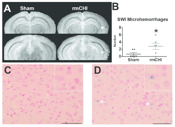
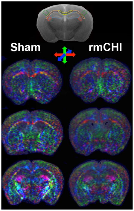
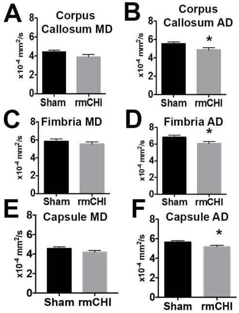
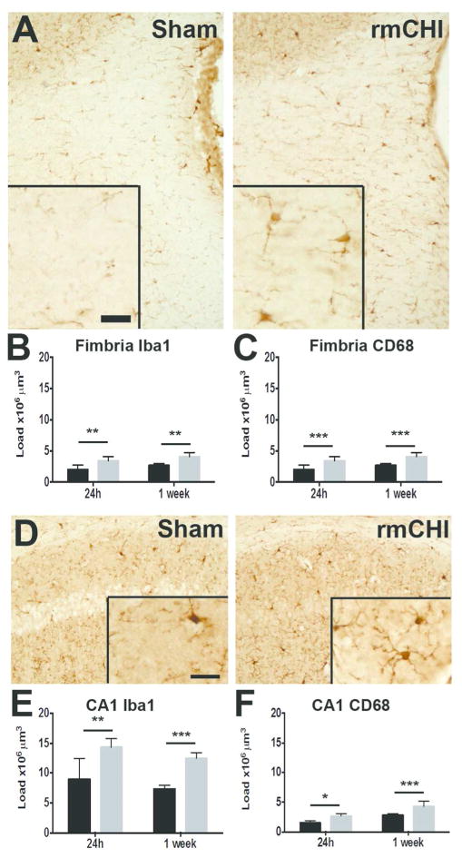
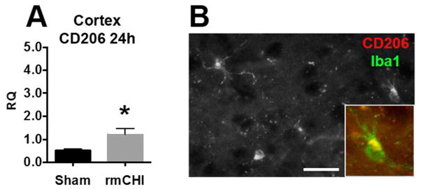
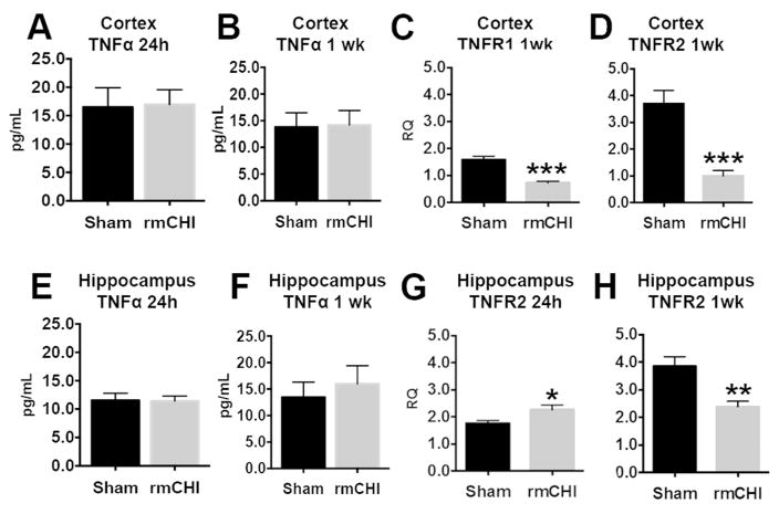
Traumatic brain injury (TBI) is a major public health issue, with recently increased awareness of the potential long-term sequelae of repetitive injury. Although TBI is common, objective diagnostic tools with sound neurobiological predictors of outcome are lacking. Indeed, such tools could help to identify those at risk for more severe outcomes after repetitive injury and improve understanding of biological underpinnings to provide important mechanistic insights. We tested the hypothesis that acute and subacute pathological injury, including the microgliosis that results from repeated mild closed head injury (rmCHI), is reflected in susceptibility-weighted magnetic resonance imaging and diffusion-tensor imaging microstructural abnormalities. Using a combination of high-resolution magnetic resonance imaging, stereology, and quantitative PCR, we studied the pathophysiology of male mice that sustained seven consecutive mild traumatic brain injuries over 9 days in acute (24 hr) and subacute (1 week) time periods. rmCHI induced focal cortical microhemorrhages and impaired axial diffusivity at 1 week postinjury. These microstructural abnormalities were associated with a significant increase in microglia. Notably, microgliosis was accompanied by a change in inflammatory microenvironment defined by robust spatiotemporal alterations in tumor necrosis factor-α receptor mRNA. Together these data contribute novel insight into the fundamental biological processes associated with repeated mild brain injury concomitant with subacute imaging abnormalities in a clinically relevant animal model of repeated mild TBI. These findings suggest new diagnostic techniques that can be used as biomarkers to guide the use of future protective or reparative interventions. © 2016 Wiley Periodicals, Inc.
Keywords: DTI; RRID:AB_2144905; RRID:AB_2314667; RRID:AB_323909; SWI; axial diffusion; inflammation; microhemorrhage.
© 2016 Wiley Periodicals, Inc.
Conflict of interest statement
Statement The authors declare no conflicts of interest related to this work.
Figures
Similar articles
-
Diffusion tensor imaging detects axonal injury in a mouse model of repetitive closed-skull traumatic brain injury.Neurosci Lett. 2012 Apr 4;513(2):160-5. doi: 10.1016/j.neulet.2012.02.024. Epub 2012 Feb 17. Neurosci Lett. 2012. PMID: 22343314 Free PMC article.
-
Microglial-derived microparticles mediate neuroinflammation after traumatic brain injury.J Neuroinflammation. 2017 Mar 15;14(1):47. doi: 10.1186/s12974-017-0819-4. J Neuroinflammation. 2017. PMID: 28292310 Free PMC article.
-
Imaging and serum biomarkers reflecting the functional efficacy of extended erythropoietin treatment in rats following infantile traumatic brain injury.J Neurosurg Pediatr. 2016 Jun;17(6):739-55. doi: 10.3171/2015.10.PEDS15554. Epub 2016 Feb 19. J Neurosurg Pediatr. 2016. PMID: 26894518 Free PMC article.
-
Traumatic brain injury: neuropathological, neurocognitive and neurobehavioral sequelae.Pituitary. 2019 Jun;22(3):270-282. doi: 10.1007/s11102-019-00957-9. Pituitary. 2019. PMID: 30929221 Review.
-
Diffusion tensor imaging and magnetic resonance spectroscopy in traumatic brain injury: a review of recent literature.Brain Imaging Behav. 2014 Dec;8(4):487-96. doi: 10.1007/s11682-013-9288-2. Brain Imaging Behav. 2014. PMID: 24449140 Review.
Cited by
-
Early onset senescence and cognitive impairment in a murine model of repeated mTBI.Acta Neuropathol Commun. 2021 May 8;9(1):82. doi: 10.1186/s40478-021-01190-x. Acta Neuropathol Commun. 2021. PMID: 33964983 Free PMC article.
-
Slow blood-to-brain transport underlies enduring barrier dysfunction in American football players.Brain. 2020 Jun 1;143(6):1826-1842. doi: 10.1093/brain/awaa140. Brain. 2020. PMID: 32464655 Free PMC article.
-
Neurological manifestations of encephalitic alphaviruses, traumatic brain injuries, and organophosphorus nerve agent exposure.Front Neurosci. 2024 Dec 13;18:1514940. doi: 10.3389/fnins.2024.1514940. eCollection 2024. Front Neurosci. 2024. PMID: 39734493 Free PMC article. Review.
-
The contribution of the meningeal immune interface to neuroinflammation in traumatic brain injury.J Neuroinflammation. 2024 May 27;21(1):135. doi: 10.1186/s12974-024-03122-7. J Neuroinflammation. 2024. PMID: 38802931 Free PMC article. Review.
-
Low Molecular Weight Dextran Sulfate (ILB®) Administration Restores Brain Energy Metabolism Following Severe Traumatic Brain Injury in the Rat.Antioxidants (Basel). 2020 Sep 10;9(9):850. doi: 10.3390/antiox9090850. Antioxidants (Basel). 2020. PMID: 32927770 Free PMC article.
References
-
- Benson RR, Gattu R, Sewick B, Kou Z, Zakariah N, Cavanaugh JM, Haacke EM. Detection of hemorrhagic and axonal pathology in mild traumatic brain injury using advanced MRI: implications for neurorehabilitation. NeuroRehabilitation. 2012;31(3):261–279. - PubMed
-
- Bruce ED, Konda S, Dean DD, Wang EW, Huang JH, Little DM. Neuroimaging and traumatic brain injury: State of the field and voids in translational knowledge. Mol Cell Neurosci. 2015;66(Pt B):103–113. - PubMed
-
- Budde MD, Janes L, Gold E, Turtzo LC, Frank JA. The contribution of gliosis to diffusion tensor anisotropy and tractography following traumatic brain injury: validation in the rat using Fourier analysis of stained tissue sections. Brain: a journal of neurology. 2011;134(Pt 8):2248–2260. - PMC - PubMed
Publication types
MeSH terms
Substances
Grants and funding
LinkOut - more resources
Full Text Sources
Other Literature Sources
Medical
