. 2016 Aug 9;7(2):236-48.
doi: 10.1016/j.stemcr.2016.06.010. Epub 2016 Jul 21.

MiR-34a Promotes Osteogenic Differentiation of Human Adipose-Derived Stem Cells via the RBP2/NOTCH1/CYCLIN D1 Coregulatory Network

Affiliations

MiR-34a Promotes Osteogenic Differentiation of Human Adipose-Derived Stem Cells via the RBP2/NOTCH1/CYCLIN D1 Coregulatory Network

Cong Fan et al. Stem Cell Reports. .

Abstract

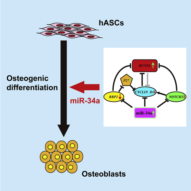
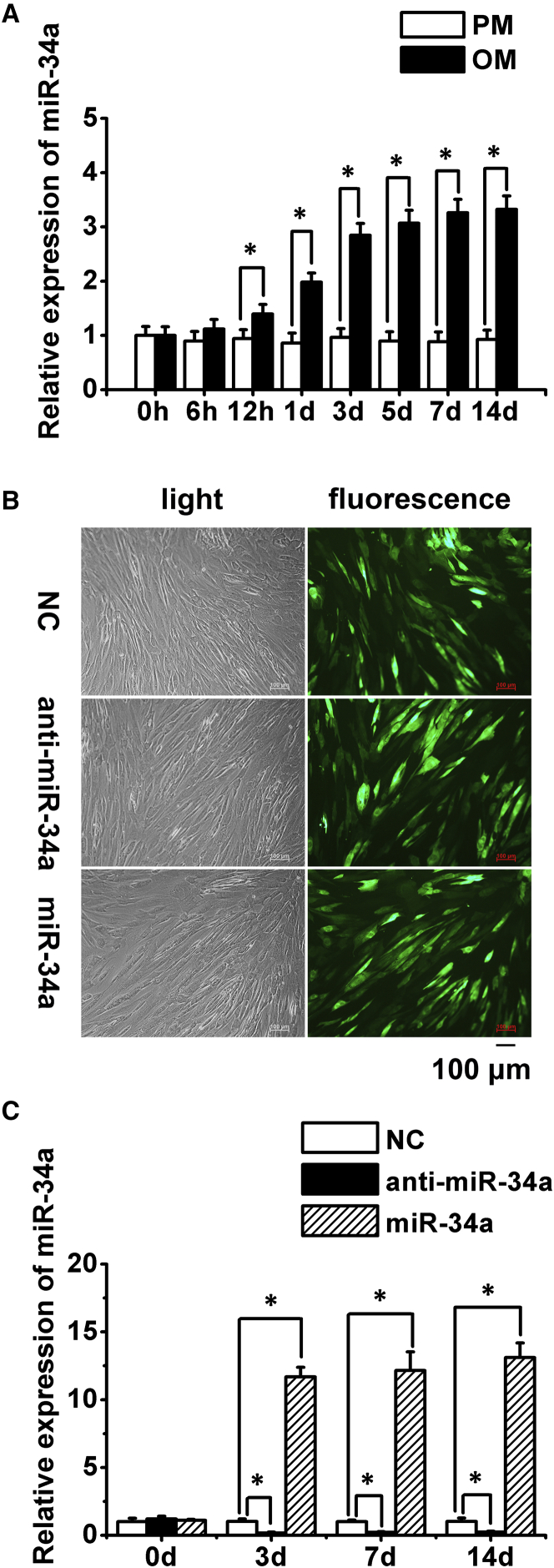
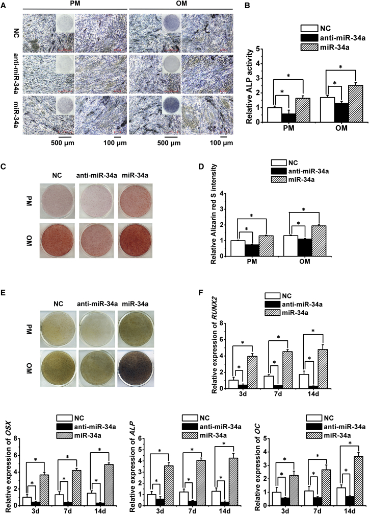
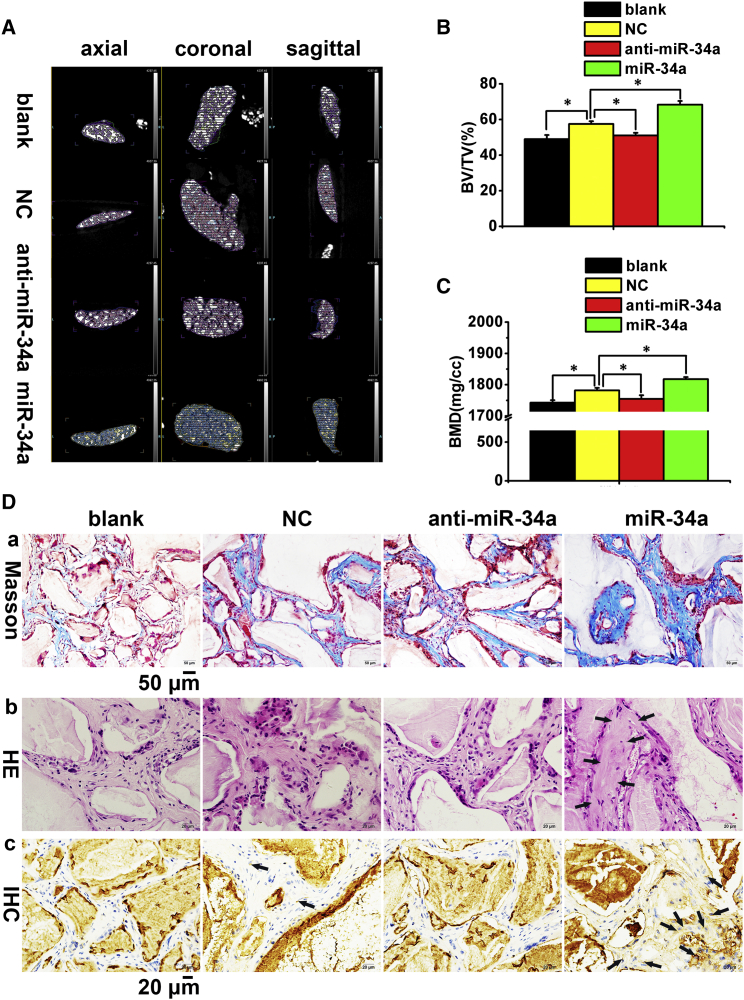
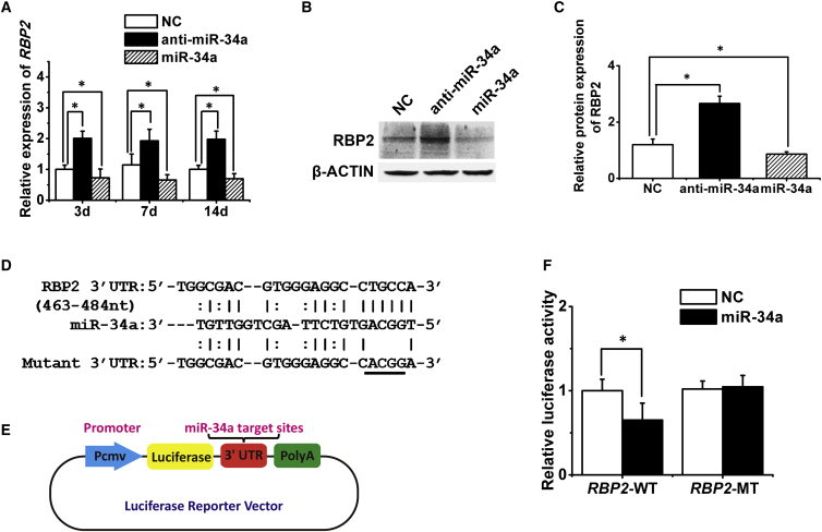
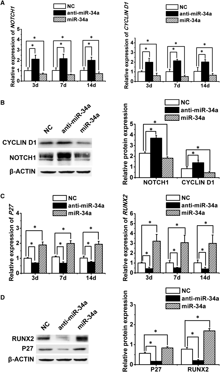
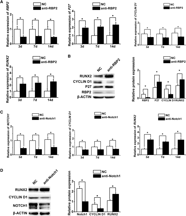
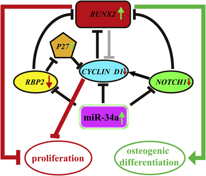
MiR-34a was demonstrated to be upregulated during the osteogenic differentiation of human adipose-derived stem cells (hASCs). Overexpression of miR-34a significantly increased alkaline phosphatase activity, mineralization capacity, and the expression of osteogenesis-associated genes in hASCs in vitro. Enhanced heterotopic bone formation in vivo was also observed upon overexpression of miR-34a in hASCs. Mechanistic investigations revealed that miR-34a inhibited the expression of retinoblastoma binding protein 2 (RBP2) and reduced the luciferase activity of reporter gene construct comprising putative miR-34a binding sites in the 3' UTR of RBP2. Moreover, miR-34a downregulated the expression of NOTCH1 and CYCLIN D1 and upregulated the expression of RUNX2 by targeting RBP2, NOTCH1, and CYCLIN D1. Taken together, our results suggested that miR-34a promotes the osteogenic differentiation of hASCs via the RBP2/NOTCH1/CYCLIN D1 coregulatory network, indicating that miR-34a-targeted therapy could be a valuable approach to promote bone regeneration.


Figures

None
Graphical abstract
Figure 1
Figure 1
Expression of Endogenous miR-34a during hASCs' Osteogenic Induction, and Determination of Lentiviral Transduction Efficiency and Effect (A) Quantitative real-time PCR analysis of miR-34a expression in hASCs cultured in PM and OM. (B) Microscopic images of GFP-positive hASCs under ordinary and fluorescent light. Scale bar, 100 μm. (C) Quantitative real-time PCR analysis of miR-34a in transduced hASCs cultured in PM. PM, proliferation medium; OM, osteogenic medium; NC, lentivirus negative control; anti-miR-34a, lentivirus anti-sense miR-34a; miR-34a, lentivirus miR-34a mimics. Data represent the means ± SD of three independent experiments. p < 0.05 versus the NC group.
Figure 2
Figure 2
Promotion of hASCs' Osteogenic Differentiation by miR-34a In Vitro (A and B) ALP staining (A) and quantification (B) of transduced hASCs. Scale bar of the left panel in PM or OM group, 500 μm; scale bar of the right panel in PM or OM group, 100 μm. (C and D) ARS staining (C) and quantification (D) of transduced hASCs. (E) von Kossa staining of transduced hASCs. (F) Quantitative real-time PCR analysis of RUNX2, OSX, ALP, and OC expression in transduced hASCs. ALP, alkaline phosphatase; ARS, Alizarin red S; RUNX2, runt-related transcription factor 2; OSX, osterix; OC, osteocalcin. Data represent the means ± SD of three independent experiments. p < 0.05 versus the NC group.
Figure 3
Figure 3
Promotion of hASCs' Osteogenic Differentiation by miR-34a In Vivo (A) Newly formed bone in Bio-Oss collagen scaffolds are indicated in different colors; scaffold remnants appear as white irregular lumps. (B and C) Quantitative analysis of BV/TV and BMD. Data represent the means ± SD of three independent experiments. p < 0.05 versus the NC group. (D) Histological assessment of ectopic bone formation. (a) Masson trichrome staining. The collagen in the bone matrix was stained blue-green. (b) H&E staining. New bone structures are indicated by black arrows. (c) IHC staining for OC. Dark-brown granules indicating positive staining are marked by black arrows. Scale bar, 50 μm in (a) and 20 μm in (b) and (c). BV/TV, percentage of new bone volume to tissue volume; BMD, bone mineral density; IHC, immunohistochemistry; blank, scaffolds without hASCs; NC/anti-miR-34a/miR-34a, scaffolds seeded with hASCs transfected by lentivirus negative control/anti-sense miR-34a/miR-34a mimics.
Figure 4
Figure 4
Validation of RBP2 as a Direct Target Gene of miR-34a (A–C) Quantitative real-time PCR (A) and western blotting (B and C) analysis of the effects of miR-34a on RBP2 expression. (D) Predicted binding sites of miR-34a in the 3′ UTR of RBP2-WT mRNA (underlined part indicates mutated base sequences in the 3′ UTR of RBP2-MT). (E) Schematic showing the constructed luciferase reporter system containing the binding sites of miR-34a. (F) Luciferase activity of cells with miR-34a overexpression in the RBP2-WT or RBP2-MT group. RBP2, retinoblastoma binding protein 2; RBP2-WT, wild-type RBP2 mRNA; RBP2-MT, mutant-type RBP2 mRNA. Data represent the means ± SD of three independent experiments. p < 0.05 versus the NC group.
Figure 5
Figure 5
miR-34a Repressed NOTCH1 and CYCLIN D1 Expression and Upregulated P27 and RUNX2 Expression (A and B) Quantitative real-time PCR (A) and western blotting (B) analysis of NOTCH1 and CYCLIN D1 with miR-34a knockdown or overexpression. (C and D) Quantitative real-time PCR (C) and western blotting (D) analysis of P27 and RUNX2 with miR-34a knockdown or overexpression. Data represent the means ± SD of three independent experiments. p < 0.05 versus the NC group.
Figure 6
Figure 6
Confirmation of the Relationships among RBP2, NOTCH1, P27, CYCLIN D1, and RUNX2 (A and B) Quantitative real-time PCR (A) and western blotting (B) analysis of RBP2, P27, CYCLIN D1, and RUNX2 expression after RBP2 knockdown. (C and D) Quantitative real-time PCR (C) and western blotting (D) analysis of NOTCH1, CYCLIN D1, and RUNX2 expression after NOTCH1 knockdown. Data represent the means ± SD of three independent experiments. p < 0.05 versus the NC group.
Figure 7
Figure 7
Schematic Representation of the RBP2/NOTCH1/CYCLIN D1 Coregulatory Network Involved in the Osteogenic Differentiation of hASCs by miR-34a MiR-34a directly targeted the RBP2, NOTCH1, and CYCLIN D1 transcripts, leading to the downregulation of these target genes and subsequent upregulation of P27 and RUNX2. The final repression of CYCLIN D1 and upregulation of RUNX2 mediated a switch from proliferation to osteogenic differentiation in hASCs.

References

    1. Ambros V. The functions of animal microRNAs. Nature. 2004;431:350–355. - PubMed
    1. Bae Y., Yang T., Zeng H.C., Campeau P.M., Chen Y., Bertin T., Dawson B.C., Munivez E., Tao J., Lee B.H. miRNA-34c regulates Notch signaling during bone development. Hum. Mol. Genet. 2012;21:2991–3000. - PMC - PubMed
    1. Bartel D.P. MicroRNAs: genomics, biogenesis, mechanism, and function. Cell. 2004;116:281–297. - PubMed
    1. Bosnakovski D., Mizuno M., Kim G., Takagi S., Okumura M., Fujinaga T. Isolation and multilineage differentiation of bovine bone marrow mesenchymal stem cells. Cell Tissue Res. 2005;319:243–253. - PubMed
    1. Chen X., Zhang T., Shi J., Xu P., Gu Z., Sandham A., Yang L., Ye Q. Notch1 signaling regulates the proliferation and self-renewal of human dental follicle cells by modulating the G1/S phase transition and telomerase activity. PLoS One. 2013;8:e69967. - PMC - PubMed

Publication types

MeSH terms