Clinical Trial
. 2016 Jul 28;166(3):609-623.
doi: 10.1016/j.cell.2016.06.043. Epub 2016 Jul 21.

Vaccine-Induced Antibodies that Neutralize Group 1 and Group 2 Influenza A Viruses

Affiliations
Clinical Trial

Vaccine-Induced Antibodies that Neutralize Group 1 and Group 2 Influenza A Viruses

M Gordon Joyce et al. Cell. .

Abstract

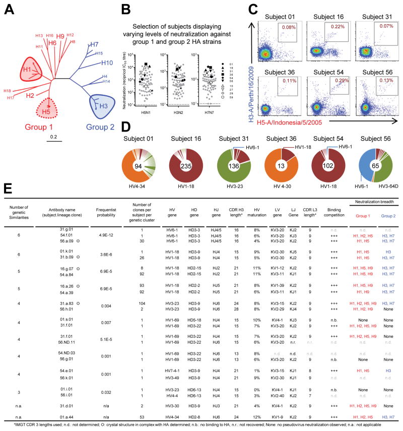
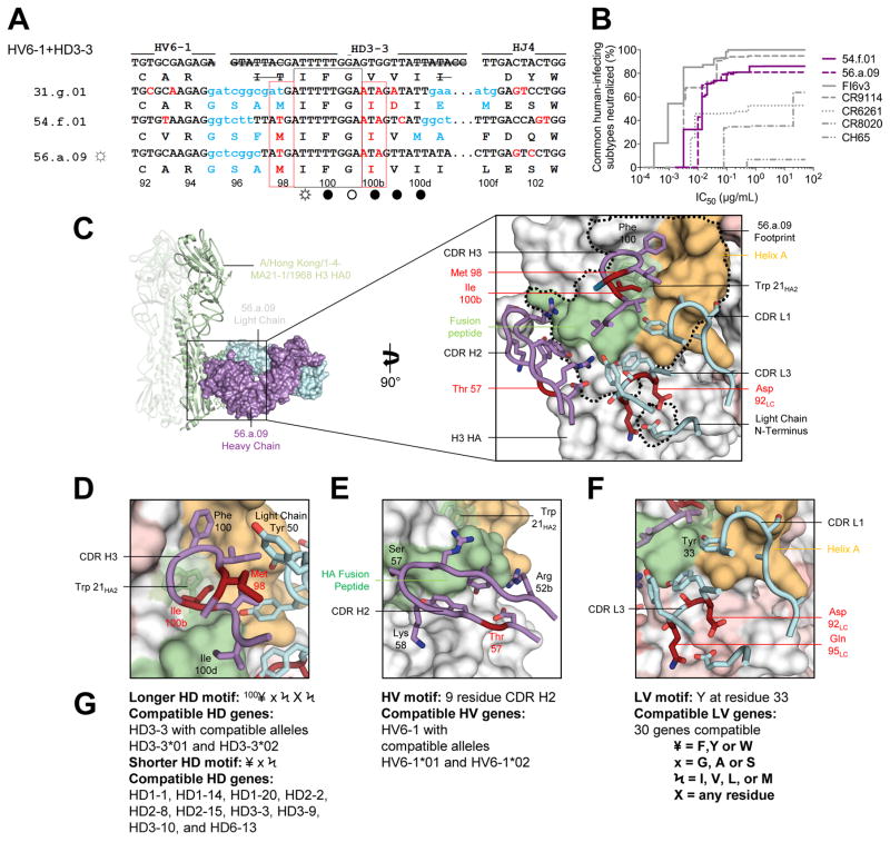
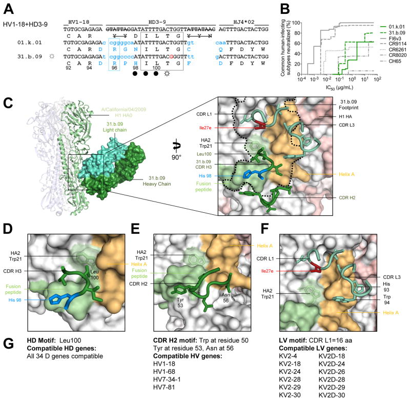
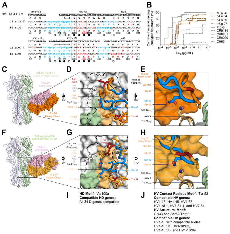
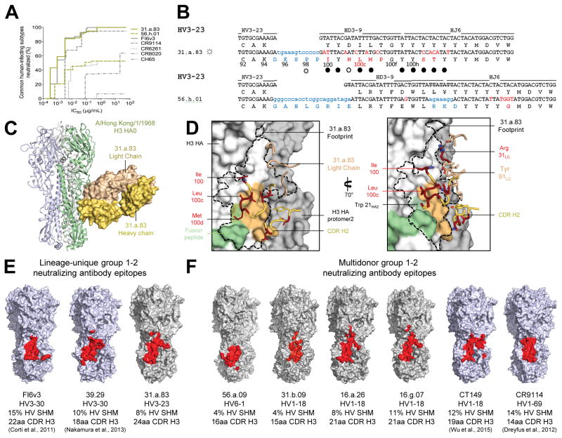
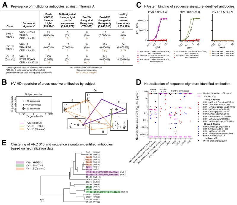
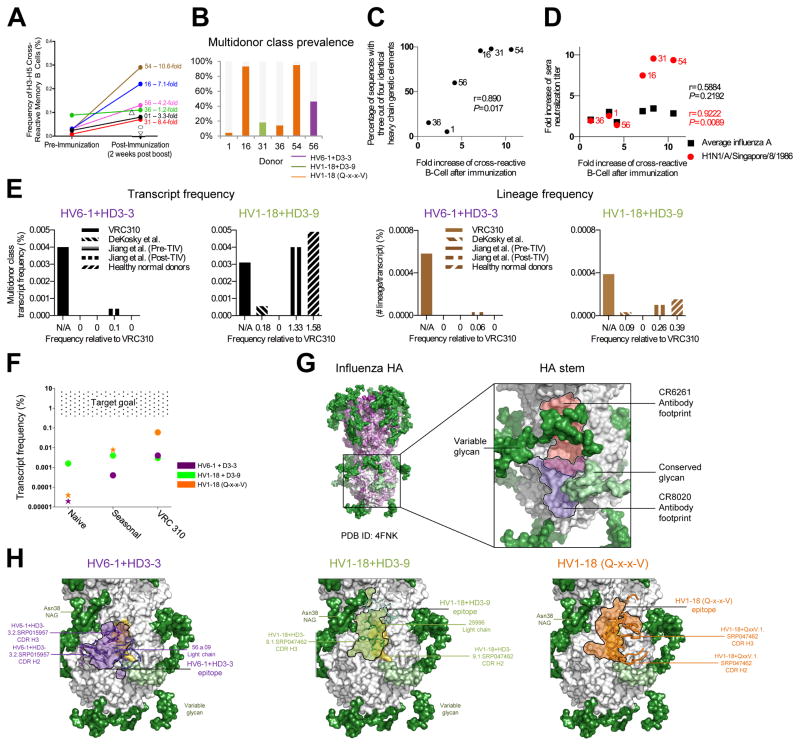
Antibodies capable of neutralizing divergent influenza A viruses could form the basis of a universal vaccine. Here, from subjects enrolled in an H5N1 DNA/MIV-prime-boost influenza vaccine trial, we sorted hemagglutinin cross-reactive memory B cells and identified three antibody classes, each capable of neutralizing diverse subtypes of group 1 and group 2 influenza A viruses. Co-crystal structures with hemagglutinin revealed that each class utilized characteristic germline genes and convergent sequence motifs to recognize overlapping epitopes in the hemagglutinin stem. All six analyzed subjects had sequences from at least one multidonor class, and-in half the subjects-multidonor-class sequences were recovered from >40% of cross-reactive B cells. By contrast, these multidonor-class sequences were rare in published antibody datasets. Vaccination with a divergent hemagglutinin can thus increase the frequency of B cells encoding broad influenza A-neutralizing antibodies. We propose the sequence signature-quantified prevalence of these B cells as a metric to guide universal influenza A immunization strategies.


Conflict of interest statement

COMPETING FINANCIAL INTERESTS

The authors declare no competing financial interests. Reprints permission information are available at www.cell.com.

Figures

Figure 1
Figure 1. H5N1-vaccine recipients have cross-reactive B cells that utilize the same genetic elements and neutralize group 1 and 2-influenza A viruses
(A) Phylogenetic tree depicting influenza A subtypes, generated using HA sequences (one per subtype) with program MEGA6. Scale bar indicates distance per fractional nucleotide change. (B) Neutralization by serum from 63 vaccinees, sampled two weeks after final H5N1 immunization and assessed against vaccine strain (A/Indonesia/5/2005) and heterologous group 2 HA strains (H3N2: A/Hong Kong/1-4-MA21-1/1968; H7N7: A/Netherlands/219/2003). Ten subjects were selected for flow cytometric (FACS) characterization, as highlighted in key. Dotted line indicates the limit of detection. (C) FACS analysis of PBMC samples isolated from H5N1-vaccine recipients two weeks after final vaccination and co-stained with HA probes H5 (A/Indonesia/5/2005) and H3 (A/Perth/16/2009). Sizable populations of H5-H3 cross-reactive memory B cells observed in six of ten subjects (Fig. S2). (D) Clonal diversity of H5-H3 cross-reactive B cells. The HV repertoire from each subject is shown as a pie chart; with each slice representing a unique HV clone or clonally related family. Total number of HV sequences recovered per subject is indicated by the number at the center of each pie chart. (E) Genetic and functional characteristics of selected antibodies recovered from H5-H3 cross-reactive B cells. Structurally characterized antibodies indicated by ☼. See also Figures S1–4 and Tables S1–S4.
Figure 2
Figure 2. A multidonor HV6-1+HD3-3 class of broadly neutralizing antibodies
(A) Immunoglobulin heavy chains utilizing germline genes HV6-1, HD3-3 and HJ4 or HJ5. Germline HV, HD, and HJ gene-encoded nucleotide and amino-acid residues are shown in black, with junction-encoded residues in light blue and residues that have undergone SHM in red. Nucleotides removed by exonuclease trimming indicated with a line through the letters. Conserved HD3-3 encoded residues (IFG) highlighted by a black box; recurrent SHM-derived Ile100bHC highlighted by a red box. HA contacts indicated with open circles (○) denoting antibody main-chain-only contacts, open circles with rays (☼) denoting antibody side-chain-only contacts, and filled circles (●) denoting both main-chain and side-chain contacts. (B) Neutralization breadth-potency curve for HV6-1+HD 3-3 antibodies, with breadth shown as percentage of pseudoviruses neutralized at each IC50 cutoff, and virus panel comprising 15 strains that includes influenza A subtypes known to infect humans (H1, H2, H3, H5, H7, H9). (C) Co-crystal structure of Fab 56.a.09 in complex with an H3 HA monomer (A/Hong Kong/1-4-MA21-1/1968). Fab heavy and light chains colored purple and cyan respectively and depicted in surface representation, while the H3 HA is depicted in ribbon and shown as a trimer (Figure S5 shows HA crystal packing). Inset: Interacting CDR loops of the 56.a.09 Fab are shown in ribbon and sticks and colored as in panel (A) with the antibody footprint outlined. (D) A five-amino acid motif within the CDR H3 inserts into the conserved Trp21 pocket of H3. (E) HV6-1encoded CDR H2 depicted with HA fusion peptide; HV6-1 is the only HV gene which encodes a nine residue CDR H2. (F) Light chain interactions that contribute to the antibody-binding surface; these are not specific to KV3-20. (G) Analysis of antibody gene compatibility, highlighting additional CDR H3 residues that may be compatible with HA binding, e.g. 100HC could be a F, Y, or W residue (¥), 101HC could be a G, A, or S residue (x). See also Figures S3–S7 and Tables S3–S7.
Figure 3
Figure 3. A multidonor HV1-18+HD3-9 class of broadly neutralizing antibodies
(A) Immunoglobulin heavy chains utilizing germline genes HV1-18, HD3-9 and HJ4, with sequences annotated as described in Figure 2A. (B) Neutralization breadth-potency curve for HV1-18+HD 3-9 antibodies on a panel of influenza A viruses that includes subtypes known to infect humans. (C) Co-crystal structure of Fab 31.b.09 in complex with an H1 trimer (A/California/04/2009). Fab heavy and light chains colored dark green and light green respectively and depicted in surface representation, while the H1 HA is depicted in ribbon and colored blue, green, and white. Inset: Interacting CDR loops of the 31.b.09 Fab are shown in ribbon and sticks and colored as in panel (A) with the antibody footprint outlined. (D) A conserved motif within the CDR H3 inserts into the highly conserved Trp21 pocket of HA while also interacting with the fusion peptide. (E) The HV1-18 encoded CDR H2 also interacts with the opposing side of the fusion peptide. (F) Light chain interactions from CDR L3 and CDR L1 also contribute to the antibody binding surface area. (G) Analysis of antibody gene compatibility. See also Figures S3–S7 and Tables S3–S7.
Figure 4
Figure 4. A multidonor HV1-18 class of broadly neutralizing antibodies with Q-x-x-V motif
(A) Immunoglobulin heavy chains utilizing germline genes HV1-18, HD2-2 or HD2-15 and HJ5 or HJ2, with sequences annotated as described in Figure 2A. (B) Neutralization breadth-potency curve for HV1-18 (Q-x-x-V) class antibodies on a panel of influenza A viruses that includes subtypes known to infect humans. (C) Co-crystal structure of Fab 16.a.26 (HV1-18, HD2-2, HJ5) in complex with H3-HK68. Fab heavy and light chains colored orange and lavender respectively and depicted in surface representation while HA is depicted in ribbon and shown as a trimer. (D) The 16.a.26 CDR H3 is depicted with junction-encoded and mutated residues colored as in panel (A) and germline-encoded residues in orange with the antibody footprint on the HA outlined in black. (E) Antibody heavy chain depicted in surface representation with CDR H3 loop in ribbon. 16.a.26 Gln98HC occupies a turn in the CDR H3, which interacts with the conserved residue Gln42HA2. (F) Co-crystal structure of Fab 16.g.07 (HV1-18, HD2-15, and HJ2) in complex with H3-HK68 depicted as in panel (C). (G) The 16.g.07 CDR H3 depicted as in panel (D) with the antibody footprint on the HA outlined in black. (H) The 16.g.07 CDR H3 is depicted as in panel (E) with Gln98HC contacting Gln42HA2. (I) Analysis of D-gene compatibility. (J) Analysis of V-gene compatibility. See also Figures S3–S7 and Tables S3–S7.
Figure 5
Figure 5. A conserved site of group 1 and 2-influenza A virus vulnerability targeted by multidonor and lineage-unique antibodies
(A) Neutralization breadth-potency curve for HV3-23-derived antibodies, on a panel of influenza A viruses that includes subtypes known to infect humans. (B) Immunoglobulin heavy chains utilizing germline genes HV3-23, HD3-9 and HJ6, with sequences annotated as described in Figure 2A. (C) Co-crystal structure of Fab 31.a.83 in complex with HA (H3-HK68). (D) Epitope for antibody 31.a.83 (outlined in black) with heavy chain depicted in yellow and junction-encoded residues (highlighted in blue), mutated residues (in red), and germline-encoded residues (in gold) and with light chain depicted in tan. (E–F) Antibodies capable of neutralizing group 1 and 2 influenza A viruses recognize overlapping epitopes within the HA stem (colored red). The HA surface is colored blue for structures determined previously (FI6v3 PDB ID:3ZTN; 39.29 PDB ID:4KVN; CT149 PDB ID:4R8W; CR9114 PDB ID:4FQI), and gray for structures determined in the current study. See also Figures S3–S6 and Tables S3–S7.
Figure 6
Figure 6. Sequence signatures of multidonor antibody lineages
(A) Multidonor class antibodies capable of neutralizing group 1 and group 2-influenza A viruses: sequence signature, number, and frequency of signature-identified class members. The number of singleton transcripts from 454-derived NGS data is reported in italicized font; these were omitted from transcript and lineage quantifications as described in Supplementary Methods. (B) HV-HD germline-gene origin plots. One box shown per subject with antibody frequencies plotted as circles at their HV (horizontal), HD (vertical) coordinates. The size of the plotted circle corresponds to the number of antibody sequences as shown in key at left. Multidonor antibodies in different subjects connected by lines colored according to each multidonor class. (C) Binding and competition MSD-ECLIA for antibodies identified in (DeKosky et al., 2015; Jiang et al., 2013) databases; competition with stem antibody F10 highlighted in red. (D) Neutralization of influenza pseudoviruses. Median IC50 for each antibody is indicated by a horizontal line with value (μg/ml) shown at the base of the graph. (E) Clustering of antibodies based on their neutralization fingerprints on a 15-virus panel. See also Figures S6 and Tables S3–S4, S7.
Figure 7
Figure 7. Vaccine induction of antibodies capable of neutralizing group 1 and 2 influenza A viruses
(A) Frequencies of H5-H3 cross-reactive memory B cells pre- and post-H5N1 vaccination. Subjects for whom pre-immunization samples were no longer available are indicated with open symbols; subject name and fold increase shown for others. (B) Frequency of multidonor class sequences by donor and multidonor class. (C) Fold-increase in cross-reactive B cells relative to prevalence of heavy chain sequences with three (out of a possible four) of the same heavy chain genetic elements in at least one sequence in any of the six analyzed subjects (Pearson correlation with the total number of sequences provided in Figure 1D). (D) Fold-increase in cross-reactive B cells relative to the fold-increase in sera neutralization titer for all tested influenza A strains (shown in black) (see Figure 1, Figure S1), or the single H1N1-A/Singapore/8/1986 strain (shown in red). (E) Bar graphs of transcript frequencies (left) and lineage frequencies (right). Left: frequency of multidonor-class transcripts by dataset; Right: frequency of multidonor class lineages for each dataset. (F) Transcript frequency versus dataset and goal. Stars depict upper-bound estimates, and circles depict frequencies confirmed by neutralization. (G) Multidonor antibodies displayed in ribbon with class-conserved contact residues shown in stick. Antibody epitopes shown in purple (HV6-1+HD3-3), green (HV1-18+HD3-9), and orange (HV1-18 with Q-x-x-V) with black outlines. Glycans shown in surface representation and colored by conservation: conserved (light green) or variable (dark green). See also Figures S3, S6 and Tables S3–S4.

Comment in

References

    1. Adderson EE, Shackelford PG, Quinn A, Wilson PM, Cunningham MW, Insel RA, Carroll WL. Restricted immunoglobulin VH usage and VDJ combinations in the human response to Haemophilus influenzae type b capsular polysaccharide. Nucleotide sequences of monospecific anti-Haemophilus antibodies and polyspecific antibodies cross-reacting with self antigens. The Journal of clinical investigation. 1993;91:2734–2743. - PMC - PubMed
    1. Boyd SD, Marshall EL, Merker JD, Maniar JM, Zhang LN, Sahaf B, Jones CD, Simen BB, Hanczaruk B, Nguyen KD, et al. Measurement and clinical monitoring of human lymphocyte clonality by massively parallel VDJ pyrosequencing. Science translational medicine. 2009;1:12ra23. - PMC - PubMed
    1. Butt KM, Smith GJ, Chen H, Zhang LJ, Leung YH, Xu KM, Lim W, Webster RG, Yuen KY, Peiris JS, et al. Human infection with an avian H9N2 influenza A virus in Hong Kong in 2003. Journal of clinical microbiology. 2005;43:5760–5767. - PMC - PubMed
    1. Corti D, Voss J, Gamblin SJ, Codoni G, Macagno A, Jarrossay D, Vachieri SG, Pinna D, Minola A, Vanzetta F, et al. A neutralizing antibody selected from plasma cells that binds to group 1 and group 2 influenza A hemagglutinins. Science. 2011;333:850–856. - PubMed
    1. DeKosky BJ, Kojima T, Rodin A, Charab W, Ippolito GC, Ellington AD, Georgiou G. In-depth determination and analysis of the human paired heavy- and light-chain antibody repertoire. Nature medicine. 2015;21:86–91. - PubMed

Publication types

MeSH terms