. 2014 Oct 5;4(19):e1254.
doi: 10.21769/bioprotoc.1254.

Hypoxia Studies with Pimonidazole in vivo

Affiliations

Hypoxia Studies with Pimonidazole in vivo

Kristina Y Aguilera et al. Bio Protoc. .

Abstract

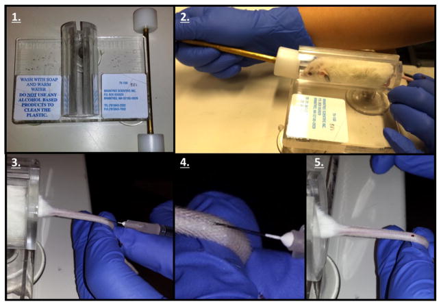
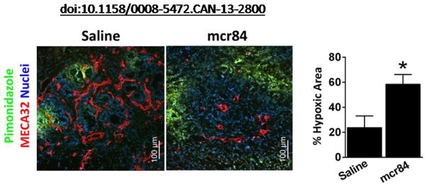
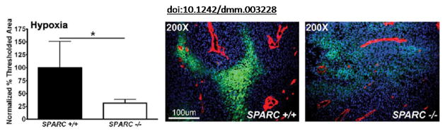
Therapy-induced hypoxia drives changes in the tumor microenvironment that contribute to the poor response to therapy. Hypoxia is capable of driving the expression and/or activation of specific signaling cascades (e.g., c-Met, Axl, CTGF), the recruitment of tumor promoting immune cells, and the induction of cell survival pathways including autophagy (Phan et al., 2013; Hu et al., 2012; Ye et al., 2010). We have recently shown that anti-VEGF therapy-induced hypoxia can result in changes in the extracellular matrix that contribute to the aggressiveness of tumors post therapy (Aguilera et al., 2014). Importantly, therapies that induce hypoxia do not always increase epithelial plasticity and tumor aggressiveness (Ostapoff et al., 2013; Cenik et al., 2013). We have used pimonidazole to evaluate hypoxia in tumors and herein provide a detailed protocol for this useful tool to interrogate the levels of hypoxia in vivo. The utility of the Hypoxyprobe (pimonidazole hydrochloride) immunohistochemical analysis approach allows for the assessment of hypoxia in different tissues as well as cell types. Pimonidazole is a 2-nitroimidazole that is reductively activated specifically in hypoxic cells and forms stable adducts with thiol groups in proteins, peptides, and amino acids (Cenik et al., 2013; Arnold et al., 2010; Raleigh and Koch, 1990; Raleigh et al., 1998). Furthermore, the amount of pimonidazole that is detected is directly proportional to the level of hypoxia within tumors.


Figures

Figure 1
Figure 1. Tail Vein Injection protocol
1. Obtain a Tailveiner Restrainer. 2. Insert mouse and push the restrainer to set the mouse in position. 3. Rotate the tail 45 degrees in order to find and inject the tail vein with the needle. 4. Close representation of the tail vein injection. 5. The appearance of blood validates a successful tail vein injection.
Figure 2
Figure 2. Representative data [taken from Aguilera et al. (2014)]
6-week-old KIC mice were treated with saline or mcr84 for 1 week. Animals were sacrificed 1 h after pimonidazole injection (i.v.). Tissue was stained for endothelial cells (MECA32, red) and pimonidazole adducts (Hypoxyprobe, green). Hypoxic area was quantified and is presented on the right. Error bars: *P<0.05, one-way ANOVA with Tukey multiple comparison test. Pancreata from 6 animals were sectioned per treatment group, an average intensity value was determined for each organ and the intensities were averaged and the SEM was calculated. Please view this reference for more information on the model and context of the representative data.
Figure 3
Figure 3. Representative data [taken from Aguilera et al. (2014)]
The amount of hypoxia within tumors was quantified using an antibody, FITC-conjugated mouse anti-pimonidazole, directed against an adduct that forms when Hypoxyprobe-1 enters hypoxic tissue. The percentage of threshold area was quantified and two separate experiments were combined by normalization to data from Sparc+/+ mice. Images display hypoxia (green) near the vasculature (red) stained with the rat antimouse endothelial cell (Meca-32) antibody within tumors. Nuclei are marked with DAPI (blue). Error bars: *P<0.05, student’s t-test. Please view this reference for more information on the model and context of the representative data.

Similar articles

Cited by

References

    1. Aguilera KY, Rivera LB, Hur H, Carbon JG, Toombs JE, Goldstein CD, Dellinger MT, Castrillon DH, Brekken RA. Collagen signaling enhances tumor progression after anti-VEGF therapy in a murine model of pancreatic ductal adenocarcinoma. Cancer Res. 2014;74(4):1032–1044. - PMC - PubMed
    1. Arnold SA, Rivera LB, Miller AF, Carbon JG, Dineen SP, Xie Y, Castrillon DH, Sage EH, Puolakkainen P, Bradshaw AD, Brekken RA. Lack of host SPARC enhances vascular function and tumor spread in an orthotopic murine model of pancreatic carcinoma. Dis Model Mech. 2010;3(1–2):57–72. - PMC - PubMed
    1. Cenik BK, Ostapoff KT, Gerber DE, Brekken RA. BIBF 1120 (nintedanib), a triple angiokinase inhibitor, induces hypoxia but not EMT and blocks progression of preclinical models of lung and pancreatic cancer. Mol Cancer Ther. 2013;12(6):992–1001. - PMC - PubMed
    1. Graves EE, Vilalta M, Cecic IK, Erler JT, Tran PT, Felsher D, Sayles L, Sweet-Cordero A, Le QT, Giaccia AJ. Hypoxia in models of lung cancer: implications for targeted therapeutics. Clin Cancer Res. 2010;16(19):4843–4852. - PMC - PubMed
    1. Hu YL, Jahangiri A, DeLay M, Aghi MK. Tumor cell autophagy as an adaptive response mediating resistance to treatments such as antiangiogenic therapy. Cancer Res. 2012;72(17):4294–4299. - PMC - PubMed