. 2016 Jul 25;11(7):e0159692.
doi: 10.1371/journal.pone.0159692. eCollection 2016.

Transcription Factors Efg1 and Bcr1 Regulate Biofilm Formation and Virulence during Candida albicans-Associated Denture Stomatitis

Affiliations

Transcription Factors Efg1 and Bcr1 Regulate Biofilm Formation and Virulence during Candida albicans-Associated Denture Stomatitis

Junko Yano et al. PLoS One. .

Abstract

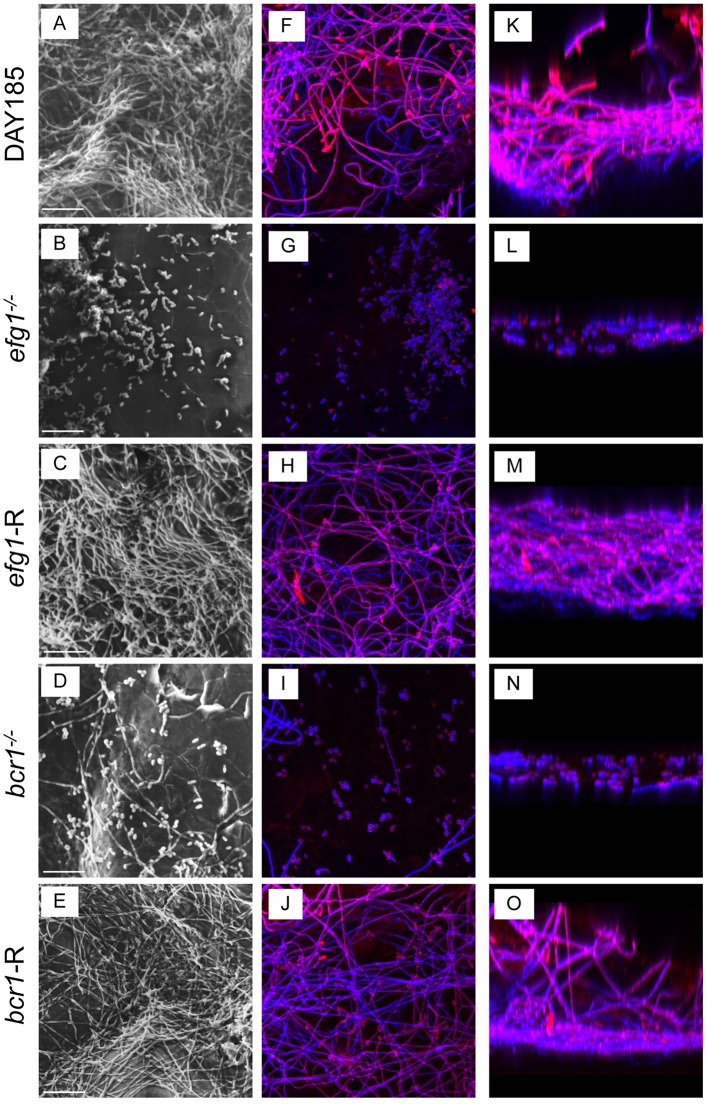
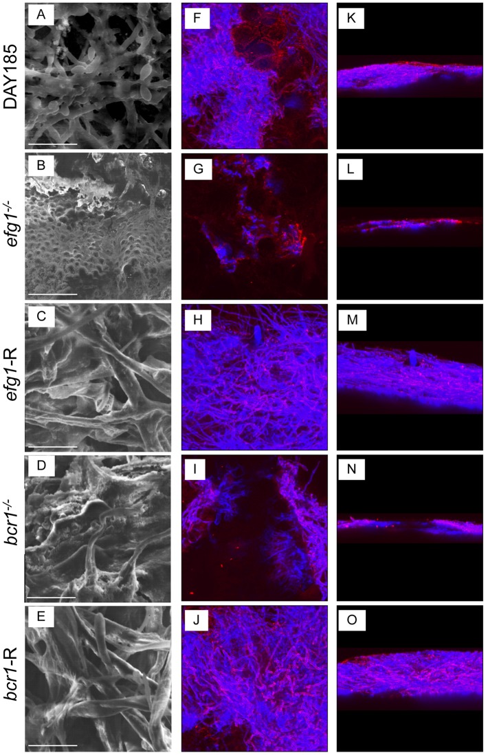
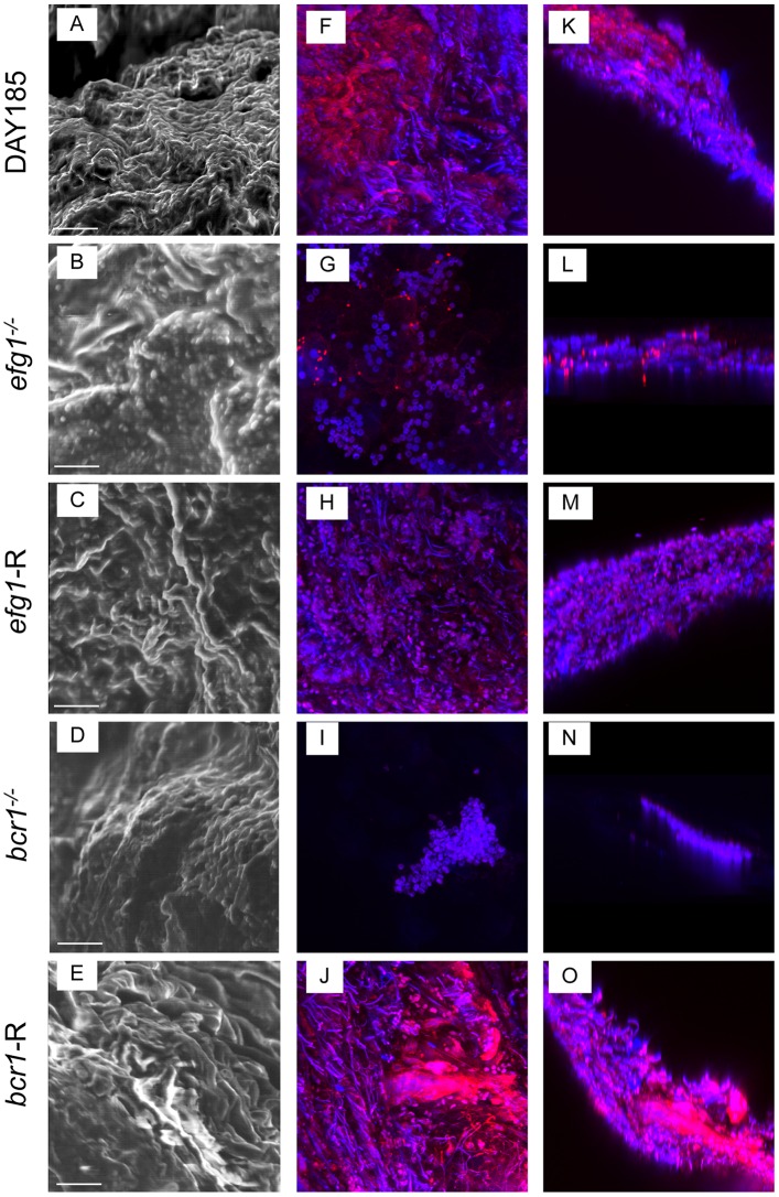
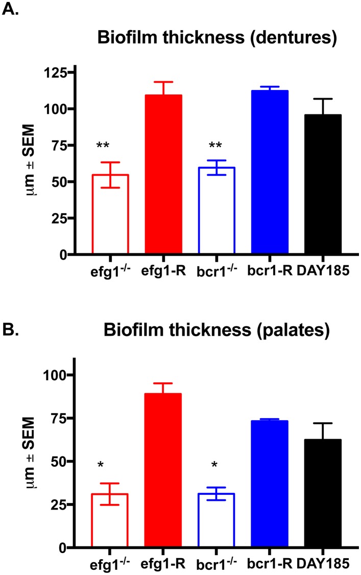
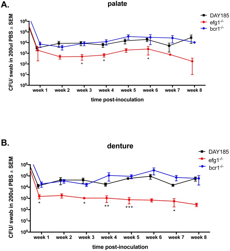
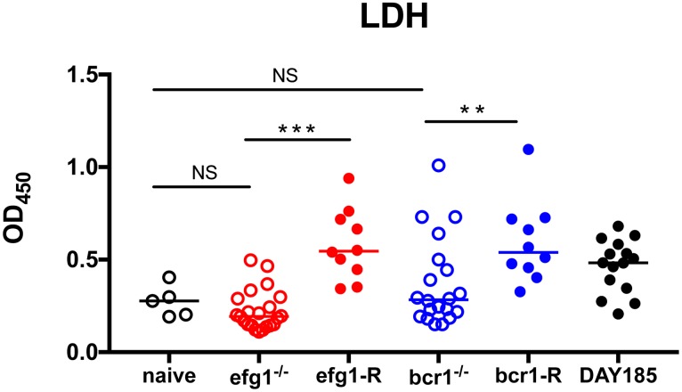
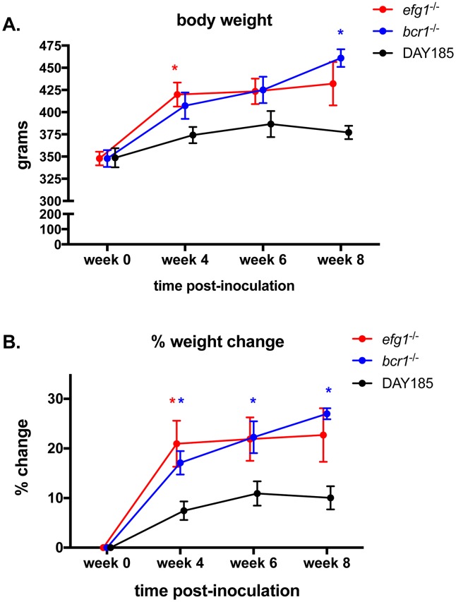
Denture stomatitis (DS) is characterized by inflammation of the oral mucosa in direct contact with dentures and affects a significant number of otherwise healthy denture wearers. The disease is caused by Candida albicans, which readily colonizes and form biofilms on denture materials. While evidence for biofilms on abiotic and biotic surfaces initiating Candida infections is accumulating, a role for biofilms in DS remains unclear. Using an established model of DS in immunocompetent animals, the purpose of this study was to determine the role of biofilm formation in mucosal damage during pathogenesis using C. albicans or mutants defective in morphogenesis (efg1-/-) or biofilm formation (bcr1-/-). For in vivo analyses, rats fitted with custom dentures, consisting of fixed and removable parts, were inoculated with wild-type C. albicans, mutants or reconstituted strains and monitored weekly for fungal burden (denture and palate), body weight and tissue damage (LDH) for up to 8 weeks. C. albicans wild-type and reconstituted mutants formed biofilms on dentures and palatal tissues under in vitro, ex vivo and in vivo conditions as indicated by microscopy demonstrating robust biofilm architecture and extracellular matrix (ECM). In contrast, both efg1-/- and bcr1-/- mutants exhibited poor biofilm growth with little to no ECM. In addition, quantification of fungal burden showed reduced colonization throughout the infection period on dentures and palates of rats inoculated with efg1-/-, but not bcr1-/-, compared to controls. Finally, rats inoculated with efg1-/- and bcr1-/- mutants had minimal palatal tissue damage/weight loss while those inoculated with wild-type or reconstituted mutants showed evidence of tissue damage and exhibited stunted weight gain. These data suggest that biofilm formation is associated with tissue damage during DS and that Efg1 and Bcr1, both central regulators of virulence in C. albicans, have pivotal roles in pathogenesis of DS.


Conflict of interest statement

Competing Interests: The authors have declared that no competing interests exist.

Figures

Fig 1
Fig 1. In vitro biofilm formation on denture materials in human saliva.
Pre-sterilized denture material was inoculated with 1 x 10⁶ C. albicans DAY185 (A, F, K), efg1-/- (B, G, L), efg1-reconstituted (C, H, M), bcr1-/- (D, I, N) or bcr1-reconstituted (E, J, O) strain and incubated in saliva for 24h at 37°C to allow biofilm growth. The denture materials were then processed for SEM (A-E) or stained with calcofluor white (blue; stains fungal chitin in the cell wall) or Concanavalin A-Texas Red conjugate (red; stains mannose in the cell wall and ECM) and examined by fluorescent confocal microscopy to visualize biofilms in XY (F-J) and XYZ (K-O) views. Each panel shows a representative image of 3 repeats. Scale bar = 50 μm.
Fig 2
Fig 2. Ex vivo biofilm formation on rat palate tissue.
Excised rat palate tissues were inoculated with 1 x 10⁶ C. albicans DAY185 (A, F, K), efg1-/- (B, G, L), efg1-reconstituted (C, H, M), bcr1-/- (D, I, N) or bcr1-reconstituted (E, J, O) strain and incubated in PBS for 24h at 37°C to allow biofilm growth. The tissues were then processed for SEM (A-E) or stained with calcofluor white (blue; stains fungal chitin in the cell wall) or Concanavalin A-Texas Red conjugate (red; stains mannose in the cell wall and ECM) and examined by fluorescent confocal microscopy to visualize biofilms in XY (F-J) and XYZ (K-O) views. Each panel shows a representative image of 2 repeats. Scale bar = 50 μm.
Fig 3
Fig 3. The role of C. albicans EFG1 and BCR1 in biofilm formation on dentures in vivo.
Equilibrated rats (n = 4/group) were weaned onto gel diet and fitted with dentures. Rats were given broad-spectrum antibiotics in the drinking water for 4 days prior to inoculation. Rats were inoculated 3x at 3-day intervals with 1x10⁹ CFU C. albicans DAY185 (A, F, K), efg1-/- (B, G, L), efg1-reconstituted (C, H, M), bcr1-/- (D, I, N) or bcr1-reconstituted (E, J, O) strain. Dentures were removed from inoculated rats at 4 weeks post-inoculation. The dentures were processed for SEM (A-E) or stained with calcofluor white (blue; stains fungal chitin in the cell wall) or Concanavalin A-Texas Red conjugate (red; stains mannose in the cell wall and ECM) and examined by fluorescent confocal microscopy to visualize biofilms in XY (F-J) and XYZ (K-O) views. Each panel shows a representative image of 2–3 animals. Scale bar = 50 μm.
Fig 4
Fig 4. The role of C. albicans EFG1 and BCR1 in biofilm formation on palate tissue in vivo.
Equilibrated rats (n = 4/group) were weaned onto gel diet and fitted with dentures. Rats were given broad-spectrum antibiotics in the drinking water for 4 days prior to inoculation. Rats were inoculated 3x at 3-day intervals with 1x10⁹ CFU C. albicans DAY185 (A, F, K), efg1-/- (B, G, L), efg1-reconstituted (C, H, M), bcr1-/- (D, I, N) or bcr1-reconstituted (E, J, O) strain. Palate tissues were excised from inoculated rats at 4 weeks post-inoculation. The tissue samples were then processed for SEM (A-E) or stained with calcofluor white (blue; C. albicans) or Concanavalin A-Texas Red conjugate (red; ECM) and examined by fluorescent confocal microscopy to visualize biofilms in XY (F-J) and XYZ (K-O) views. Each panel shows a representative image of 2–3 animals. Scale bar = 50 μm.
Fig 5
Fig 5. Quantification of biofilm thickness on dentures and palate tissue in vivo.
Equilibrated rats were weaned onto gel diet and fitted with dentures. Rats were given broad-spectrum antibiotics in the drinking water for 4 days prior to inoculation. Rats were inoculated 3x at 3-day intervals with 1 x 10⁹ C. albicans DAY185, efg1-/- or bcr1-/- strain. (A) Dentures and (B) palate tissues were removed from inoculated rats at 4 weeks post-inoculation. Samples were stained with calcofluor white (blue; stains fungal chitin in the cell wall) or Concanavalin A-Texas Red conjugate (red; stains mannose in the cell wall and ECM) and examined by fluorescent confocal microscopy to visualize biofilms. Cross-sectional images of biofilms were visualized by confocal microscopy at 600X magnification, and the depths of biofilms were measured using the Fluoview software. Figure represents cumulative results from 2 independent experiments with 2–3 animals per group and assessment of 5 random areas per animal. Data were analyzed using a one-way ANOVA followed by the Tukey’s post hoc multiple comparison test. *, P < 0.05; **, P < 0.01 compared to the WT control.
Fig 6
Fig 6. Fungal colonization levels on dentures and palate tissue in rats inoculated with C. albicans biofilm deficient strains.
Equilibrated rats were weaned onto gel diet and fitted with dentures. Rats were given broad-spectrum antibiotics in the drinking water for 4 days prior to inoculation. Rats were inoculated 3x at 3-day intervals with 1 x 10⁹ C. albicans DAY185, efg1-/- or bcr1-/- strain. Swab samples of both the palate (A) and denture (B) were taken weekly for a period of 8 weeks post-inoculation. Fungal burdens were assessed from swab suspension fluid. Figure represents cumulative results from 2 independent experiments with 4–5 animals per group. Data were analyzed using Repeated Measures ANOVA (longitudinal data for each group) and the unpaired Student’s t test (individual time points (weeks), experimental vs. control). *, P < 0.05; **, P < 0.01; ***, P < 0.001.
Fig 7
Fig 7. Mucosal damage in rats fitted with dentures inoculated with C. albicans biofilm deficient strains.
Equilibrated rats were fitted with dentures, weaned onto gel diet, given antibiotics in the drinking water, and inoculated 3 times at 3 day intervals with 1 x 10⁹ C. albicans DAY185, efg1-/- or bcr1-/- or reconstituted mutants. Swab samples of the palate were taken weekly for a period of 4 weeks post-inoculation. LDH levels were assessed from swab suspension fluid. The results represent cumulative data from 2 independent experiments at 1–4 weeks post-inoculation with 4–5 animals per group. Data were analyzed using the Kruskal-Wallis test followed by the post hoc Mann-Whitney U test. n.s., not significant; **, P < 0.01; ***, P < 0.001.
Fig 8
Fig 8. Body weight change over time in rats fitted with dentures inoculated with C. albicans biofilm deficient strains.
Equilibrated rats were fitted with dentures, weaned onto gel diet, given antibiotics in the drinking water, and inoculated 3x with 1 x 10⁹ C. albicans DAY185, efg1-/- or bcr1-/-. Rats were weighed bi-weekly for a period of 8 weeks post-inoculation using 3–4 animals per group and data are shown as (A) absolute weights and (B) % weight change (% weight change = weight at time point/weight at week 0 prior to inoculation). Data for individual weight changes per group were analyzed using the Repeated Measures ANOVA. Weight changes between groups at specific time points (experimental vs. control, actual weight or % change) were analyzed by the unpaired Student’s t test. * P < 0.05.

Similar articles

Cited by

References

    1. Budtz-Jorgensen E, Stenderup A, Grabowski M. An epidemiologic study of yeasts in elderly denture wearers. Community Dent Oral Epidemiol. 1975;3(3):115–9. Epub 1975/05/01. . - PubMed
    1. Arendorf TM, Walker DM. Denture stomatitis: a review. J Oral Rehabil. 1987;14(3):217–27. Epub 1987/05/01. . - PubMed
    1. Cumming CG, Wight C, Blackwell CL, Wray D. Denture stomatitis in the elderly. Oral Microbiol Immunol. 1990;5(2):82–5. Epub 1990/04/01. . - PubMed
    1. Budtz-Jorgensen E. Oral mucosal lesions associated with the wearing of removable dentures. J Oral Pathol. 1981;10(2):65–80. Epub 1981/04/01. . - PubMed
    1. Pires FR, Santos EB, Bonan PR, De Almeida OP, Lopes MA. Denture stomatitis and salivary Candida in Brazilian edentulous patients. J Oral Rehabil. 2002;29(11):1115–9. Epub 2002/11/28. 947 [pii]. . - PubMed