. 2016 Aug 1;126(8):3130-44.
doi: 10.1172/JCI83092. Epub 2016 Jul 25.

Human CAR T cells with cell-intrinsic PD-1 checkpoint blockade resist tumor-mediated inhibition

Human CAR T cells with cell-intrinsic PD-1 checkpoint blockade resist tumor-mediated inhibition

Leonid Cherkassky et al. J Clin Invest. .

Abstract

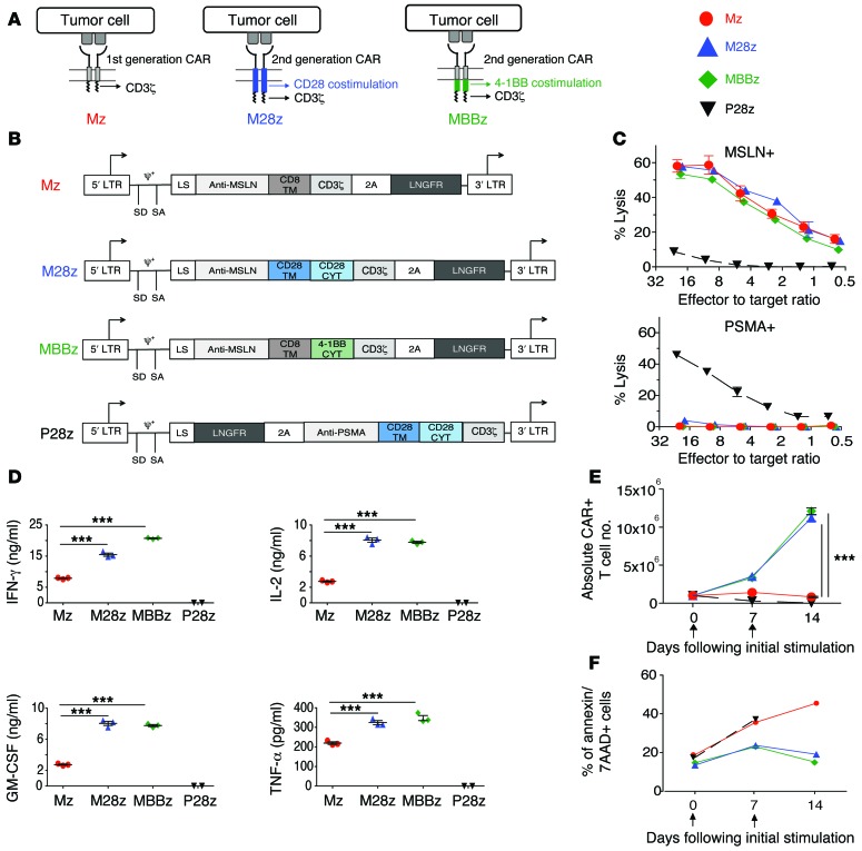
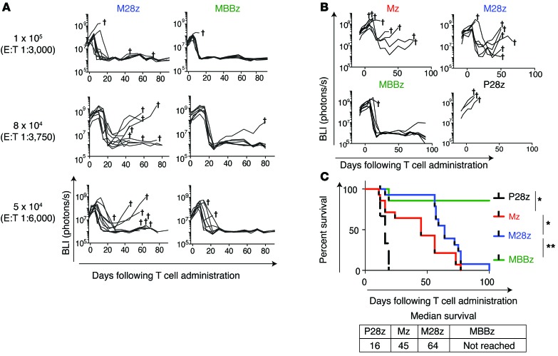
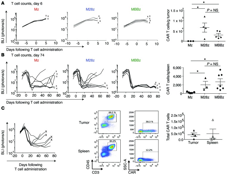
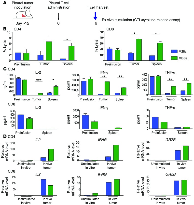
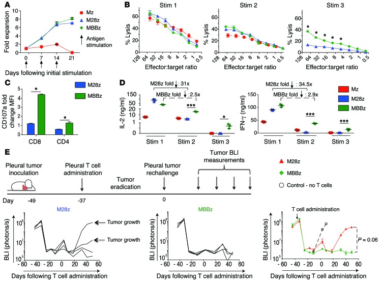
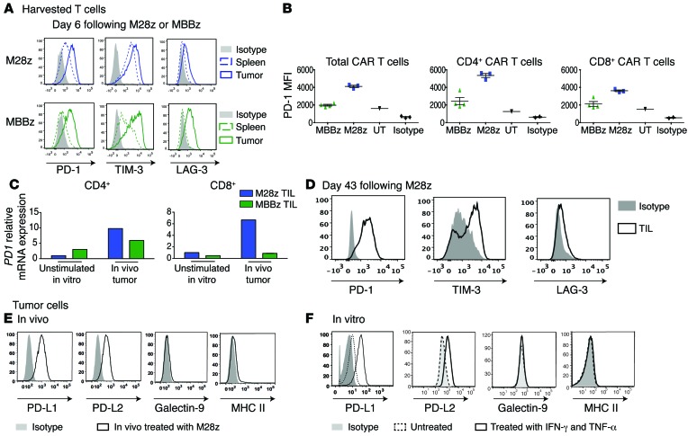
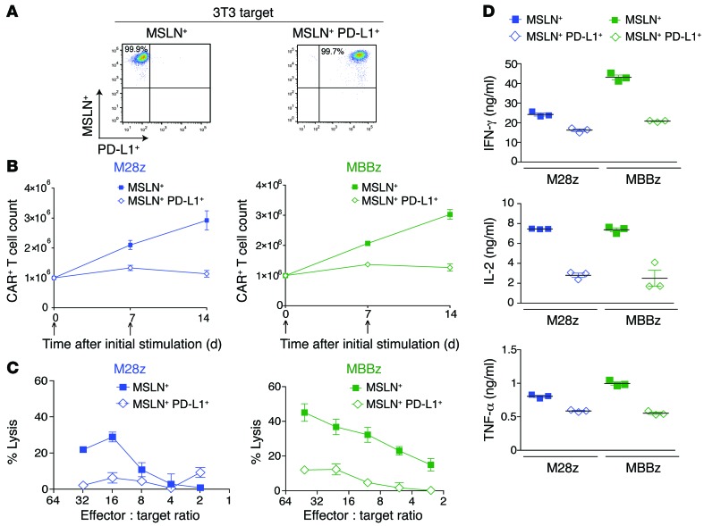
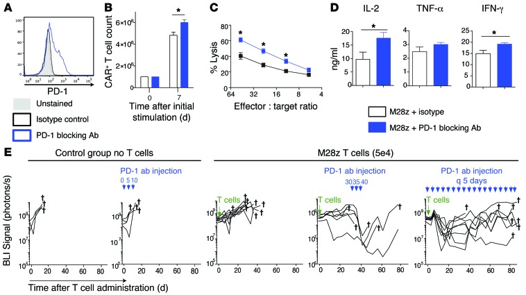
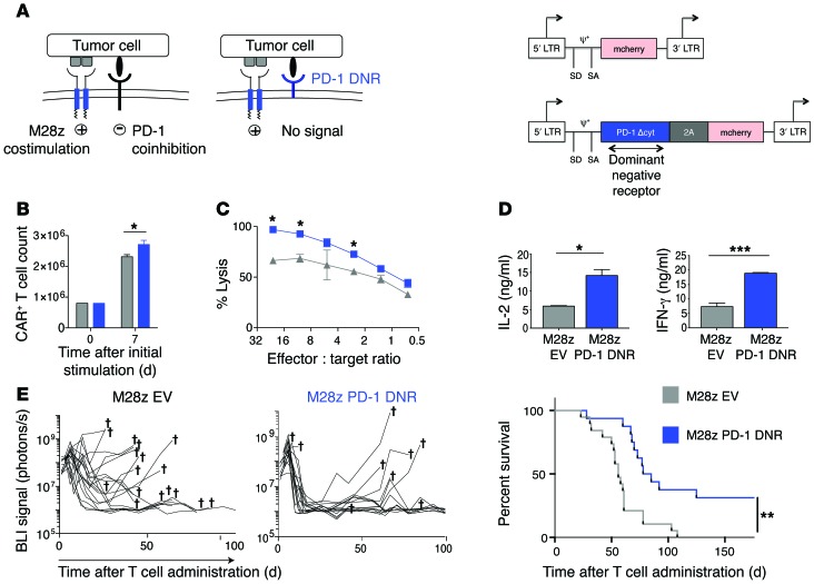
Following immune attack, solid tumors upregulate coinhibitory ligands that bind to inhibitory receptors on T cells. This adaptive resistance compromises the efficacy of chimeric antigen receptor (CAR) T cell therapies, which redirect T cells to solid tumors. Here, we investigated whether programmed death-1-mediated (PD-1-mediated) T cell exhaustion affects mesothelin-targeted CAR T cells and explored cell-intrinsic strategies to overcome inhibition of CAR T cells. Using an orthotopic mouse model of pleural mesothelioma, we determined that relatively high doses of both CD28- and 4-1BB-based second-generation CAR T cells achieved tumor eradication. CAR-mediated CD28 and 4-1BB costimulation resulted in similar levels of T cell persistence in animals treated with low T cell doses; however, PD-1 upregulation within the tumor microenvironment inhibited T cell function. At lower doses, 4-1BB CAR T cells retained their cytotoxic and cytokine secretion functions longer than CD28 CAR T cells. The prolonged function of 4-1BB CAR T cells correlated with improved survival. PD-1/PD-1 ligand [PD-L1] pathway interference, through PD-1 antibody checkpoint blockade, cell-intrinsic PD-1 shRNA blockade, or a PD-1 dominant negative receptor, restored the effector function of CD28 CAR T cells. These findings provide mechanistic insights into human CAR T cell exhaustion in solid tumors and suggest that PD-1/PD-L1 blockade may be an effective strategy for improving the potency of CAR T cell therapies.


Figures

Figure 1
Figure 1. CARs with CD28 or 4-1BB costimulation exhibit similar cytolytic functions, effector cytokine secretion, and proliferation in vitro upon initial antigen stimulation.
(A) First- and second-generation CARs. (B) MSLN–targeted CARs contain the CD3ζ endodomain either alone (Mz, first-generation CAR) or in combination with the CD28 (M28z) or 4-1BB (MBBz) costimulatory domain (second-generation CAR). PSMA-directed CARs with CD28 costimulation (P28z) as well as PSMA-expressing targets (PSMA+) are included in experiments as negative controls. CYT, cytoplasmic domain; LS, leader sequence; LTR, long terminal repeat; SA, splice acceptor; SD, splice donor; TM, transmembrane. (CE) Antigen-specific effector functions of CAR-transduced T cells. (C) Lysis of MSLN-expressing targets (MSLN+), but not PSMA+ targets, as measured by chromium-release assays. (D) 4-1BB and CD28 costimulations enhance cytokine secretion, as assessed by Luminex assay, after coculture of CAR T cells with MSLN+ cells. (E) M28z and MBBz CARs facilitate robust T cell accumulation after stimulation with MSLN+ cells. (F) 4-1BB and CD28 costimulations decrease the rate of apoptosis as assessed by annexin V/7-AAD+ staining every 7 days after coculture with MSLN+ target. (D and E) ***P < 0.001, comparing costimulated CAR T cells (M28z or MBBz) with the first-generation receptor (Mz), by Student’s t test; significance was determined using the Sidak-Bonferonni correction for multiple comparisons. Data are representative of at least 3 independent experiments and represent the mean ± SEM (C and E) of 3 replicates or are plotted as individual points.
Figure 2
Figure 2. Mice treated with M28z and MBBz CAR T cells demonstrate tumor eradication at a higher dose, whereas treatment with lower doses results in higher rate of tumor relapse with M28z.
(A) In vivo BLI was used to monitor tumor burden (ffLuc+MSLN+) in NOD/SCID/γcnull mice. Mice with established pleural tumor were treated with a single dose of 1 × 10⁵ (E:T 1:3,000), 8 × 10⁴ (E:T1:3,750), or 5 × 10⁴ (E:T 1:6,000) M28z or MBBz CAR T cells. Daggers indicate the deaths of mice. For A and B, 2 similar experiments with the same donor are combined for the illustration. n = 7–9 mice for each group treated with MSLN-targeted CAR T cells. (B) Mice were treated with 4 × 10⁴ CAR T cells (E:T 1:7,500). The first generation Mz CAR and negative control P28z are included. (C) Kaplan-Meier survival analysis comparing the in vivo efficacy of intrapleural administration of 4 × 10⁴ Mz (n = 13, red), M28z (n = 15, blue), MBBz (n = 8, green), and P28z (n = 3, black) CAR T cells. Two independent experiments performed under similar conditions were combined. Median survival in days following T cell administration. The survival curve was analyzed using the log-rank test. *P < 0.05; **P < 0.01. All data are representative of multiple experiments performed with multiple donors.
Figure 3
Figure 3. M28z- and MBBz-treated mice demonstrate similar early and long-term CAR T cell accumulation, and M28z-treated mice with progressing tumors contain persisting CAR T cells.
(A) CD28 and 4-1BB costimulation enhance intratumoral CAR T cell accumulation to equal extents. The left panels show the results of tumor BLI after administration of a single dose of 8 × 10⁴ CAR T cells. After 6 days, T cells were harvested from the tumor; x’s denote mice whose T cell counts are represented as data points. The right panel shows absolute CAR T cells per gram of tumor tissue. *P < 0.05. Student’s t tests were performed, and statistical significance was determined using the Sidak-Bonferonni correction for multiple comparisons. (B) CD28 and 4-1BB costimulation enhance CAR T cell persistence, as measured in the spleen, to equal extents. Absolute CAR T cells per spleen are shown 74 days after intrapleural administration of CAR T cells (8 × 10⁴). The left panels show the results of tumor BLI; x’s denote mice whose T cell counts are represented as data points. *P < 0.05. Student’s t tests were performed, and statistical significance was determined using the Sidak-Bonferonni correction for multiple comparisons. (C) Mice treated with a low dose of M28z T cells (4 × 10⁴) display tumor recurrence with persisting CAR T cells in the spleen and tumor. The left panel shows the results of tumor BLI. Spleen and tumor from mice denoted by an x were harvested and used for FACS analysis (middle panel) and T cell quantification (right panel). Data are representative of multiple tested mice of at least 3 independent experiments.
Figure 4
Figure 4. CAR T cells become exhausted following in vivo antigen exposure, although MBBz CAR T cells preferentially retain effector cytokine secretion and cytotoxicity.
(A) Six days after intrapleural administration of CAR T cells, M28z and MBBz CAR T cells were isolated from the tumor and spleen and subjected to ex vivo antigen stimulation. (B) Chromium-release assay upon ex vivo stimulation demonstrates a decrease in M28z but persistent MBBz cytolytic function (E:T ratio, 5:1). (C) Cytokine secretion measurements demonstrate decreases in effector cytokine secretion by CAR T cells, although MBBz CAR T cells are better able to retain secretion. (D) RT-PCR measurements of GRZB, IFNG, and IL2 expression by harvested CAR T cells correlate well with protein level measurements in panels A and B. Data represent the fold change relative to the mRNA expression of unstimulated M28z CAR T cell in vitro. Student’s t tests were performed, and statistical significance was determined using the Sidak-Bonferonni correction for multiple comparisons. *P < 0.05; **P < 0.01; ***P < 0.001. Data represent the mean ± SEM of 3 individual wells per condition. Results are reproduced in 2 separate cohorts of mice used for each of the 2 experiments.
Figure 5
Figure 5. CAR T cells become exhausted upon repeated antigen stimulation in vitro, although MBBz CAR T cells preferentially retain effector cytokine secretion and cytotoxicity in vitro and upon tumor rechallenge in vivo.
(A) Both M28z and MBBz CAR T cells retain proliferative capacity in vitro upon repeated antigen stimulation. T cells were also tested for cytotoxicity by chromium-release assay and for cytokine secretion by Luminex assay (BD). (B) (Left) CAR T cells demonstrate equal killing at the first stimulation and loss of cytolytic function upon repeated antigen stimulation, although MBBz CAR T cells are better able to retain cytolytic function as measured by chromium-release assay. (C) Cytotoxic granule release as measured by CD107a expression correlates with chromium-release assay (B). Data represent the mean ± SD (triplicates) of the fold-change relative to the CD107a mean fluorescence intensity (MFI) of unstimulated CD8+ CAR T cells. (D) Cytokine secretion measurements similarly demonstrate loss of CAR T cell effector function upon repeated antigen encounter; again, MBBz CAR T cells are better able to preserve their function. (E) Persisting MBBz CAR T cells demonstrate superior efficacy in vivo and eradicate MSLN+ tumor cells following tumor rechallenge. Twenty-eight days after pleural tumor eradication (following a single dose of 1 × 10⁵ CAR T cells), 1 × 10⁶ MSLN+ tumor cells were injected into the pleural cavity (tumor rechallenge). Control (white circle) represents mice without any previous injections of tumor or T cells. MBBz CAR T cells prevented tumor growth in all mice, whereas tumor growth and death were observed in 2 of 4 mice initially treated with M28z CAR T cells. Student’s t tests were performed, and statistical significance was determined using the Sidak-Bonferonni correction. *P < 0.05; ***P < 0.001. Data represent the mean ± SEM of 3 replicates or are plotted as individual points and are representative of at least 3 independent experiments.
Figure 6
Figure 6. PD1 receptor and its ligands are upregulated in vivo.
(A) Tumor-infiltrating M28z and MBBz CAR T cells overexpressed inhibitory receptors 6 days after their administration. (B) MBBz CAR T cells express lower levels of PD-1 compared with M28z CAR T cells as shown by MFI of PD1 receptor expression of tumor-infiltrating CAR T cells (TIL) 6 days after intrapleural administration. Unstransduced tumor-infiltrating T cells (UT) express a low baseline level of PD1. (C) Relative expression of PD1 mRNA in CD4 and CD8 subsets of tumor-infiltrating CAR T cells 6 days after intrapleural administration. Data are represented as fold change relative to the PD1 mRNA expression of unstimulated M28z CAR+ T cells. (D) Tumor-infiltrating M28z CAR T cells isolated from progressing tumors express inhibitory receptors PD1, TIM-3, and LAG-3. (E) Single-cell tumor suspensions harvested from mice treated with M28z CAR T cells express high levels of PD-1–binding ligands. (F) In vitro–cultured mesothelioma tumor cells express the ligands (PD-L1, PD-L2) for the PD1 receptor, and expression is further upregulated following incubation for 24 hours with IFN-γ and TNF-α. Data are representative of at least 2 to 3 independent experiments.
Figure 7
Figure 7. PD-L1 inhibits CAR T cell effector function.
(A) 3T3 fibroblasts were transduced to either express MSLN alone (MSLN+, left) or coexpress MSLN in addition to PD-L1 (MSLN+ PD-L1+, right). (BD) M28z and MBBz CAR T cell effector functions were assessed after stimulation with 3T3 MSLN+ or MSLN+ PD-L1+ targets. PD-L1 inhibits M28z and MBBz CAR T cell accumulation upon repeated antigen stimulation (B), cytolytic function following 2 stimulations with MSLN+ PD-L1+ tumor cells (C), and Th1 effector cytokine secretion upon the first stimulation (D). Data represent the mean ± SEM of 3 replicates or are plotted as individual points and are representative of at least 2 independent experiments.
Figure 8
Figure 8. PD-1–blocking antibody restores the effector function of exhausted M28z CAR T cells in vitro and in vivo.
(A) M28z CAR T cells were stained with the PD-1–blocking antibody used for functional assays. For in vitro experiments (BD), M28z CAR T cells were stimulated with MSLN+ tumor cells that had been treated with IFN-γ and TNF-α to upregulate PD-1 ligands. M28z CAR T cells treated with PD-1–blocking antibody (10 μg/ml) demonstrated a small enhancement in accumulation (B), an increase in cytolytic function upon the third stimulation (C), and enhanced effector cytokine secretion (D). Student’s t tests were performed for statistical significance. Data represent the mean ± SEM of triplicates and are representative of 2–3 independent experiments. (E) Injection of PD-1–blocking antibody rescues M28z CAR T cells in vivo as shown by tumor BLI in a model treating established high tumor burdens with a single low dose of intrapleurally administered M28z CAR T cells. PD-1–blocking antibody (10 mg/kg) was injected intraperitoneally 3 times on days 30, 35, and 40 or continuously injected every 5 days from day 0 to day 85 following T cell injection. n = 6–9 mice per group. *P < 0.05.
Figure 9
Figure 9. Cotransduction of a PD-1 DNR rescues M28z CAR T cells from PD-1 ligand–mediated inhibition in vitro and in vivo.
(A) (Left) Schematic representations of CD28-costimulated T cells binding tumor ligand via the endogenous PD-1 receptor (transmitting a coinhibitory signal) or a cotransduced PD-1 DNR lacking an inhibitory signaling domain. (Right) For in vitro and in vivo experiments, M28z CAR T cells were cotransduced with either EV (SFG-mCherry) or PD-1 DNR (SFG-2A-PD-1 DNR). CAR T cells sorted for mCherry expression were then incubated for 24 hours with MSLN+ tumor cells that had been treated with IFN-γ and TNF-α to upregulate PD-1 ligands. M28z PD-1 DNR CAR T cells demonstrated a small but statistically significant enhancement in accumulation (B), an enhanced cytolytic function, as measured by chromium-release assay upon the third stimulation with MSLN+ PD-L1+ tumor cells (C), and an increased expression of Th1 cytokine secretion (D). Student’s t tests were performed, and statistical significance was determined using the Sidak-Bonferonni correction for multiple comparisons. *P < 0.05; **P < 0.01; ***P < 0.001. Data represent the mean ± SEM of triplicates and are representative of at least 3 independent experiments. (E) Tumor BLI (left) and Kaplan-Meier survival analysis (right) comparing the in vivo efficacy of a single dose of 5 × 10⁴ M28z EV (n = 19; grey) or M28z PD-1 DNR (n = 16; blue) pleurally administered. Data shown are a combination of 2 independent experiments. Daggers indicate deaths. Median survival is shown in days following T cell administration. The survival curve was analyzed using the log-rank test (P = 0.001). The log-rank test for each independent experiment was significant at the P < 0.05 level; 2 experiments are combined for illustration.

Comment in

References

    1. Sadelain M, Rivière I, Brentjens R. Targeting tumours with genetically enhanced T lymphocytes. Nat Rev Cancer. 2003;3(1):35–45. doi: 10.1038/nrc971. - DOI - PubMed
    1. Sadelain M, Brentjens R, Rivière I. The basic principles of chimeric antigen receptor design. Cancer Discov. 2013;3(4):388–398. doi: 10.1158/2159-8290.CD-12-0548. - DOI - PMC - PubMed
    1. Brentjens RJ, et al. CD19-targeted T cells rapidly induce molecular remissions in adults with chemotherapy-refractory acute lymphoblastic leukemia. Sci Transl Med. 2013;5(177): - PMC - PubMed
    1. Brentjens RJ, et al. Safety and persistence of adoptively transferred autologous CD19-targeted T cells in patients with relapsed or chemotherapy refractory B-cell leukemias. Blood. 2011;118(18):4817–4828. doi: 10.1182/blood-2011-04-348540. - DOI - PMC - PubMed
    1. Davila ML, et al. Efficacy and toxicity management of 19-28z CAR T cell therapy in B cell acute lymphoblastic leukemia. Sci Transl Med. 2014;6(224): doi: 10.1126/scitranslmed.3008226. - DOI - PMC - PubMed