Review
. 2016 Jul 23;14(8):139.
doi: 10.3390/md14080139.

The Role of Spongia sp. in the Discovery of Marine Lead Compounds

Affiliations
Review

The Role of Spongia sp. in the Discovery of Marine Lead Compounds

Patrícia Máximo et al. Mar Drugs. .

Abstract

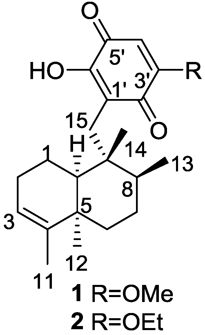
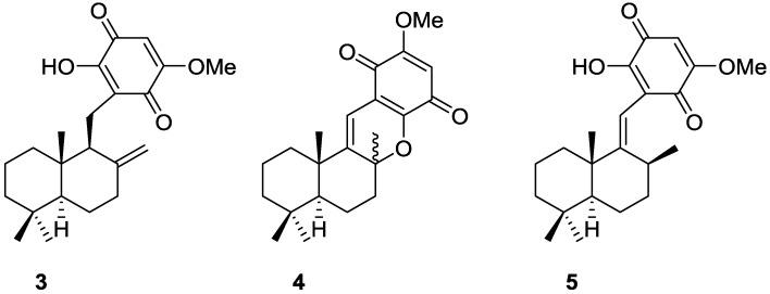
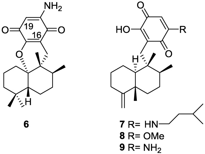
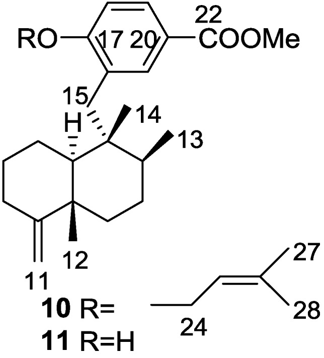
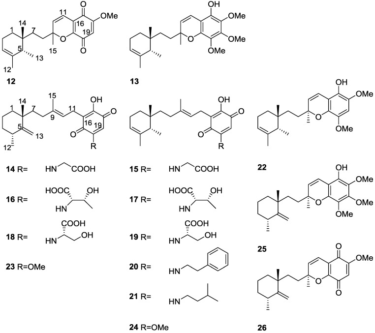
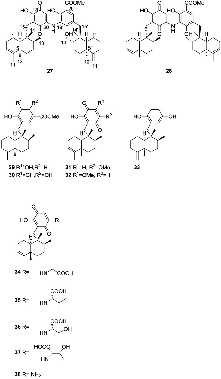
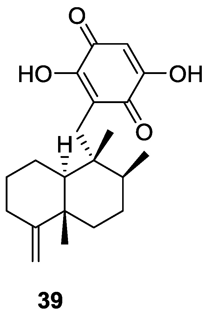
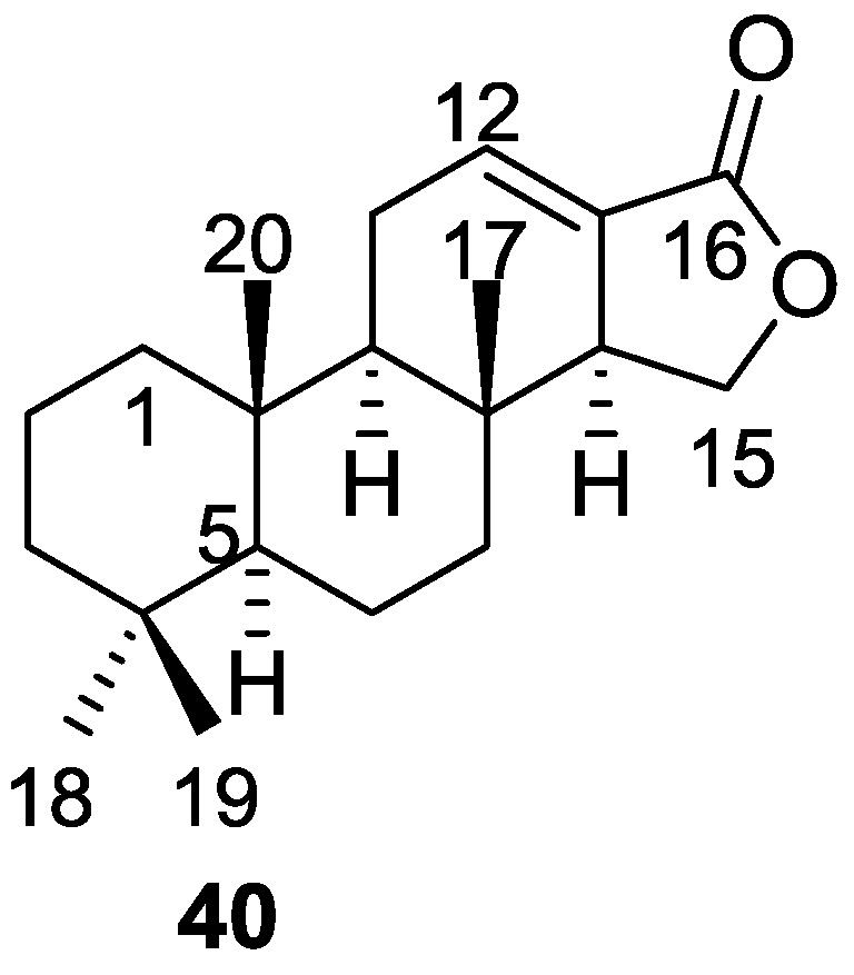
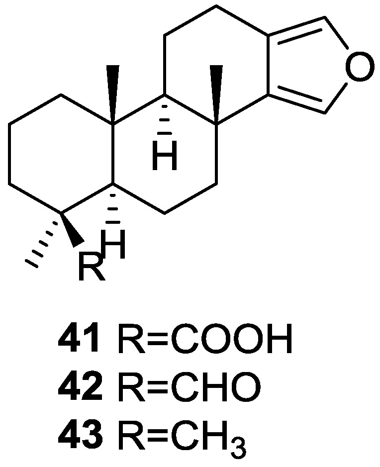
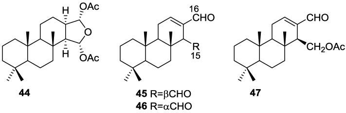
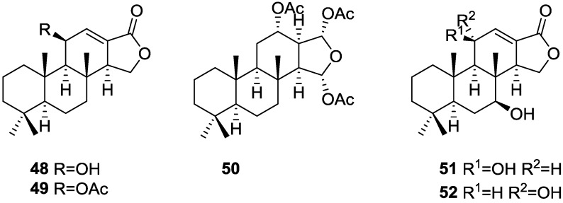
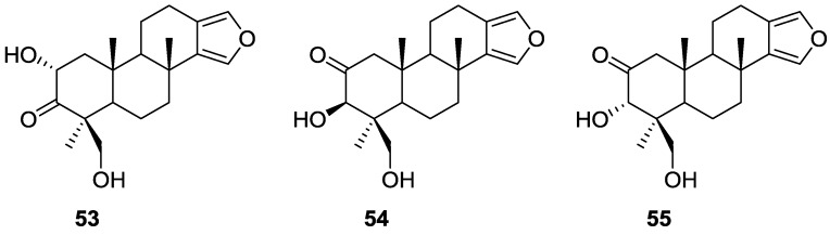
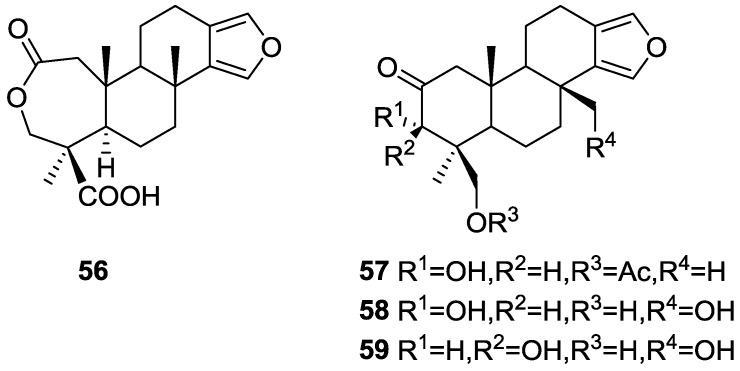
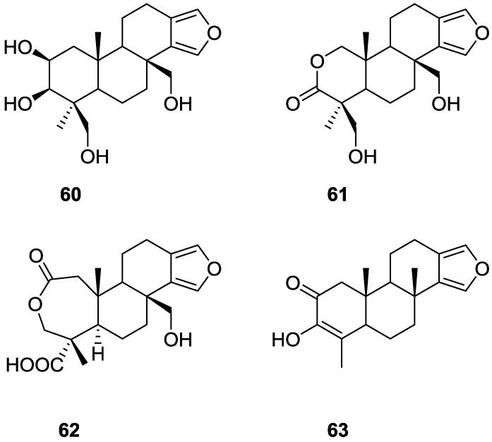
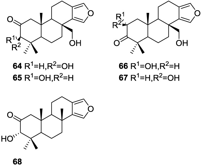
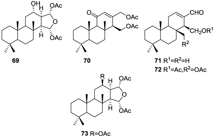
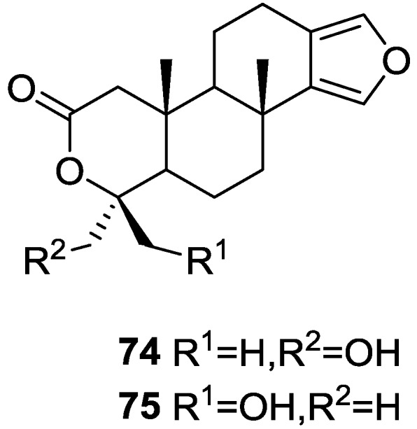
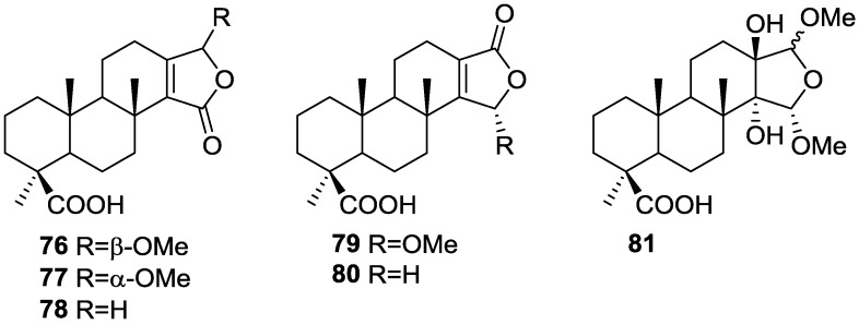
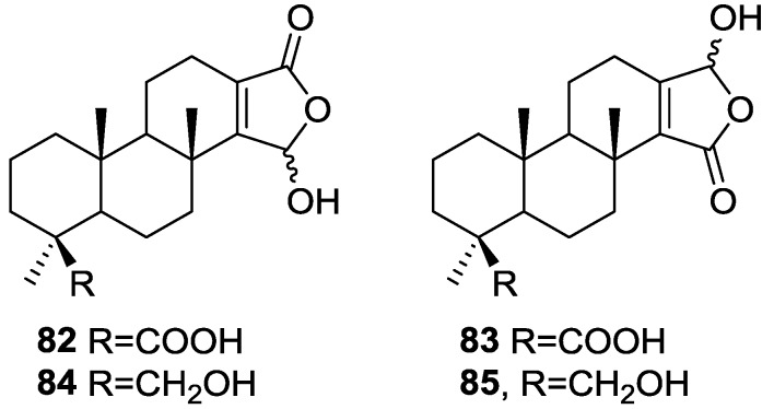
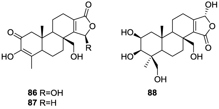
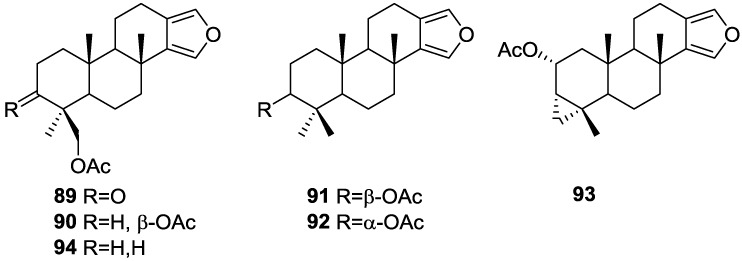
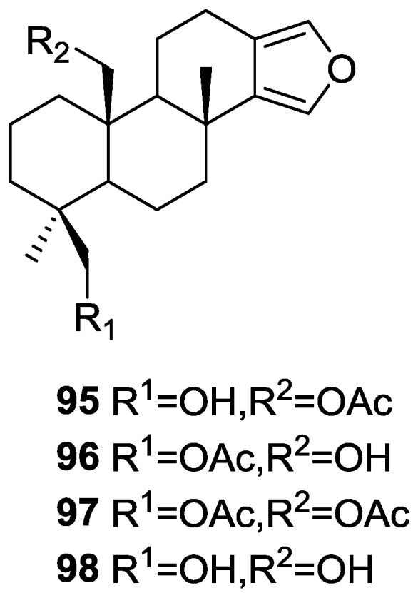
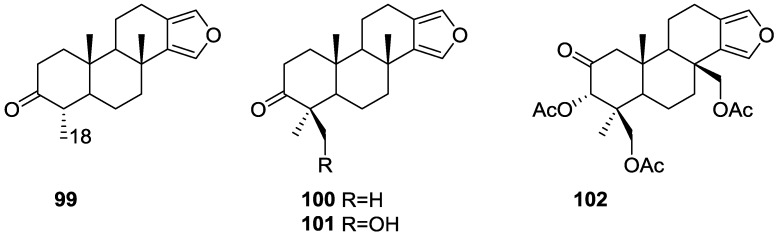
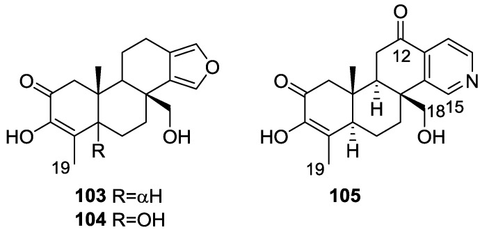
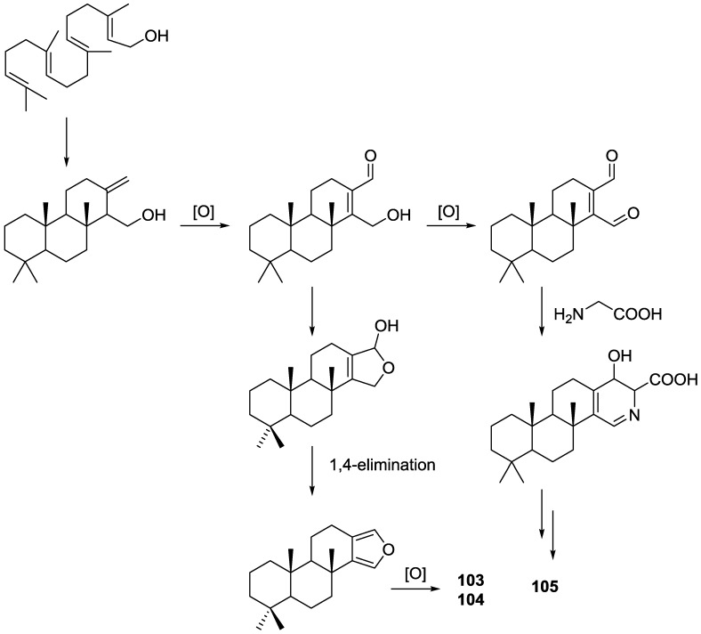
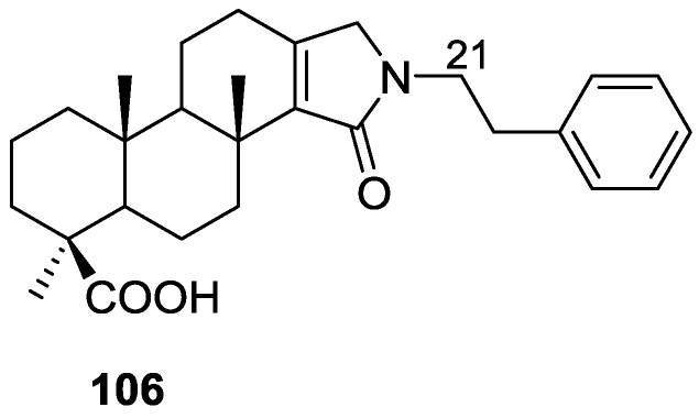
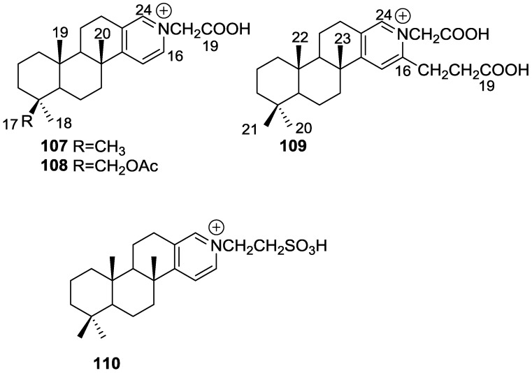
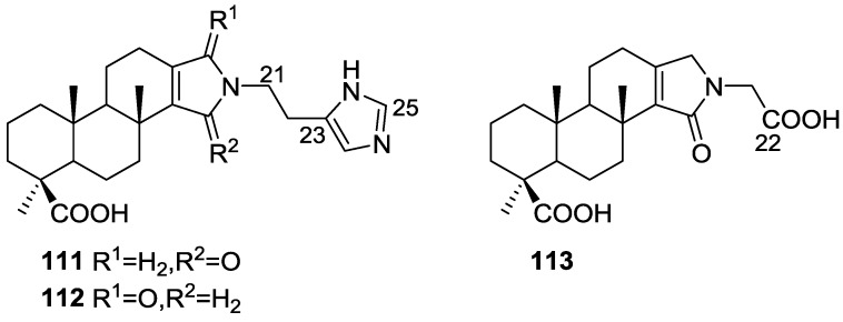
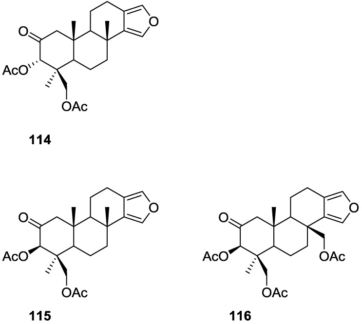
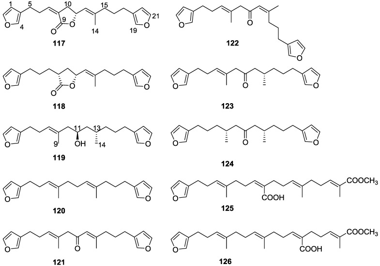
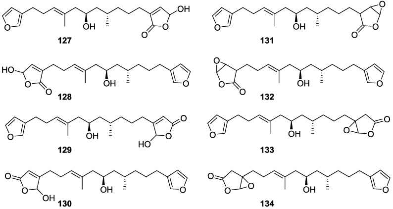
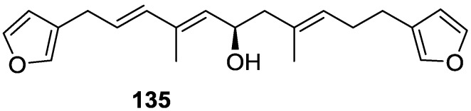
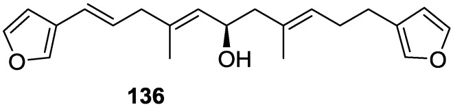
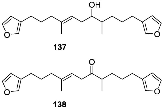
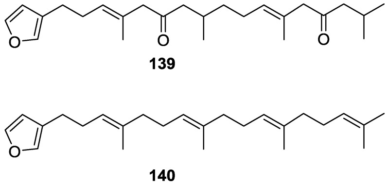
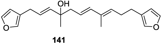
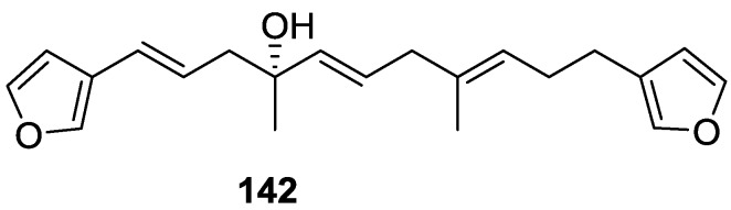
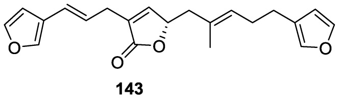
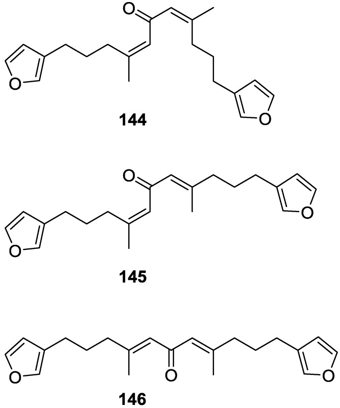
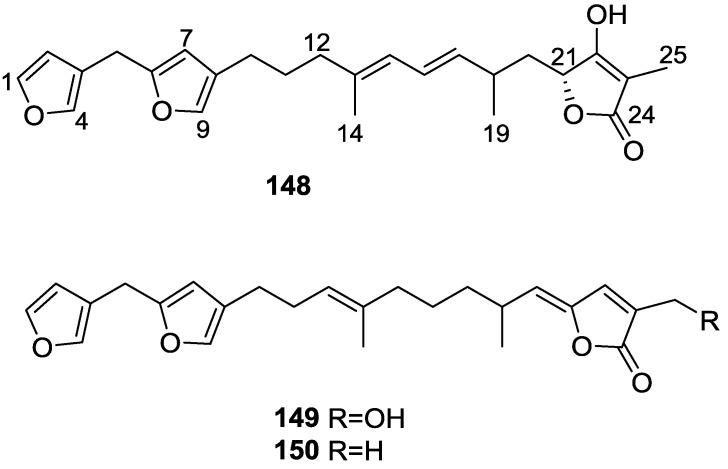
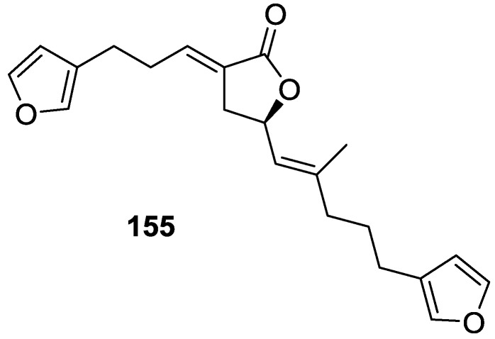
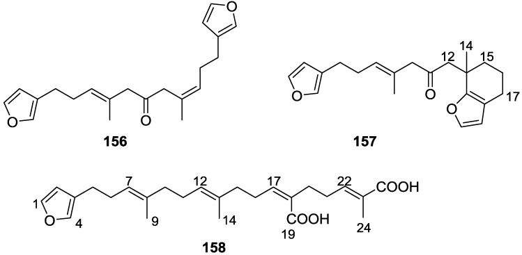
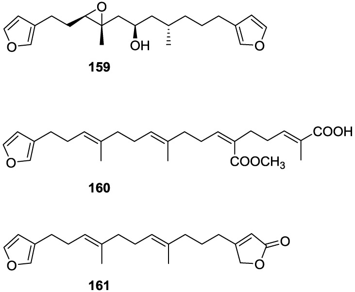
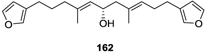
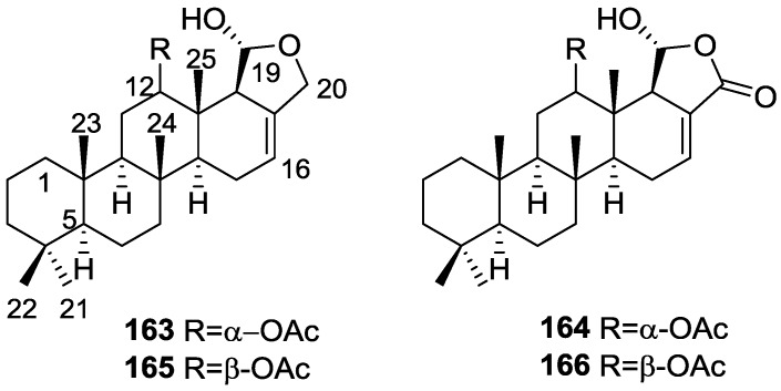
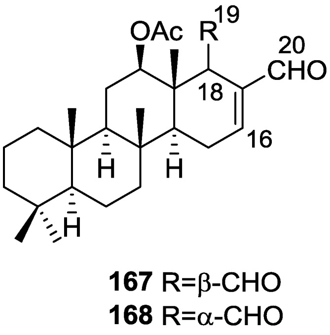
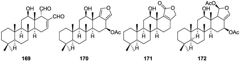
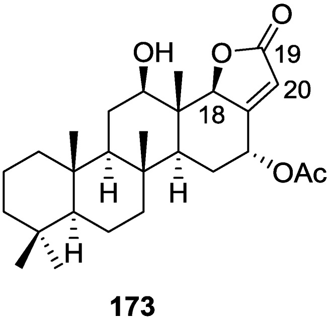
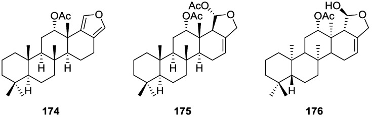
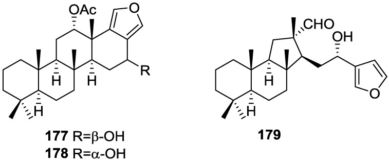
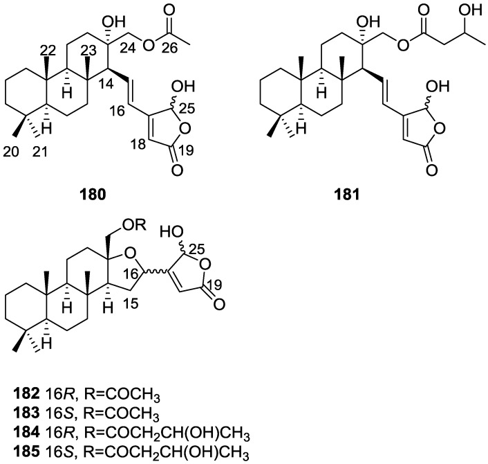
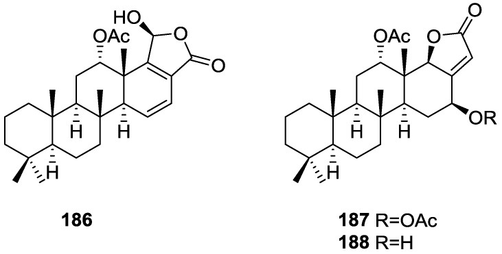
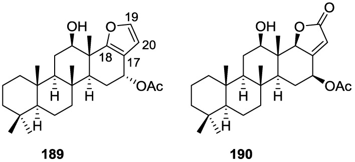
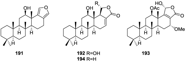
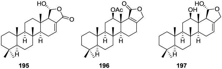
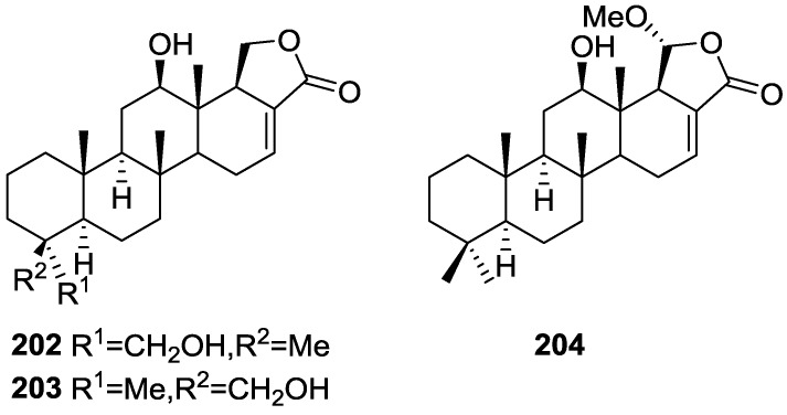
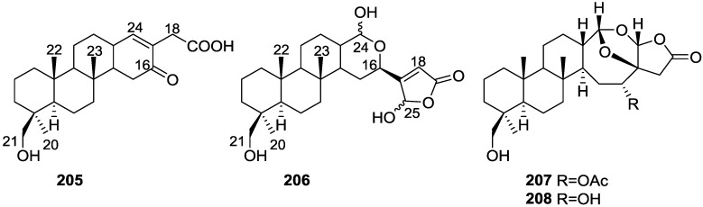
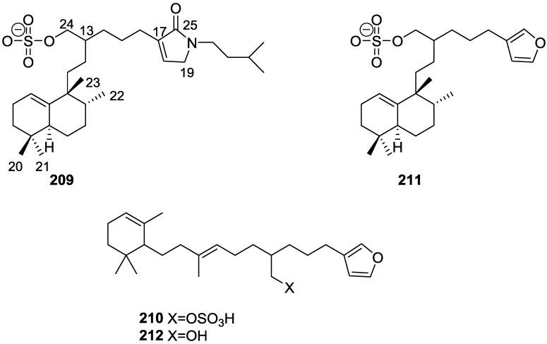
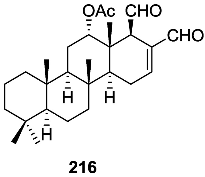
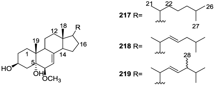
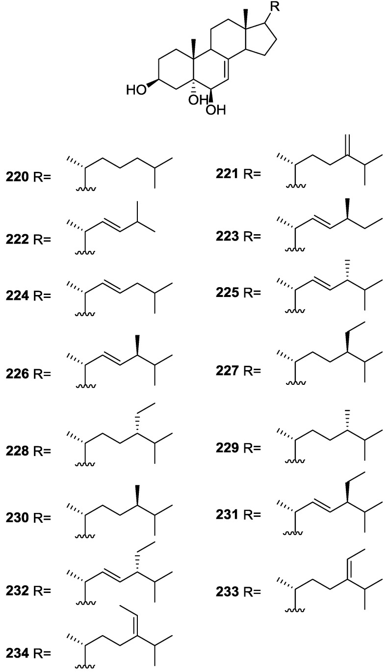
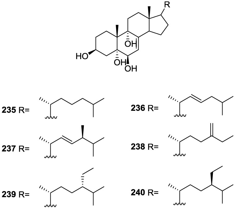
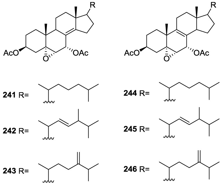
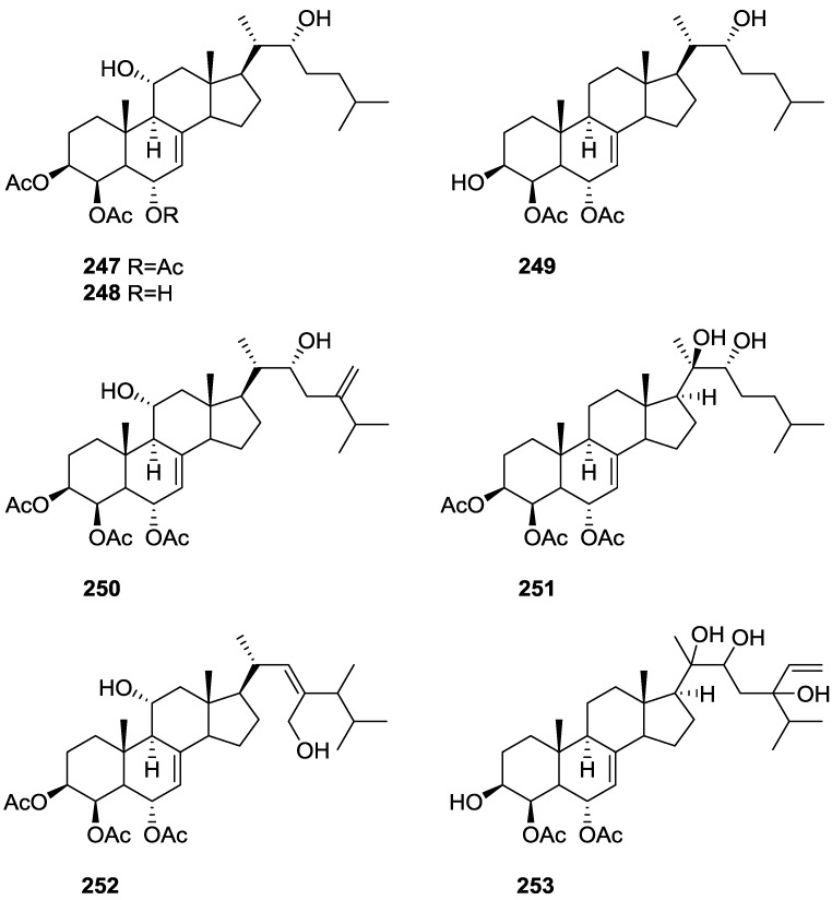
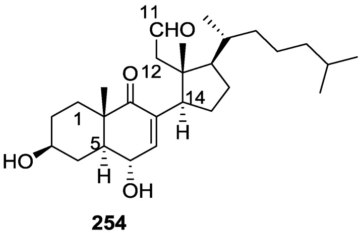
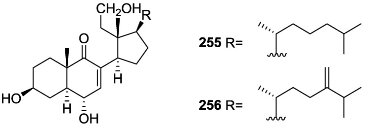
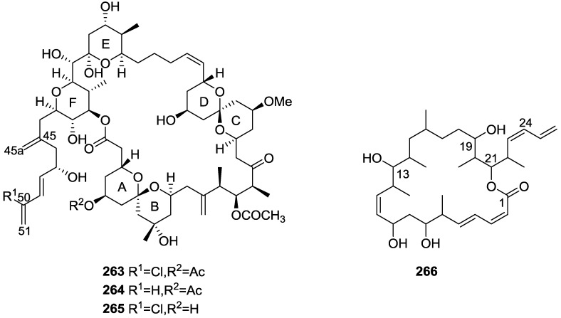
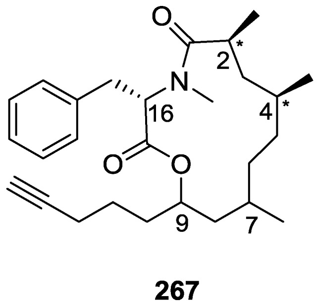
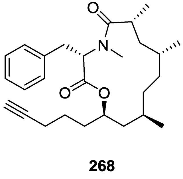
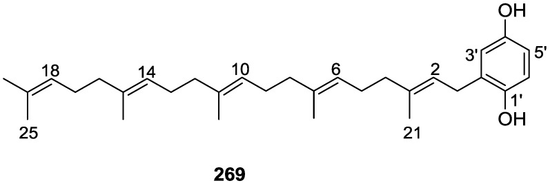
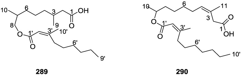
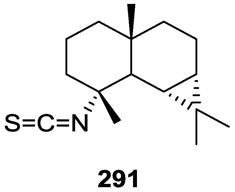
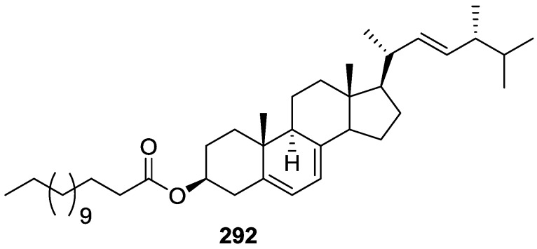
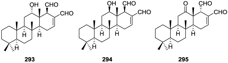
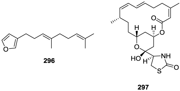
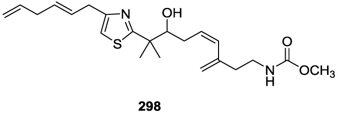
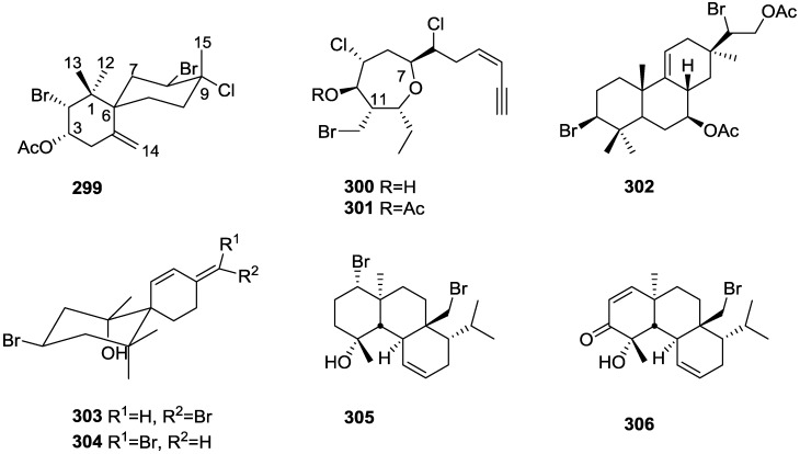
A comprehensive review on the chemistry of Spongia sp. is here presented, together with the biological activity of the isolated compounds. The compounds are grouped in sesquiterpene quinones, diterpenes, C21 and other linear furanoterpenes, sesterterpenes, sterols (including secosterols), macrolides and miscellaneous compounds. Among other reports we include studies on the intraspecific diversity of a Mediterranean species, compounds isolated from associated sponge and nudibranch and compounds isolated from S. zimocca and the red seaweed Laurentia microcladia. Under biological activity a table of the reported biological activities of the various compounds and the biological screening of extracts are described. The present review covers the literature from 1971 to 2015.

Keywords: C21 furanoterpenes; Spongia sp.; biological activity; diterpenes; macrolides; sesquiterpene quinones; sesterterpenes; sterols.


Figures

Figure 1
Figure 1
Structures of 5-epi-isospongiaquinone 1 and 5-epi-homoisospongiaquinone 2.
Figure 2
Figure 2
Structures of compound 3, dehydrocyclospongiaquinone-1 4 and spongiaquinone 5.
Figure 3
Figure 3
Structures of cyclosmenospongine 6, smenospongiarine 7, ilimaquinone 8 and smenospongine 9.
Figure 4
Figure 4
Structures of 17-O-isoprenyldictyoceratin-C 10 and dictyoceratin-C 11.
Figure 5
Figure 5
Structures of metachromins J 12 and K 13, L–T, 1422, A 23, and C–E 2426.
Figure 6
Figure 6
Structures of nakijiquinone E 27 and F 28, dictyoceratins A–C, 29, 30, isospongiaquinone 31, 6′-hydroxy-4′-methoxyavarone 32, neoavarol 33, nakijiquinones A–D 3437, and an endo olefin isomer at C-3 of smenospongine 38.
Figure 7
Figure 7
Structure of smenoquinone 39.
Figure 8
Figure 8
Structure of isoagatholactone 40.
Figure 9
Figure 9
Structures of spongia-13(16),14-dien-19-oic acid 41, spongia-13(16),14-dien-19-al 42 and spongia-13(16),14-diene 43.
Figure 10
Figure 10
Structures of 15α,16α-diacetoxyspongian 44, ent-isocopal-12-en-15,16-dial 45, 14-iso-ent-isocopal-12-en-15,16-dial 46 and 15-acetoxy-ent-isocopal-12-en-16-al 47.
Figure 11
Figure 11
Structures of 11β-hydroxyspongi-12-en-16-one 48, 11β-acetoxyspongi-12-en-16-one 49, aplysillin 50, 7β,11β-dihydroxyspongi-12-en-16-one 51, and 7β,11α-dihydroxyspongi-12-en-16-one 52.
Figure 12
Figure 12
Structures of 2α,19-dihydroxyspongia-13(16),14-dien-3-one (isospongiadiol) 53, 54 (epispongiadiol) and 55 (spongiadiol).
Figure 13
Figure 13
Structures of spongialactone A 56, 19-acetoxy-3α-hydroxyspongia-13(16),14-dien-2-one 57, 3α-17,19-trihydroxyspongia-13(16),14-dien-2-one 58, and 3β,17,19-trihydroxyspongia-13(16),14-dien-2-one 59.
Figure 14
Figure 14
Structures of 2β,3β,17,19-tetrahydroxyspongia-13(16),14-diene 60, 2-oxa-17,19-dihydroxyspongia 13(16),14-dien-3-one 61, 17-hydroxy-4-epi-spongialactone A 62, and 19-nor-3-hydroxyspongia-3,13(16),14-trien-2-one 63.
Figure 15
Figure 15
Structures of 3β,17-dihydroxyspongia-13(16),14-dien-2-one 64, 3α,17-dihydroxyspongia-13(16),14-dien-2-one 65, 2α,17-dihydroxyspongia-13(16),14-dien-3-one 66, 2β,17-dihydroxyspongia-13(16),14-dien-3-one 67, and 3α-hydroxyspongia-13(16),14-dien-3-one 68.
Figure 16
Figure 16
Structures of 12-deacetyl-aplysillin 69, 15,16-diacetoxy-11-oxo-ent-isocopal-12ene 70, 15-hydroxy-ent-isocopal-12-en-16-al 71, 15,17-diacetoxy-ent-isocopal-12-en-16-al 72, and compound 73.
Figure 17
Figure 17
Strutures of furanoterpenes 74 and 75.
Figure 18
Figure 18
Strutctures of 16β-methoxy-15-oxospongi-13-en-19-oic-acid 76, 16α-methoxy-15-oxospongi-13-en-19-oic-acid 77, 15-oxospongi-13-en-19-oic acid 78, 15α-methoxy-16-oxospongi-13-en-19-oic-acid 79, 16-oxospongi-13-en-19-oic acid 80, and 13β,14α-dihydroxy-15α,16ξ-dimethoxyspongian-19-oic-acid 81.
Figure 19
Figure 19
Structures of spongiabutenolides A–D, 8285.
Figure 20
Figure 20
Structures of zimoclactone A 86, zimoclactone B 87, and zimoclactone C 88.
Figure 21
Figure 21
Structures of 19-acetoxyspongia-13(16),14-dien-3-one 89, 3β,19-diacetoxyspongia-13(16),14-diene 90, 3β-acetoxyspongia-13(16),14-diene 91, 3α-acetoxyspongia-13(16),14-diene 92, 2(R),3(S),4(S)-3,18-methylene-2α-acetoxyspongia-13(16),14-diene 93, and 19-acetoxyspongia-13(16),14-diene 94.
Figure 22
Figure 22
Structures of 20-acetoxy-19-hydroxyspongia-13(16),14-diene 95, 19-acetoxy-20-hydroxyspongia-13(16),14-diene 96, 19,20-diacetoxyspongia-13(16),14-diene 97, and 19,20-dihydroxyspongia-13(16),14-diene 98.
Figure 23
Figure 23
Structures of 19-norspongia-13(16),14-dien-3-one 99, and compounds 100102.
Figure 24
Figure 24
Structures of 18-nor-3,17-dihydroxyspongia-3,13(16),14-trien-2-one 103, 18-nor-3,5,17-trihydroxyspongia-3,13(16),14-trien-2-one 104, and spongiapyridine 105.
Figure 25
Figure 25
Proposed biosynthesis route for compounds 103, 104 and 105.
Figure 26
Figure 26
Structure of haumanamide 106.
Figure 27
Figure 27
Structures of spongidines A–D 107110.
Figure 28
Figure 28
Structures of spongolactams A–C, 111113.
Figure 29
Figure 29
Structures of 3α,19-diacetoxyspongia-13(16),14-dien-2-one 114, 3β,19-diacetoxyspongia-13(16),14-dien-2-one 115, and 3β,17,19-triacetoxyspongia-13(16),14-dien-2-one 116.
Figure 30
Figure 30
Structures of nitenin 117, dihydronitenin 118, furospongin-1 119, anhydrofurospongin-1 120, furospongin-2 121, isofurospongin-2 122, dihydrofurospongin-2 123, tetrahydrofurospongin-2 124, furospongin-3 125, and furospongin-4 126.
Figure 31
Figure 31
Structures of furospongin-1 119 related compounds with γ-hydroxy-α,β-butenolide and β,γ-epoxybutenolide rings, 127134.
Figure 32
Figure 32
Structure of tetradehydrofurospongin-1 135.
Figure 33
Figure 33
Structure of tetradehydrofurospongin-1 136.
Figure 34
Figure 34
Structures of furospongenol 137 and furospongenone 138.
Figure 35
Figure 35
Structures of idiadione 139 and furospinulosin-1 140.
Figure 36
Figure 36
Structure of C-21 furanoterpene 141.
Figure 37
Figure 37
Structure of (−)-isotetradehydrofurospongin-1 142.
Figure 38
Figure 38
Structure of kurospongin 143.
Figure 39
Figure 39
Structures of furospongin-2 121 isomers 144146.
Figure 40
Figure 40
Structure of tetronic acid 147.
Figure 41
Figure 41
Structures of cometins A–C 148150.
Figure 42
Figure 42
Structures of ambliofuran 151, (S)-12-hydroxyambliofuran 152, (S)-12-acetoxyambliofuran 153, and compound 154.
Figure 43
Figure 43
Structure of isonitenin 155.
Figure 44
Figure 44
Structures of furospongin-5 156, cyclofurospongin-2 157, and demethylfurospongin-4 158.
Figure 45
Figure 45
Structures of 7,8-epoxy-furospongin-1 159, isofurospongin-4 160, and compound 161.
Figure 46
Figure 46
Structure of compound 162.
Figure 47
Figure 47
Structures of deoxoscalarin 163, scalarin 164, 12-epi-deoxoscalarin 165, and 12-epi-scalarin 166.
Figure 48
Figure 48
Structures of 12-epi-scalaradial 167 and 12,18-di-epi-scalaradial 168.
Figure 49
Figure 49
Structures of 12-deacetyl-12,18-di-epi-scalaradial 169, scalarafuran 170, scalarolide 171, and 172.
Figure 50
Figure 50
Structure of scalarolbutenolide 173.
Figure 51
Figure 51
Structures of 16-deacetoxy-12-epi-scalarafuran acetate 174, deoxoscalarin acetate 175, and (−)-12-epi-deoxoscalarin 176.
Figure 52
Figure 52
Structures of isoscalarafuran A 177, isoscalarafuran B 178, and hyrtiosal 179.
Figure 53
Figure 53
Structures of spongianolides A–F 180185.
Figure 54
Figure 54
Structures of 12α-acetoxy-19β-hydroxyscalara-15,17-dien-20,19-olide 186, 12α,16β-diacetoxyscalarolbutenolide 187, and 12α-acetoxy-16β-hydroxyscalarolbutenolide 188.
Figure 55
Figure 55
Structures of 12,16-di-epi-12-O-deacetyl-16-O-acetylfuroscalarol 189 and 16-epi-scalarolbutenolide 190.
Figure 56
Figure 56
Structures of 12-O-deacetylscalafuran 191, 12-O-deacetyl-12-epi-scalarin 192, 12-O-acetyl-16-O-methylhyrtiolide 193, and 12-O-deacetyl-12-epi-19-deoxyscalarin 194.
Figure 57
Figure 57
Structures of deacetoxy scalarin 195, and compounds 196 and 197.
Figure 58
Figure 58
Structures of 12,24-diacetoxy-deoxoscalarin 198, 12-O-deacetoxyl-24-hydroxyl-deoxoscalarin 199, and 12-O-deacetoxyl-19-O-methyldeoxoscalarin 200, and compound 201.
Figure 59
Figure 59
Structures of 12-O-deacetyl-12-epi-19-deoxy-21-hydroxyscalarin 202, 12-O-deacetyl-12-epi-19-deoxy-22-hydroxyscalarin 203, and 12-O-deacetyl-12-epi-19-O-methylscalarin 204.
Figure 60
Figure 60
Structures of 21-hydroxy petrosaspongiolide K 205, 21-hydroxy petrosaspongiolide P 206, petrosapongiolides D 207, and G 208.
Figure 61
Figure 61
Strutures of irregularasulfate 209, hipposulfate C 210, halisulfate-7 211, and igernellin 212.
Figure 62
Figure 62
Structures of petrosaspongiolides A 213, B 214, and I 215.
Figure 63
Figure 63
Structure of scalaradial 216.
Figure 64
Figure 64
Structures of 3β,5α-dihydroxy-6β-methoxycholest-7-enes 217, 218, and 219.
Figure 65
Figure 65
Structures of 3β,5α,6β-trihydroxycholest-7-enes 220234.
Figure 66
Figure 66
Structures of 5α-cholest-7-ene-3β,5,6β,9-tetraol 235, (22E)-5α-cholest-7,22-diene-3β,5,6β,9-tetraol 236, (22E,24S)-24-methyl-5α-cholest-7,22-diene-3β,5,6β,9-tetraol 237, 24-methylene-5α-cholest-7-ene-3β,5,6β,9-tetraol 238, (24S)-24-ethyl-5α-cholest-7-ene-3β,5,6β,9-tetraol 239, and (24R)-24-ethyl-5α-cholest-7-ene-3β,5,6β,9-tetraol 240.
Figure 67
Figure 67
Structures of 5α,6α-epoxycholest-8(14)-ene-3β,7α-diol 3,7-diacetate 241, (22E,24ξ)-5α,6α-epoxy-24-methylcholesta-8(14),22-diene-3β,7α-diol 3,7-diacetate 242, 5α,6α-epoxy-24-methylcholesta-8(14),24(28)-diene-3β,7α-diol 3,7-diacetate 243, 5α,6α-epoxy-cholest-8-ene-3β,7α-diol 3,7-diacetate 244, (22E,24ξ)-5α,6α-epoxy-24-methylcholesta-8,22-diene-3β,7α-diol 3,7-diacetate 245, and 5α,6α-epoxy-24-methylcholesta-8,24(28)-diene-3β,7α-diol 3,7-diacetate 246.
Figure 68
Figure 68
Structures of agosterol A 247, B 248, C 249, A4 250, D2 251, A5 252 and C6 253.
Figure 69
Figure 69
Structure of 9,11-secosterol, 3β,6α-dihydroxy-9-oxo-9,11-seco-5α-cholest-7-en-11-al 254.
Figure 70
Figure 70
Structures of 9,11-seco-3β,6α,11-trihydroxy-5α-cholest-7-en-9-one 255, and 9,11-seco-3β,6α,11-trihydroxy-24-methylene-5α-cholest-7-en-9-one 256.
Figure 71
Figure 71
Structure of 3β-acetoxy-5,6β-dihydroxy-9-oxo-9,11-seco-5α-cholest-7-en-11-al 257.
Figure 72
Figure 72
Structure of 3β-hydroxy-5α,6α-epoxy-9-oxo-9,11-seco-5α-cholest-7-en-11-al 258.
Figure 73
Figure 73
Structures of 3-O-deacetylluffasterol B 259 and 3-O-deacetyl-22,23-dihydro-24,28-dehydroluffasterol B 260.
Figure 74
Figure 74
Structures of fijianolides A 261 and B 262.
Figure 75
Figure 75
Structures of spongistatins 1–3 263265 and dictyostatin 1 266.
Figure 76
Figure 76
Structure of spongidepsin 267.
Figure 77
Figure 77
Structure of (2R,4R,7R,9R,16S)-spongidepsin 268.
Figure 78
Figure 78
Structure of p-quinol 269.
Figure 79
Figure 79
Structure of pokepola ester 270.
Figure 80
Figure 80
Structure of spongilipid 271.
Figure 81
Figure 81
Structure of the alkaloids 272274.
Figure 82
Figure 82
Structure of spongiacysteine 275.
Figure 83
Figure 83
Structures of ceramide 276, compound 277 and the guanidine acetic salt 278.
Figure 84
Figure 84
Structures of the ceramides 2-hydroxy-N-(1,3,4-trihydroxy-17-methyloctadecan-2-yl)-18-methylarachidamide 279, 2-hydroxy-N-(1,3,4-trihydroxy-17-methyloctadecan-2-yl)-19-methyl-henicosanamide 280, and 2-hydroxy-N-(1,3,4-trihydroxy-17-methyloctadecan-2-yl)-20-methyl-behenamide 281.
Figure 85
Figure 85
Structures of N-palmitoyl-heptacosane-1,3,5-triol 282.
Figure 86
Figure 86
Structures of heterofibrins A1 283, A2 284, A3 285, B1 286, B2 287 and B3 288.
Figure 87
Figure 87
Structures of officinoic acid A 289 and officinoic acid B 290.
Figure 88
Figure 88
Structure of compound 291.
Figure 89
Figure 89
Structure of ergosteryl myristate 292.
Figure 90
Figure 90
Structures of 12-deacetylscalaradial 293, 12-deacetyl-12-epi-scalaradial 294, and 12-deacetyl-18-epi-12-oxoscalaradial 295.
Figure 91
Figure 91
Structures of dendrolasin 296 and latrunculin A 297.
Figure 92
Figure 92
Structure of mycothiazole 298.
Figure 93
Figure 93
Structures of rogiolol acetate 299, rogiolenyne B 300, rogiolenyne C 301, isopimarane 302, chamigrene 4E 303, chamigrene 4Z 304, bromosphaerol 305, and sphaerococcenol A 306.

References

    1. Blunt J.W., Copp B.R., Keyzers R.A., Munroa M.H.G., Prinsepd M.R. Marine natural products. Nat. Prod. Rep. 2015;32:116–211. doi: 10.1039/C4NP00144C. - DOI - PubMed
    1. Perdicaris S., Vlachogianni T., Valavanidis A. Bioactive Natural Substances from Marine Sponges: New Developments and Prospects for Future Pharmaceuticals. Nat. Prod. Chem. Res. 2013;1 doi: 10.4172/2329-6836.1000114. - DOI
    1. Pronzato R., Manconi R. Mediterranean Commercial Sponges: Over 5000 Years of Natural History and Cultural Heritage. Mar. Ecol. 2008;29:146–166. doi: 10.1111/j.1439-0485.2008.00235.x. - DOI
    1. Voultsiadou E., Dailianis T., Antoniadou C., Vafidis D., Dounas C., Chintiroglou1 C.C. Aegean Bath Sponges: Historical Data and Current Status. Rev. Fish. Sci. 2011;19:34–51. doi: 10.1080/10641262.2010.531794. - DOI
    1. Noyer C., Agell G., Pascual M., Becerro M.A. Isolation and Characterization of Microsatellite Loci from the Endangered Mediterranean Sponge Spongia agaricina (Demospongiae: Dictyoceratida) Conserv. Genet. 2009;10:1895–1898. doi: 10.1007/s10592-009-9848-6. - DOI

MeSH terms

LinkOut - more resources