Review
. 2016 Jul 23;8(8):230.
doi: 10.3390/toxins8080230.

Colubrid Venom Composition: An -Omics Perspective

Affiliations
Review

Colubrid Venom Composition: An -Omics Perspective

Inácio L M Junqueira-de-Azevedo et al. Toxins (Basel). .

Abstract

Snake venoms have been subjected to increasingly sensitive analyses for well over 100 years, but most research has been restricted to front-fanged snakes, which actually represent a relatively small proportion of extant species of advanced snakes. Because rear-fanged snakes are a diverse and distinct radiation of the advanced snakes, understanding venom composition among "colubrids" is critical to understanding the evolution of venom among snakes. Here we review the state of knowledge concerning rear-fanged snake venom composition, emphasizing those toxins for which protein or transcript sequences are available. We have also added new transcriptome-based data on venoms of three species of rear-fanged snakes. Based on this compilation, it is apparent that several components, including cysteine-rich secretory proteins (CRiSPs), C-type lectins (CTLs), CTLs-like proteins and snake venom metalloproteinases (SVMPs), are broadly distributed among "colubrid" venoms, while others, notably three-finger toxins (3FTxs), appear nearly restricted to the Colubridae (sensu stricto). Some putative new toxins, such as snake venom matrix metalloproteinases, are in fact present in several colubrid venoms, while others are only transcribed, at lower levels. This work provides insights into the evolution of these toxin classes, but because only a small number of species have been explored, generalizations are still rather limited. It is likely that new venom protein families await discovery, particularly among those species with highly specialized diets.

Keywords: Colubridae; evolution; proteins; snake; toxins; transcriptomics.


Figures

Figure 1
Figure 1
Schematic cladograms showing the phylogenetic relationships among families and species of snakes discussed in this work (colored branches). The cladogram was based on the phylogenetic tree proposed by Pyron et al. [34]. Dashed lines in Philodryas indicate the presumed placement of P. chamissonis.
Figure 2
Figure 2
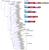
Maximum likelihood tree showing the relationship among representative SVMPs from different snake families. Bootstrap values are plotted close to the internal nodes. Colors in the terminal nodes indicate the types of the precursors, and their domain arrangements are depicted on the right. Abbreviated domains are: S, signal peptide; Pro, prodomain; Catalytic, metalloproteinase; D-like, disintegrin-like; Dis, disintegrin; Cys, cysteine rich; TM, transmembrane; EGF, epidermal growth factor; and Cytopl, cytoplasmic. The protein sequences are referred to by their accession numbers in GenBank, except those initiated by the codes EMILISO, OGUIISO, PMERREF, TSTRCLU and XMERCLU, which are mentioned in the “definition” field of sequence files deposited in the Transcriptome Shotgun Assembly (TSA) database.
Figure 3
Figure 3
Maximum likelihood circular cladogram showing the relationship among representative CTLs from different snake families. Bootstrap values are plotted close to internal nodes. Colors at the terminal nodes (circles) indicate typical vs. atypical venom proteins and the evidence of occurrence in the venoms. Colors in the diagram surrounding the cladogram indicate the taxonomic groups. The carbohydrate binding motifs, as discussed in the text (EPN, QPD, etc.), are indicated by red type. The protein sequences are referred by their GenBank accession numbers, except those initiated by the codes EMILISO, OGUIISO, PMERREF and XMERCLU, which are mentioned in the “definition” field of sequence files deposited in TSA.
Figure 4
Figure 4
Schematic organization of CNP (and BPP) precursors in the different snake families and in other vertebrates. The precursor of P. mertensi [57] exhibits a Pro-rich insertion in the linker region (detached at the bottom), which includes a BPP-like segment that may generate a BPP after processing.
Figure 5
Figure 5
Maximum likelihood tree showing the relationship among svMMPs from different snake families and MMPs from other vertebrate groups. Bootstrap values are plotted close to internal nodes. The domain arrangement of each precursor type is depicted on the right. The types of evidence for the occurrence in venoms are indicated by “T” (transcribed) and “V” (detected in venom). The protein sequences are labeled by their accession numbers in GenBank, except those initiated by the codes EMILREF, OGUIISO, and PMERREF, which are mentioned in the “definition” field of sequence files deposited in TSA.
Figure 6
Figure 6
Maximum likelihood tree showing the relationship among lipocalin proteins from different snake families and from other vertebrate groups. Note that transcripts highly expressed in venom glands are all in the same clade. Bootstrap values are plotted close to internal nodes. The protein sequences are labeled by their accession numbers in GenBank, except those initiated by the code EMIL, which is mentioned in the “definition” field of sequence files deposited in TSA and the sequence OGUIREF_Lipo1 (Accession Number KX450875). Sequences labeled GAMF from A aterrima were translated from the original nucleotide contigs retrieved from TSA.

References

    1. Calvete J.J. Snake venomics: From the inventory of toxins to biology. Toxicon. 2013;75:44–62. doi: 10.1016/j.toxicon.2013.03.020. - DOI - PubMed
    1. Ducancel F., Durban J., Verdenaud M. Transcriptomics and venomics: Implications for medicinal chemistry. Future Med. Chem. 2014;15:1629–1643. doi: 10.4155/fmc.14.95. - DOI - PubMed
    1. Brahma R.K., McCleary R.J., Kini R.M., Doley R. Venom gland transcriptomics for identifying, cataloging, and characterizing venom proteins in snakes. Toxicon. 2015;93:1–10. doi: 10.1016/j.toxicon.2014.10.022. - DOI - PubMed
    1. Gilles N., Servent D. The European FP7 Venomics Project. Future Med. Chem. 2014;15:1611–1612. doi: 10.4155/fmc.14.85. - DOI - PubMed
    1. Aird S.D., Watanabe Y., Villar-Briones A., Roy M.C., Terada K., Mikheyev A.S. Quantitative high-throughput profiling of snake venom gland transcriptomes and proteomes (Ovophis okinavensis and Protobothrops flavoviridis) BMC Genom. 2013;14 doi: 10.1186/1471-2164-14-790. - DOI - PMC - PubMed

Publication types

LinkOut - more resources