. 2016 Sep 6;55(35):4949-59.
doi: 10.1021/acs.biochem.6b00637. Epub 2016 Aug 23.

Nuclear Magnetic Resonance Observation of α-Synuclein Membrane Interaction by Monitoring the Acetylation Reactivity of Its Lysine Side Chains

Affiliations

Nuclear Magnetic Resonance Observation of α-Synuclein Membrane Interaction by Monitoring the Acetylation Reactivity of Its Lysine Side Chains

Jung Ho Lee et al. Biochemistry. .

Abstract

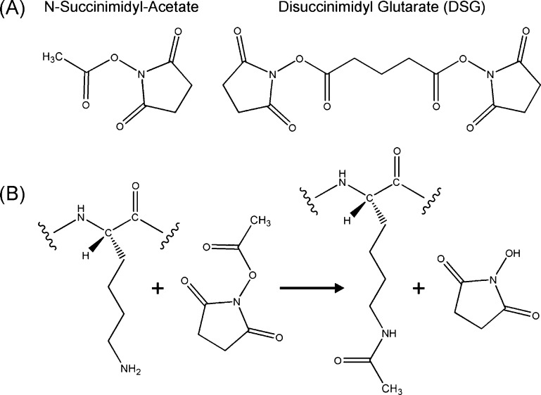
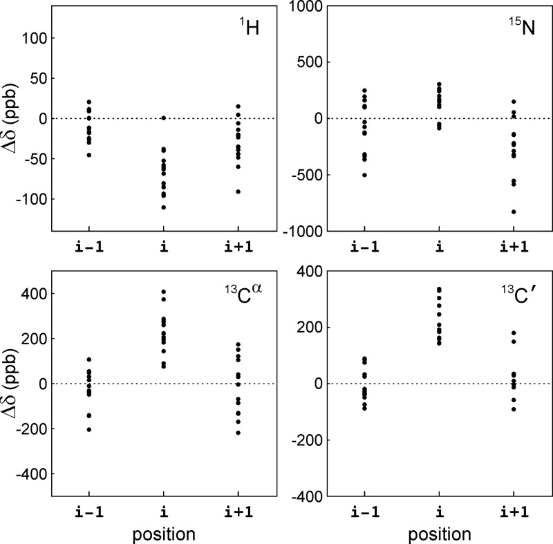
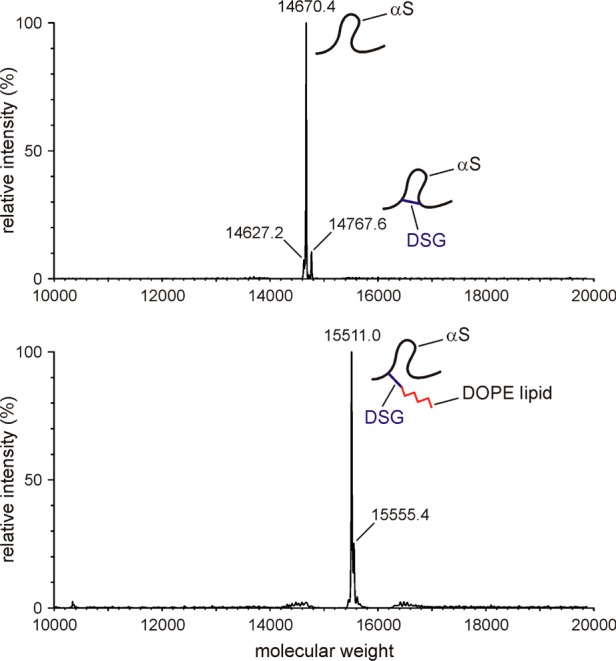
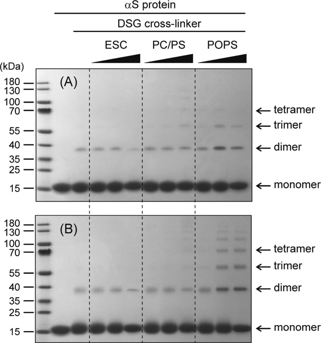
The interaction between α-synuclein (αS) protein and lipid membranes is key to its role in synaptic vesicle homeostasis and plays a role in initiating fibril formation, which is implicated in Parkinson's disease. The natural state of αS inside the cell is generally believed to be intrinsically disordered, but chemical cross-linking experiments provided evidence of a tetrameric arrangement, which was reported to be rich in α-helical secondary structure based on circular dichroism (CD). Cross-linking relies on chemical modification of the protein's Lys C(ε) amino groups, commonly by glutaraldehyde, or by disuccinimidyl glutarate (DSG), with the latter agent preferred for cellular assays. We used ultra-high-resolution homonuclear decoupled nuclear magnetic resonance experiments to probe the reactivity of the 15 αS Lys residues toward N-succinimidyl acetate, effectively half the DSG cross-linker, which results in acetylation of Lys. The intensities of both side chain and backbone amide signals of acetylated Lys residues provide direct information about the reactivity, showing a difference of a factor of 2.5 between the most reactive (K6) and the least reactive (K102) residue. The presence of phospholipid vesicles decreases reactivity of most Lys residues by up to an order of magnitude at high lipid:protein stoichiometries (500:1), but only weakly at low ratios. The decrease in Lys reactivity is found to be impacted by lipid composition, even for vesicles that yield similar αS CD signatures. Our data provide new insight into the αS-bilayer interaction, including the pivotal state in which the available lipid surface is limited. Protection of Lys C(ε) amino groups by αS-bilayer interaction will strongly impact quantitative interpretation of DSG cross-linking experiments.


Conflict of interest statement

The authors declare no competing financial interest.

Figures

Figure 1
Figure 1
Chemicals used and their reaction with lysine side chains. (A) N-Succinimidyl acetate acetylates one Lys, while DSG can cross-link two Lys side chains. (B) Chemistry of Lys side chain acetylation reaction by N-succinimidyl acetate.
Figure 2
Figure 2
Reaction kinetics of 0.25 mM N-succinimidyl acetate monitored by the intensity decay of its methyl NMR resonance and the intensity increase of the product’s methylene NMR resonance (red arrows), in samples consisting of PBS buffer (circles), 10 mM PC/PS SUVs (triangles), 50 μM αS (diamonds), or 10 mM ESC SUVs (squares), all at 20 °C in PBS buffer.
Figure 3
Figure 3
Assignment of acetylated αS lysine side chains, illustrated for K6. Initially, backbone 15N–1H chemical shifts of acetylated Lys residues and their neighbors were assigned using conventional triple-resonance experiments at very high resolution, using NUS data acquisition. The bottom left panel shows backbone 15N–1H correlations of unmodified and acetylated K6. The backbone chemical shifts of αS containing acetylated Lys at position i progressively converge to that of unmodified αS beyond position i ± 1. Next, a high-resolution 3D NUS 1H(t1)–TOCSY–15N(t2)–1H(t3) experiment permits the connection of backbone (red) and side chain (blue) amides to the aliphatic protons. By matching the chemical shifts of these aliphatic protons, we linked amide chemical shifts of the backbone and side chain for acetylated Lys residues. The TOCSY spectrum (120 ms mixing time) was recorded at 700 MHz using 1.5 mM αS.
Figure 4
Figure 4
Distribution of backbone chemical shift perturbations caused by Lys acetylation. Differences between the 1H, 15N, 13Cα, and 13C′ chemical shifts of acetylated Lys residues and corresponding values of the unmodified counterpart, for the 15 Lys residues in αS (i) and their flanking residues (i – 1 and i + 1).
Figure 5
Figure 5
Small regions of the 2D 1H–15N HSQC NMR spectra, showing the Lys side chain amide signals of chemically acetylated αS. To obtain a low level (≤∼7%) of side chain acetylation, reactions with 0.25 mM N-succinimidyl acetate were quenched after 5 min by addition of an equal volume of 50 mM l-lysine. Spectra were recorded at 900 MHz and 10 °C in 10 mM sodium phosphate buffer and 10 mM NaCl (pH 6.0), with an αS concentration of 0.2 mM. Spectra were acquired using (A) a standard 15N–1H HSQC pulse scheme, utilizing a single 1H pulse for t1 decoupling and no homonuclear decoupling during detection, and (B) 1H composite pulse decoupling in the t1 dimension and 1H-BASH homonuclear decoupling during t2. Acquisition times were 830 ms for 15N and 200 ms for 1H in panel A and 830 ms for 15N and 300 ms for 1H in panel B.
Figure 6
Figure 6
Reaction of αS Lys residues (50 μM protein) with 250 μM N-succinimidyl acetate in the presence of different ESC (5:3:2 DOPE:DOPS:DOPC) SUV concentrations. (A) High-resolution 2D 1H–15N NMR spectra of the acetylated side chain region in the absence (top) and presence (bottom) of a 200-fold molar excess of ESC SUVs. Lower contour levels were used for the bottom panel for better visibility. (B) Change of the second-order rate constants with increasing lipid:αS ratio, L, illustrated for K6, K32, and K102. Dashed lines correspond to Kn(L) = AneBnL + Cn, where Kn is the second-order rate constant for the reaction of N-succinimidyl acetate with any given Lys in αS, L is the lipid:αS molar ratio, and An, Bn, and Cn are the fitted parameters. (C) Designation of different regions in the primary structure of αS. At low lipid:αS ratios, two binding modes are believed to exist, SL1 and SL2, where the first ∼25 and ∼100 N-terminal residues are NMR-invisible, respectively. αS consists of a positively charged N-terminal region, a hydrophobic NAC (non-amyloid-β component, residues 61–95) region, and an acidic C-terminal region. (D) Reaction rate, An + Cn, as a function of residue number in the absence of lipids. (E) Bn as a function of residue number, which reflects the sensitivity of the reaction rate constant to lipid concentration, at a low lipid:αS ratio. (F) Cn/(An + Cn) as a function of Lys residue number, n, which is a measure for the attenuation of Lys reactivity in the high lipid/αS limit.
Figure 7
Figure 7
Mass characterization of αS/SUV samples after DSG cross-linking. A mixture of 50 μM 15N-enriched N-terminally acetylated αS with 2.5 mM ESC SUV (1:50 αS:ESC SUV) in PBS buffer was reacted with 125 μM DSG for 5 min at room temperature, followed by LC–MS to characterize the mass. The main peaks with molecular masses of 14670, 14768, and 15511 correspond to αS, intramolecularly cross-linked αS, and αS–DOPE cross-linked species, respectively. No separate peaks for αS–DOPS or αS–Lys cross-linked species were observed. Top and bottom panels represent different LC fractions. Note that all measurements were taken on NMR samples containing uniformly 15N-enriched αS, increasing its mass by ∼168 Da over that of the natural abundance protein.
Figure 8
Figure 8
Overlay of the 2D BASH-decoupled 1H–15N HSQC NMR spectra of αS Lys side chains reacted with 250 μM N-succinimidyl acetate (black) or 125 μM disuccinimidyl glutarate (DSG) cross-linker (orange). The reaction conditions are the same as those described in the legend of Figure 6 (1:50 αS:ESC SUV). The DSG-reacted spectrum (orange) is displayed at a 2-fold lower contour level compared with that of the black spectrum for better visibility of the broad heterogeneous resonances.
Figure 9
Figure 9
Comparison of αS Lys reactivity with N-succinimidyl acetate in the presence of (A) moderate (200:1) and (B) near-saturating (500:1) quantities of different SUVs. Shown are the apparent second-order rate constants in the presence of ESC SUVs (5:3:2 DOPE:DOPS:DOPC) and PC/PS SUVs (7:3 POPC:POPS). Note that the acetylation rate of K102 is minimally impacted by the amount or type of lipids, whereas for all other residues, the ESC SUVs are more protective than PC/PS SUVs.
Figure 10
Figure 10
Effect of lipid vesicle composition on αS oligomerization analyzed by DSG cross-linking. N-Terminally acetylated 100 μM αS was reacted with (A) 0.2 and (B) 1.0 mM DSG cross-linker for 10 min at room temperature in the presence of increasing amounts (1:5, 1:50, and 1:500 αS:SUV) of ESC, PC/PS, and POPS SUVs. The gels were stained with Coomassie blue.

References

    1. Sinz A. (2006) Chemical cross-linking and mass spectrometry to map three-dimensional protein structures and protein-protein interactions. Mass Spectrom. Rev. 25, 663–682. 10.1002/mas.20082. - DOI - PubMed
    1. Avrameas S.; Ternynck T. (1969) Cross-Linking of Proteins with Glutaraldehyde and Its Use for Preparation of Immunoadsorbents. Immunochemistry 6, 53–66. 10.1016/0019-2791(69)90178-5. - DOI - PubMed
    1. Dettmer U.; Newman A. J.; Luth E. S.; Bartels T.; Selkoe D. (2013) In Vivo Cross-linking Reveals Principally Oligomeric Forms of alpha-Synuclein and beta-Synuclein in Neurons and Non-neural Cells. J. Biol. Chem. 288, 6371–6385. 10.1074/jbc.M112.403311. - DOI - PMC - PubMed
    1. Fauvet B.; Mbefo M. K.; Fares M.-B.; Desobry C.; Michael S.; Ardah M. T.; Tsika E.; Coune P.; Prudent M.; Lion N.; Eliezer D.; Moore D. J.; Schneider B.; Aebischer P.; El-Agnaf O. M.; Masliah E.; Lashuel H. A. (2012) alpha-Synuclein in Central Nervous System and from Erythrocytes, Mammalian Cells, and Escherichia coli Exists Predominantly as Disordered Monomer. J. Biol. Chem. 287, 15345–15364. 10.1074/jbc.M111.318949. - DOI - PMC - PubMed
    1. Bartels T.; Choi J. G.; Selkoe D. J. (2011) alpha-Synuclein occurs physiologically as a helically folded tetramer that resists aggregation. Nature 477, 107–U123. 10.1038/nature10324. - DOI - PMC - PubMed

Publication types

LinkOut - more resources