

Evaluation of a combined respiratory-gating system comprising the TrueBeam linear accelerator and a new real-time tumor-tracking radiotherapy system: a preliminary study
- PMID: 27455483
- PMCID: PMC5690064
- DOI: 10.1120/jacmp.v17i4.6114
Evaluation of a combined respiratory-gating system comprising the TrueBeam linear accelerator and a new real-time tumor-tracking radiotherapy system: a preliminary study
Erratum in
-
"Evaluation of a combined respiratory-gating system comprising the TrueBeam linear accelerator and a new real-time tumor-tracking radiotherapy system: A preliminary study" [JACMP, 17(4), 2016].J Appl Clin Med Phys. 2017 Jul;18(4):238. doi: 10.1002/acm2.12125. Epub 2017 Jun 26. J Appl Clin Med Phys. 2017. PMID: 28681447 Free PMC article. No abstract available.
Abstract
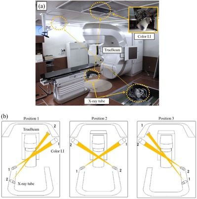
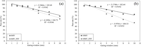
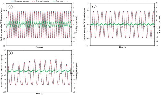
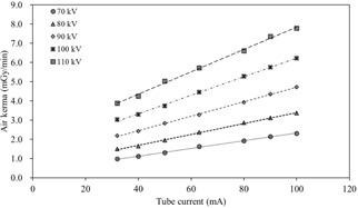
A combined system comprising the TrueBeam linear accelerator and a new real-time, tumor-tracking radiotherapy system, SyncTraX, was installed in our institution. The goals of this study were to assess the capability of SyncTraX in measuring the position of a fiducial marker using color fluoroscopic images, and to evaluate the dosimetric and geometric accuracy of respiratory-gated radiotherapy using this combined system for the simple geometry. For the fundamental evaluation of respiratory-gated radiotherapy using SyncTraX, the following were performed:1) determination of dosimetric and positional characteristics of sinusoidal patterns using a motor-driven base for several gating windows; 2) measurement of time delay using an oscilloscope; 3) positional verification of sinusoidal patterns and the pattern in the case of a lung cancer patient; 4) measurement of the half-value layer (HVL in mm AL), effective kVp, and air kerma, using a solid-state detector for each fluoroscopic condition, to determine the patient dose. The dose profile in a moving phantom with gated radiotherapy having a gating window ≤ 4 mm was in good agreement with that under static conditions for each photon beam. The total time delay between TrueBeam and SyncTraX was < 227 ms for each photon beam. The mean of the positional tracking error was < 0.4 mm for sinusoidal patterns and for the pattern in the case of a lung cancer patient. The air-kerma rates from one fluoroscopy direction were 1.93 ± 0.01, 2.86 ± 0.01, 3.92 ± 0.04, 5.28 ± 0.03, and 6.60 ± 0.05 mGy/min for 70, 80, 90, 100, and 110 kV X-ray beams at 80 mA, respectively. The combined system comprising TrueBeam and SyncTraX could track the motion of the fiducial marker and control radiation delivery with reasonable accuracy; therefore, this system provides significant dosimetric improvement. However, patient exposure dose from fluoroscopy was not clinically negligible.
© 2016 The Authors.
Figures


Similar articles
-
Technical aspects of real time positron emission tracking for gated radiotherapy.Med Phys. 2016 Feb;43(2):783-95. doi: 10.1118/1.4939664. Med Phys. 2016. PMID: 26843241
-
Verification of respiratory-gated radiotherapy with new real-time tumour-tracking radiotherapy system using cine EPID images and a log file.Phys Med Biol. 2017 Feb 21;62(4):1585-1599. doi: 10.1088/1361-6560/aa587d. Epub 2017 Jan 10. Phys Med Biol. 2017. PMID: 28072584
-
Clinical commissioning of a new patient positioning system, SyncTraX FX4, for intracranial stereotactic radiotherapy.J Appl Clin Med Phys. 2018 Nov;19(6):149-158. doi: 10.1002/acm2.12467. Epub 2018 Oct 1. J Appl Clin Med Phys. 2018. PMID: 30273444 Free PMC article.
-
Particle therapy of moving targets-the strategies for tumour motion monitoring and moving targets irradiation.Br J Radiol. 2016 Oct;89(1066):20150275. doi: 10.1259/bjr.20150275. Epub 2016 Jul 19. Br J Radiol. 2016. PMID: 27376637 Free PMC article. Review.
-
Technologies of image guidance and the development of advanced linear accelerator systems for radiotherapy.Front Radiat Ther Oncol. 2011;43:132-164. doi: 10.1159/000322414. Epub 2011 May 20. Front Radiat Ther Oncol. 2011. PMID: 21625152 Review.
Cited by
-
Clinical application of real-time tumor-tracking for stereotactic volumetric modulated arc therapy for liver tumors.Phys Imaging Radiat Oncol. 2024 Aug 5;31:100623. doi: 10.1016/j.phro.2024.100623. eCollection 2024 Jul. Phys Imaging Radiat Oncol. 2024. PMID: 39224689 Free PMC article.
-
Biomedical advances and future prospects of high-precision three-dimensional radiotherapy and four-dimensional radiotherapy.Proc Jpn Acad Ser B Phys Biol Sci. 2023 Nov 10;99(9):389-426. doi: 10.2183/pjab.99.024. Epub 2023 Oct 12. Proc Jpn Acad Ser B Phys Biol Sci. 2023. PMID: 37821390 Free PMC article.
-
"Evaluation of a combined respiratory-gating system comprising the TrueBeam linear accelerator and a new real-time tumor-tracking radiotherapy system: A preliminary study" [JACMP, 17(4), 2016].J Appl Clin Med Phys. 2017 Jul;18(4):238. doi: 10.1002/acm2.12125. Epub 2017 Jun 26. J Appl Clin Med Phys. 2017. PMID: 28681447 Free PMC article. No abstract available.
-
Enhanced analysis of gating latency in 0.35T MR-linac through innovative time synchronization of a motion phantom and plastic scintillation detector.J Appl Clin Med Phys. 2025 Jul;26(7):e70116. doi: 10.1002/acm2.70116. Epub 2025 May 13. J Appl Clin Med Phys. 2025. PMID: 40358922 Free PMC article.
-
Feasibility of a real-time dual energy markerless monitoring of lung tumors using a clinical room-mounted stereoscopic and monoscopic x-ray imaging system.Med Phys. 2025 Jul;52(7):e17966. doi: 10.1002/mp.17966. Med Phys. 2025. PMID: 40665506 Free PMC article.
References
-
- Langen KM and Jones DTL. Organ motion and its management. Int J Radiat Oncol Biol Phys. 2001;50(1):265–78. - PubMed
-
- Nakamura M, Shibuya K, Shiinoki T et al. Positional reproducibility of pancreatic tumors under end‐exhalation breath‐hold conditions using a visual feedback technique. Int J Radiat Oncol Biol Phys. 2011;79(5):1565–71. - PubMed
-
- Berson AM, Emery R, Rodriguez L et al. Clinical experience using respiratory gated radiation therapy: comparison of free‐breathing and breath‐hold techniques. Int J Radiat Oncol Biol Phys. 2004;60(2):419–26. - PubMed
-
- Ford EC, Mageras GS, Yorke E, Rosenzweig KE, Wagman R, Ling CC. Evaluation of respiratory movement during gated radiotherapy using film and electronic portal imaging. Int J Radiat Oncol Biol Phys. 2002;52(2):522–31. - PubMed
-
- Ramsey CR, Cordrey IL, and Oliver AL. A comparison of beam characteristics for gated and nongated clinical x‐ray beams. Med Phys. 1999;26(10):2086–91. - PubMed
Publication types
MeSH terms
LinkOut - more resources
Full Text Sources
Other Literature Sources
Medical
