. 2016 Jul 26;4(1):75.
doi: 10.1186/s40478-016-0344-1.

Role of IL-33 and ST2 signalling pathway in multiple sclerosis: expression by oligodendrocytes and inhibition of myelination in central nervous system

Affiliations

Role of IL-33 and ST2 signalling pathway in multiple sclerosis: expression by oligodendrocytes and inhibition of myelination in central nervous system

Debbie Allan et al. Acta Neuropathol Commun. .

Abstract

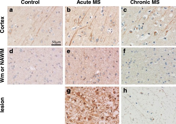
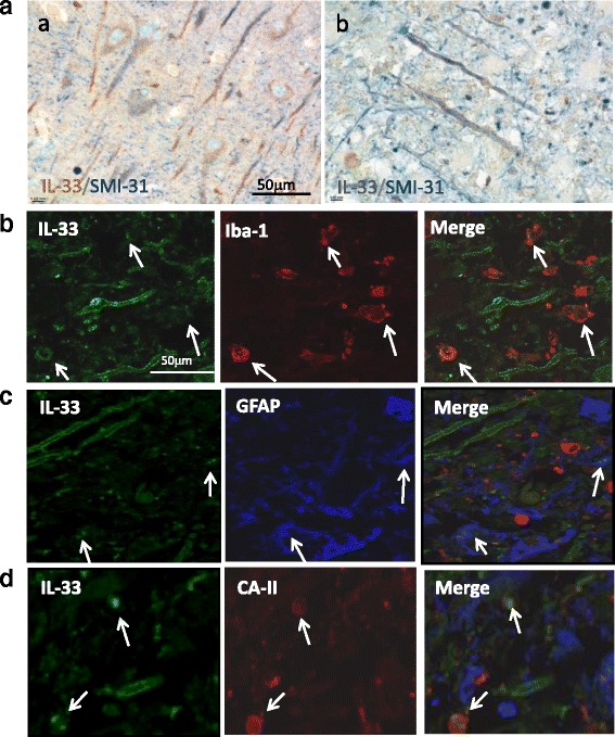
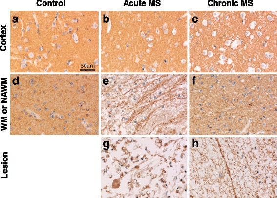
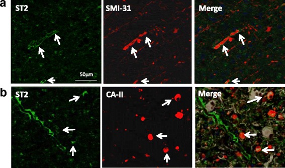
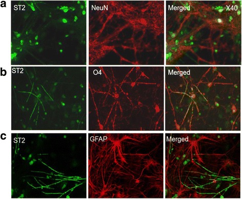
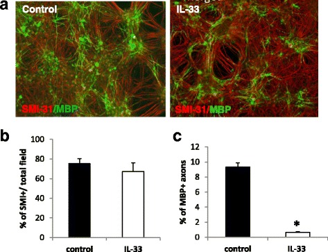
Recent research findings have provided convincing evidence indicating a role for Interleukin-33 (IL-33) signalling pathway in a number of central nervous system (CNS) diseases including multiple sclerosis (MS) and Alzheimer's disease. However, the exact function of IL-33 molecule within the CNS under normal and pathological conditions is currently unknown. In this study, we have mapped cellular expression of IL-33 and its receptor ST2 by immunohistochemistry in the brain tissues of MS patients and appropriate controls; and investigated the functional significance of these findings in vitro using a myelinating culture system. Our results demonstrate that IL-33 is expressed by neurons, astrocytes and microglia as well as oligodendrocytes, while ST2 is expressed in the lesions by oligodendrocytes and within and around axons. Furthermore, the expression levels and patterns of IL-33 and ST2 in the lesions of acute and chronic MS patient brain samples are enhanced compared with the healthy brain tissues. Finally, our data using rat myelinating co-cultures suggest that IL-33 may play an important role in MS development by inhibiting CNS myelination.

Keywords: IL-33; Multiple sclerosis; Myelination; Oligodendrocyte; ST2.


Figures

Fig. 1
Fig. 1
Expression of IL-33 (brown) in healthy and MS brain patient samples. Immunohistochemical staining was used to determine the expression of IL-33 in the cortex and WM of healthy brain samples, and in the cortex, NAWM and lesion of acute and chronic MS patients. a cortex and d white matter of healthy brain tissues; b cortex, e NAWM and g lesion of acute MS brain sample; c cortex, f NAWM and h lesion of chronic MS brain sample
Fig. 2
Fig. 2
Expression of IL-33 by CNS resident cells in human. a Co-staining of IL-33 (brown) with SMI-31 (blue), a marker for neuron and axons, in cortex of normal human brain (a) and MS acute lesion (b). b Co-staining of IL-33 with Iba1, a marker for microglia cells, in human brain samples. Arrows indicate colocalization of IL-33 with Iba-1. c Co-staining of IL-33 with GFAP, a marker for astrocytes (arrows) in human brain samples. d Co-staining of IL-33 with CA-II, a marker for oligodendrocytes (arrows) in human brain samples
Fig. 3
Fig. 3
Expression of ST2 (brown) in healthy and MS patient brain samples. Immunohistochemical staining was used to determine the expression of ST2 in the cortex and WM of healthy brain samples, and in the cortex, NAWM and lesion of acute and chronic MS patients. a cortex and d white matter of healthy brain tissues; b cortex, e NAWM and g lesion of acute MS brain sample; c cortex, f NAWM and h lesion of chronic MS brain sample
Fig. 4
Fig. 4
Expression of ST2 by CNS cells in human. a Co-staining of ST2 (green) with SMI-31 (red), a marker for axons, in human brain samples. Arrows indicate expression of ST2 around SMI-31 expressing axons. b Co-staining of ST2 (green) with CA-II (red) positive oligodendrocytes in human brain samples. Arrows indicate colocalization of ST2 with CA-II expressing oligodendrocytes
Fig. 5
Fig. 5
Expression of ST2 by CNS cells in rat melinating co-cultures. Double immunofluorescence staining of ST2 (green) and antibodies against NeuN (red, marker for neuron cells) and GFAP (red, astrocyte marker), and the O4 antibody (red, a marker for immature oligodendrocytes and mature progenitor cells) in the cultured CNS cells at 12 DIV
Fig. 6
Fig. 6
Recombinant IL-33 inhibits axon myelination in rat myelinating co-cultures. The cell cultures were treated with or without recombinant IL-33 from DIV 12 to 28, a and the cells immuno-labelled with SMI-31 and anti-MBP, markers for axons and mature myelin respectively. b Graph depicting the percentage of SMI-31+ cells in total field of view. c Graph depicting the percentage of myelination measured by MBP+ and SMI-31+ overlapping pixels/total SMI31 pixels. * P < 0.05. Data are presented as Mean + SEM, and were compiled from three independent experiments

References

    1. Molofsky AB, Savage AK, Locksley RM. Interleukin-33 in Tissue Homeostasis, Injury, and Inflammation. Immunity. 2015;42:1005–19. doi: 10.1016/j.immuni.2015.06.006. - DOI - PMC - PubMed
    1. Rostan O, Arshad MI, Piquet-Pellorce C, Robert-Gangneux F, Gangneux J-P, Samson M. Crucial and diverse role of the interleukin-33/ST2 axis in infectious diseases. Infect Immun. 2015;83:1738–48. doi: 10.1128/IAI.02908-14. - DOI - PMC - PubMed
    1. Pei C, Barbour M, Fairlie-Clarke KJ, Allan D, Mu R, Jiang H-R. Emerging role of interleukin-33 in autoimmune diseases. Immunol. 2014;141:9–17. doi: 10.1111/imm.12174. - DOI - PMC - PubMed
    1. Kaczmarek A, Vandenabeele P, Krysko DV. Necroptosis: the release of damage-associated molecular patterns and its physiological relevance. Immunity. 2013;38:209–23. doi: 10.1016/j.immuni.2013.02.003. - DOI - PubMed
    1. Gadani SP, Walsh JT, Smirnov I, Zheng J, Kipnis J. The glia-derived alarmin IL-33 orchestrates the immune response and promotes recovery following CNS injury. Neuron. 2015;85:703–9. doi: 10.1016/j.neuron.2015.01.013. - DOI - PubMed

MeSH terms