. 2016 Jul 26:6:30093.
doi: 10.1038/srep30093.

Immune infiltration and PD-L1 expression in the tumor microenvironment are prognostic in osteosarcoma

Affiliations

Immune infiltration and PD-L1 expression in the tumor microenvironment are prognostic in osteosarcoma

Pratistha Koirala et al. Sci Rep. .

Abstract

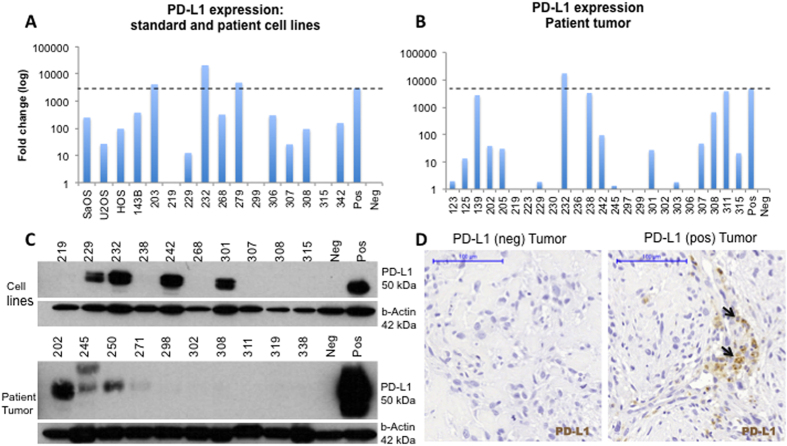
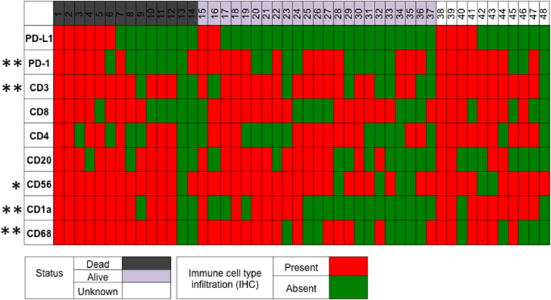
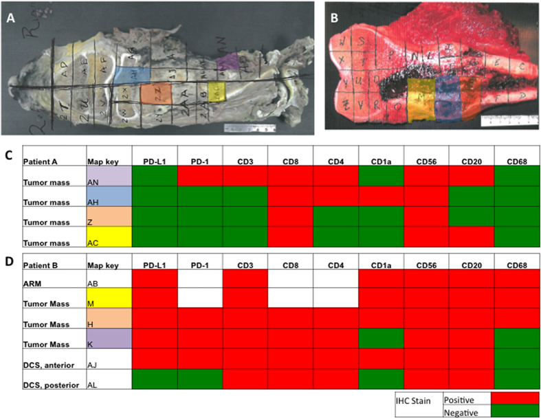
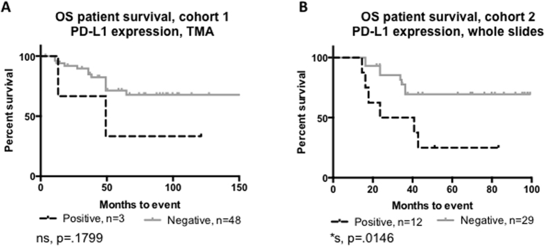
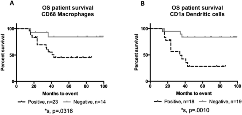
Osteosarcoma patient survival has remained stagnant for 30 years. Novel therapeutic approaches are needed to improve outcomes. We examined the expression of Programmed Death Ligand 1 (PD-L1) and defined the tumor immune microenvironment to assess the prognostic utility in osteosarcoma. PD-L1 expression in osteosarcoma was examined in two patient cohorts using immunohistochemistry (IHC) (n = 48, n = 59) and expression was validated using quantitative real time PCR (n = 21) and western blotting (n = 9). IHC was used to determine the presence of tumor infiltrating lymphocytes and antigen-presenting cells (APCs) in the tumor. Expression of PD-L1 was correlated with immune cell infiltration and event-free-survival (EFS). The 25% of primary osteosarcoma tumors that express PD-L1 were more likely to contain cells that express PD-1 than PD-L1 negative tumors (91.7% vs 47.2%, p = 0.002). Expression of PD-L1 was significantly associated with the presence of T cells, dendritic cells, and natural killer cells. Although all immune cell types examined were present in osteosarcoma samples, only infiltration by dendritic cells (28.3% vs. 83.9%, p = 0.001) and macrophages (45.5% vs. 84.4%, p = 0.031) were associated with worse five-year-EFS. PD-L1 expression was significantly associated with poorer five-year-EFS (25.0%. vs. 69.4%, p = 0.014). Further studies in osteosarcoma are needed to determine if targeting the PD-L1:PD-1 axis improves survival.


Figures

Figure 1
Figure 1. PD-L1 is expressed in some primary osteosarcoma tumors.
(A) PD-L1 mRNA expression in cell lines derived from primary osteosarcoma tumors. The PD-L1 expressing breast cancer cell line MDA-MB-231 was used as the positive control. (B) PD-L1 mRNA expression in osteosarcoma tumor specimens. Dashed lines represent the level of PD-L1 expression in the positive control. (C) Western blot validation confirms that PD-L1 is expressed in primary osteosarcoma tumors (30%) (D) IHC on selected osteosarcoma tumor specimens show areas of heterogeneous PD-L1 expression. Scale bar represents 100 μm. Neg = negative, pos = positive.
Figure 2
Figure 2. Expression of the immune checkpoint, PD-L1, is associated with the presence of tumor infiltrating lymphocytes and antigen presenting cells.
Each individual column represents a unique patient; of the 28 patients included in the study, 37 patients had known clinical outcomes, and 29 patients had both clinical outcome data and as well as demographic data available. Rows represent either PD-L1 expression or tumor infiltration by an immune cell type, as determined by immunohistochemistry. PD-L1 expression was significantly associated with infiltration by PD-1+ immune cells, CD3+ T cells, CD56+ natural killer cells, CD68+ cells, and CD1a+ dendritic cells. Presence of immune cells or positive PD-L1 expression was defined as IHC staining of >1% the tumor volume. * p < 0.05, **p < 0.01.
Figure 3
Figure 3. PD-L1 expression is consistent throughout the tumor mass.
(A,C) All four slides from the tumor mass of patient A did not demonstrate PD-L1 staining. Few immune cells are present in this tumor map. (B,D) All of the slides from the tumor mass of patient B stained PD-L1 positive with the exception of one section outside the tumor mass (not shown). Numerous immune cells are present throughout all sections of this tumor map. ARM = anterior resection margin, DCS = distal cross section.
Figure 4
Figure 4. PD-L1 expression is prognostic in osteosarcoma.
(A) PD-L1 expression in the TMA trends with poorer EFS. (B) PD-L1 expression in the whole slide specimens is significantly associated with poorer EFS.
Figure 5
Figure 5. APC infiltration is prognostic in osteosarcoma.
Presence of APCs, specifically (A) CD68 positive macrophages or (B) CD1a positive dendritic cells are significantly associated with poorer EFS in osteosarcoma. Over 50% of osteosarcoma samples have APCs present.

References

    1. Damron T. A., Ward W. G. & Stewart A. Osteosarcoma, chondrosarcoma, and Ewing’s sarcoma: National Cancer Data Base Report. Clin Orthop Relat Res. 459, 40–47, 10.1097/BLO.0b013e318059b8c9 (2007). - DOI - PubMed
    1. Sweetnam R. Osteosarcoma. Br J Hosp Med. 28(112), 116–121 (1982). - PubMed
    1. Ottaviani G. & Jaffe N. The epidemiology of osteosarcoma. Cancer Treat Res. 152, 3–13, 10.1007/978-1-4419-0284-9_1 (2009). - DOI - PubMed
    1. Carrle D. & Bielack S. Osteosarcoma lung metastases detection and principles of multimodal therapy. Cancer Treat Res. 152, 165–184, 10.1007/978-1-4419-0284-9_8 (2009). - DOI - PubMed
    1. Harting M. T. & Blakely M. L. Management of osteosarcoma pulmonary metastases. Semin Pediatr Surg. 15, 25–29, 10.1053/j.sempedsurg.2005.11.005 (2006). - DOI - PubMed

Publication types