. 2016 Sep;186(9):2500-14.
doi: 10.1016/j.ajpath.2016.05.016. Epub 2016 Jul 25.

Increased Pyruvate Dehydrogenase Kinase 4 Expression in Lung Pericytes Is Associated with Reduced Endothelial-Pericyte Interactions and Small Vessel Loss in Pulmonary Arterial Hypertension

Affiliations

Increased Pyruvate Dehydrogenase Kinase 4 Expression in Lung Pericytes Is Associated with Reduced Endothelial-Pericyte Interactions and Small Vessel Loss in Pulmonary Arterial Hypertension

Ke Yuan et al. Am J Pathol. 2016 Sep.

Abstract

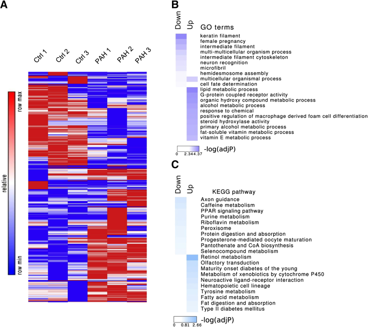
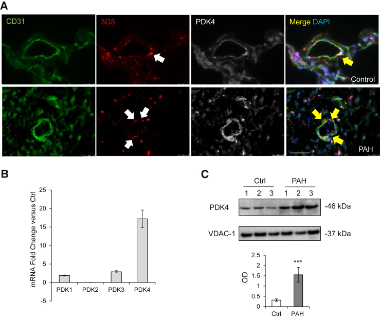
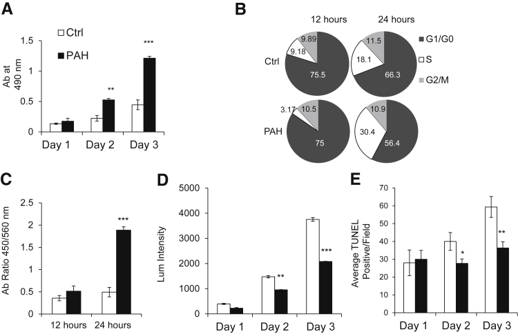
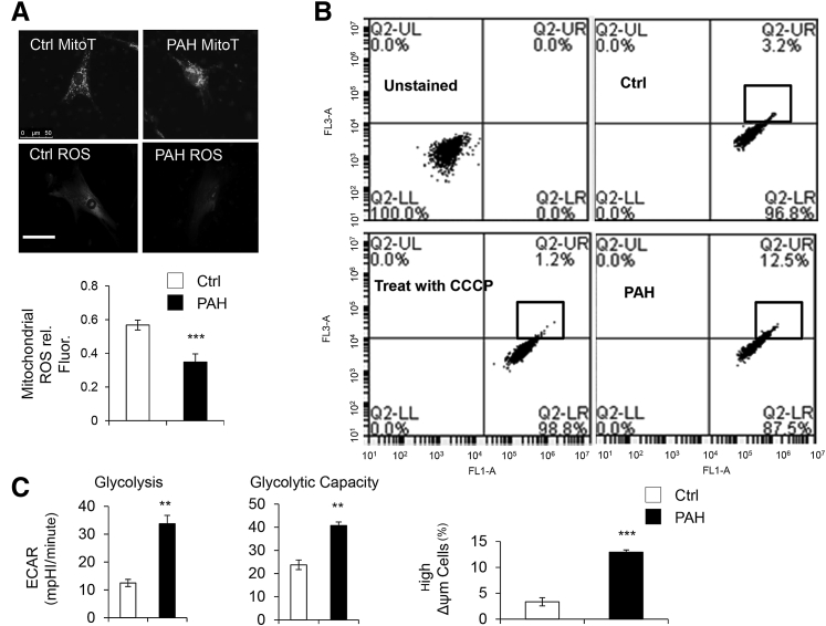
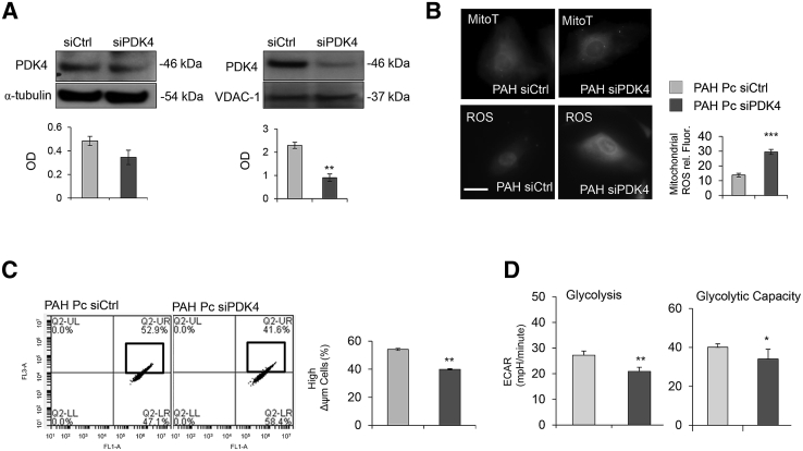
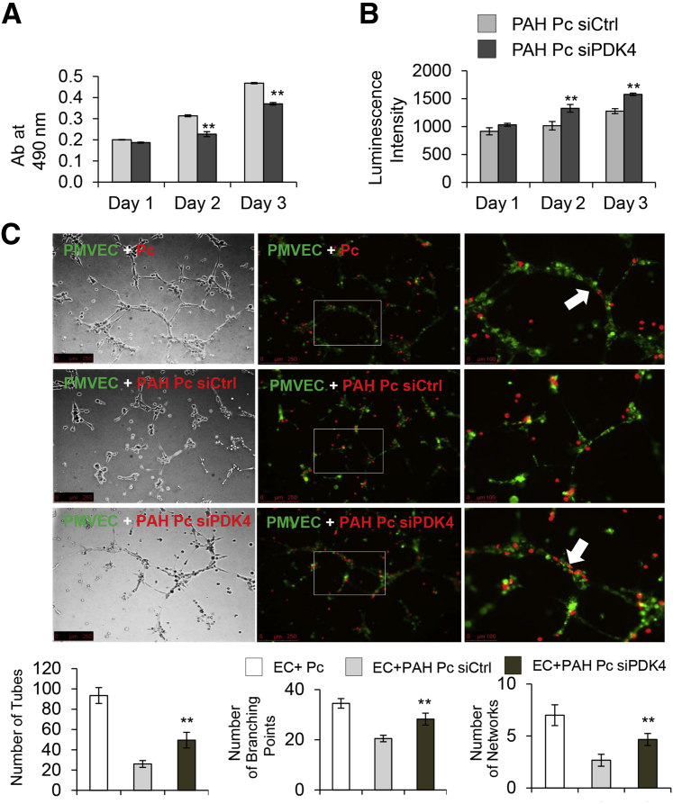
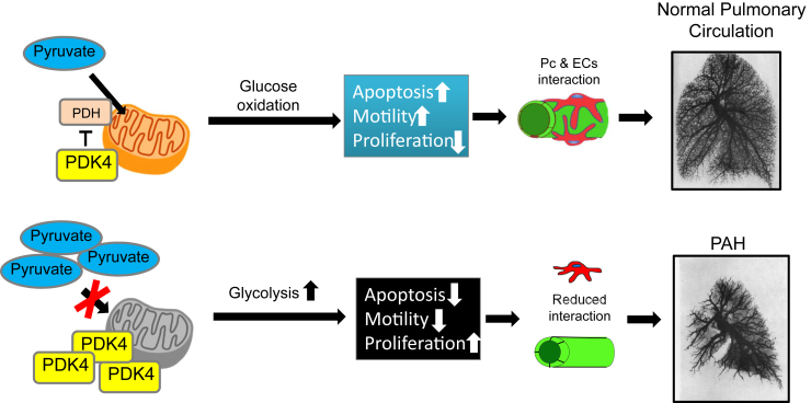
Reduced endothelial-pericyte interactions are linked to progressive small vessel loss in pulmonary arterial hypertension (PAH), but the molecular mechanisms underlying this disease remain poorly understood. To identify relevant gene candidates associated with aberrant pericyte behavior, we performed a transcriptome analysis of patient-derived donor control and PAH lung pericytes followed by functional genomics analysis. Compared with donor control cells, PAH pericytes had significant enrichment of genes involved in various metabolic processes, the top hit being PDK4, a gene coding for an enzyme that suppresses mitochondrial activity in favor of glycolysis. Given reports that link reduced mitochondrial activity with increased PAH cell proliferation, we hypothesized that increased PDK4 is associated with PAH pericyte hyperproliferation and reduced endothelial-pericyte interactions. We found that PDK4 gene and protein expression was significantly elevated in PAH pericytes and correlated with reduced mitochondrial metabolism, higher rates of glycolysis, and hyperproliferation. Importantly, reducing PDK4 levels restored mitochondrial metabolism, reduced cell proliferation, and improved endothelial-pericyte interactions. To our knowledge, this is the first study that documents significant differences in gene expression between human donor control and PAH lung pericytes and the link between mitochondrial dysfunction and aberrant endothelial-pericyte interactions in PAH. Comprehensive characterization of these candidate genes could provide novel therapeutic targets to improve endothelial-pericyte interactions and prevent small vessel loss in PAH.


Figures

Figure 1
Figure 1
Comparison of gene expression patterns of healthy donor control (Ctrl) and pulmonary arterial hypertension (PAH) pericytes by microarray analysis. A: The heat map of differently expressed genes (P < 0.05, fold change >2.0) between three PAH versus three donor control pericytes. B: Gene Ontology (GO) enrichment analyses of significantly up-regulated/down-regulated genes in PAH. C: Kyoto Encyclopedia of Genes and Genomes (KEGG) pathway enrichment analyses of significantly up-regulated/down-regulated genes in PAH.
Figure 2
Figure 2
PDK4 increases in pulmonary arterial hypertension (PAH) pericytes. A: Confocal images of PDK4 lung sections. Endothelium was stained with CD31 (green), pericytes with 3G5 (red), and lung tissue with PDK4 (white). White arrows indicate pericyte-positive staining, and yellow arrows indicate co-staining of 3G5 and PDK4. DAPI labels cell nuclei (blue). B: TaqMan quantitative PCR for the four PDK family proteins in donor control and PAH pericytes. The expression of each gene is shown relative to that in donor control pericytes. C: Representative Western immunoblot images of PDK4 in enriched mitochondria of pericytes from three donor control (Ctrl) versus three patients with PAH. ∗∗∗P < 0.001 versus unstimulated controls using the unpaired t-test. Scale bar = 50 μm. OD, optical density.
Figure 3
Figure 3
Pulmonary arterial hypertension (PAH) pericytes have greater proliferation and survival compared with healthy donor cells. A: Cell growth was evaluated by the MTS assay on days 1, 2, and 3 of culture. All values were normalized to the cells after 48 hours of starvation medium. B: Serum-starved cells were analyzed by flow cytometry on day 3 after the addition of serum of 12 and 24 hours. The percentage of cells in the G1, S, and G2/M phases is indicated. C: DNA analysis of control and PAH pericytes in 12 hours and 24 hours by BrdU. D: Cell apoptosis was evaluated by caspase 3/7 assay on days 1, 2, and 3 of culture. All values were normalized to the cells after 48 hours of starvation medium. E: Cell apoptosis level was further evaluated by the terminal deoxynucleotidyl transferase-mediated dUTP nick-end labeling (TUNEL) assay on days 1, 2, and 3 of culture. All values were normalized after 48 hours of starvation. Average TUNEL-positive cells per field obtained from six independent microscopic fields. All experiments performed were repeated three times P < 0.05, ∗∗P < 0.01, and ∗∗∗P < 0.001 versus control using one-way analysis of variance with Dunnett's posttest. Ctrl, control.
Figure 4
Figure 4
Increased PDK4 expression correlates with reduced mitochondrial activity in pulmonary arterial hypertension (PAH) pericytes. A: Reactive oxygen species (ROS) production in PAH versus donor control (Ctrl) cells. Fluorescence intensity (rel. Fluor.) was normalized against MitoTracker (MitoT). B: Fluorescence activated cell sorting of healthy donor and PAH pericytes using mitochondrial membrane potential (MMP). FL1-A: fluorescein isothiocyanate; FL3-A: Texas Red. The percentage of high MMP is found in the block of Q2-UR. C: Aerobic glycolysis and glycolytic capacity in cells as measured by Seahorse analysis. ∗∗P < 0.01, ∗∗∗P < 0.001 compared with controls using unpaired t-test. Scale bar = 50 μm. CCCP, Carbonyl cyanide 3-chlorophenylhydrazone; ECAR, extracellular acidification rate; LL, lower left; LR, lower right; mpH, mili pH per minute; UL, up-left; UR, up-right.
Figure 5
Figure 5
Knockdown of PDK4 in pulmonary arterial hypertension (PAH) pericytes restores mitochondrial activity. A: Western immunoblot for PDK4 in total cell lysate (α-tubulin as an internal control) and mitochondria lysate (VDAC-1 as an internal control) from PAH pericytes transfected with either nontargeting (siCtrl) or PDK4-specific (siPDK4) siRNAs. B: Reactive oxygen species (ROS) production in PAH pericytes treated with either siCtrl or siPDK4. Fluorescence intensity (rel. Fluor.) was normalized against MitoTracker (MitoT). Mitochondrial membrane potential (MMP) (C) and aerobic glycolysis and glycolytic capacity (D) in donor control versus PAH with PDK4 siRNA treatment. P < 0.05, ∗∗P < 0.01, and ∗∗∗P < 0.001 compared with controls using the unpaired t-test. Scale bar = 25 μm. ECAR, extracellular acidification rate; LL, lower left; LR, lower right; mpH, mili pH per minute; OD, optical density; Pc, pericyte; UL, up-left; UR, up right.
Figure 6
Figure 6
Knockdown of PDK4 reduces proliferation and survival of pulmonary arterial hypertension (PAH) pericytes and restores capacity to enhance endothelial tube formation. Cell growth by MTS (A) and cell apoptosis by caspase 3/7 (B) were evaluated on days 1, 2, and 3 of culture after 48 hours siRNA posttransfection. C: Pericytes (Pcs) were transfected with nontargeting siRNA control (siCtrl) or PDK4-specific (siPDK4) for 48 hours when compared with healthy pericytes. Before seeded on Matrigel, cells were stained with PKHs. Pulmonary microvascular endothelial cells (PMVECs) were stained in PKH67 (green) and PAH Pcs in PKH26 (red). The white boxes indicate the enlarged areas of magnification in the right column. The white arrows indicate EC-Pc interaction. The numbers of tubes, branching points, and networks were assessed after six hours. ∗∗P < 0.01 compared with control (unpaired t-test, one-way analysis of variance with Bonferroni post-test). Scale bars: 250 μm (C, left and middle columns); 100 μm (C, right column). EC, endothelial cell.
Figure 7
Figure 7
Proposed model. Elevated PDK4 in pulmonary arterial hypertension (PAH) pericytes (Pcs) reduces mitochondrial metabolism in favor of glycolysis. Although pericytes exhibit heightened proliferation and survival, they have limited capacity to establish communication with pulmonary microvascular endothelial cells (ECs), resulting in small vessel loss and impaired angiogenesis.

References

    1. Humbert M., Morrell N.W., Archer S.L., Stenmark K.R., MacLean M.R., Lang I.M., Christman B.W., Weir E.K., Eickelberg O., Voelkel N.F., Rabinovitch M. Cellular and molecular pathobiology of pulmonary arterial hypertension. J Am Coll Cardiol. 2004;43:13S–24S. - PubMed
    1. Rich S., Dantzker D.R., Ayres S.M., Bergofsky E.H., Brundage B.H., Detre K.M. Primary pulmonary hypertension: a national prospective study. Ann Intern Med. 1987;107:216–223. - PubMed
    1. Tuder R.M., Voelkel N.F. Angiogenesis and pulmonary hypertension: a unique process in a unique disease. Antioxid Redox Signal. 2002;4:833–843. - PubMed
    1. Tuder R.M., Cool C.D., Yeager M., Taraseviciene-Stewart L., Bull T.M., Voelkel N.F. The pathobiology of pulmonary hypertension: endothelium. Clin Chest Med. 2001;22:405–418. - PubMed
    1. Voelkel N.F., Gomez-Arroyo J. The role of vascular endothelial growth factor in pulmonary arterial hypertension: the angiogenesis paradox. Am J Respir Cell Mol Biol. 2014;51:474–484. - PubMed

MeSH terms

Substances