Randomized Controlled Trial
. 2016 Sep 1;197(5):1801-8.
doi: 10.4049/jimmunol.1501330. Epub 2016 Jul 25.

Mucosal Inducible NO Synthase-Producing IgA+ Plasma Cells in Helicobacter pylori-Infected Patients

Affiliations
Randomized Controlled Trial

Mucosal Inducible NO Synthase-Producing IgA+ Plasma Cells in Helicobacter pylori-Infected Patients

Laura Neumann et al. J Immunol. .

Abstract

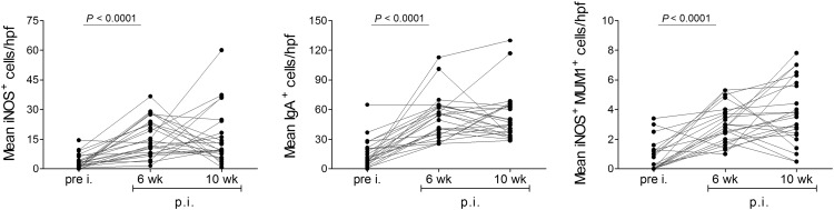
The mucosal immune system is relevant for homeostasis, immunity, and also pathological conditions in the gastrointestinal tract. Inducible NO synthase (iNOS)-dependent production of NO is one of the factors linked to both antimicrobial immunity and pathological conditions. Upregulation of iNOS has been observed in human Helicobacter pylori infection, but the cellular sources of iNOS are ill defined. Key differences in regulation of iNOS expression impair the translation from mouse models to human medicine. To characterize mucosal iNOS-producing leukocytes, biopsy specimens from H. pylori-infected patients, controls, and participants of a vaccination trial were analyzed by immunohistochemistry, along with flow cytometric analyses of lymphocytes for iNOS expression and activity. We newly identified mucosal IgA-producing plasma cells (PCs) as one major iNOS(+) cell population in H. pylori-infected patients and confirmed intracellular NO production. Because we did not detect iNOS(+) PCs in three distinct infectious diseases, this is not a general feature of mucosal PCs under conditions of infection. Furthermore, numbers of mucosal iNOS(+) PCs were elevated in individuals who had cleared experimental H. pylori infection compared with those who had not. Thus, IgA(+) PCs expressing iNOS are described for the first time, to our knowledge, in humans. iNOS(+) PCs are induced in the course of human H. pylori infection, and their abundance seems to correlate with the clinical course of the infection.


Figures

FIGURE 1.
FIGURE 1.
The antral mucosa of H. pylori–infected patients contains enhanced numbers of iNOS+, CD68+, PAX5+, and IgA+ cells as well as iNOS+MUM1+ PCs. (A) Representative immunohistochemically stained paraffin sections of mucosal biopsy specimens of H. pylori–negative controls (H. pylori) and H. pylori–infected patients (H. pylori+). iNOS-expressing cells were identified by anti-iNOS Ab, macrophages by anti-CD68 Ab, B cells by anti-PAX5 Ab, and PCs by anti-IgA Ab. iNOS-producing PCs were visualized by double staining of anti-MUM1 and anti-iNOS Ab. Scale bar, 20 μm. (B) Quantitative analysis of iNOS+ cells, CD68+ cells, Pax5+ cells, IgA+ cells, and iNOS+MUM1+ cells in the antral mucosa of uninfected controls (n = 16, black) and H. pylori+ patients (n = 38, gray). Positive cells were counted per hpf and depicted as median plus interquartile ranges (Mann–Whitney test).
FIGURE 2.
FIGURE 2.
Early mucosal influx of iNOS+ cells, IgA+ cells, and iNOS+MUM1+ PCs in experimental human H. pylori infection. Quantitative analysis of iNOS+ cells, IgA+ cells, and iNOS+MUM1+ cells in the antral mucosa of study participants (n = 24) at preinfection (pre i.) as well as 6 and 10 wk (wk) p.i. with H. pylori. Immunohistochemical staining was performed on paraffin sections. Positive cells per hpf are depicted as associated values over time. Data were analyzed with the Friedman test with Dunn’s post hoc analysis.
FIGURE 3.
FIGURE 3.
Infiltration of B cells and iNOS-expressing PCs in H. pylori+ patients. (A) Representative gating strategy for analysis of LPLs by flow cytometry: 1) gating on lymphocytes by forward (FSC) and side scatter (SSC) properties; 2) exclusion of T cells (CD3+) and monocytes (CD14+) and gating on CD19+ B cells; 3) detection of PCs (CD20CD27++), mBCs (CD20+CD27+), and naive B cells (CD20+CD27); 4) detection of iNOS+CD38++ PCs; and 5) detection of IgA+ PCs within iNOS+CD38++ PCs. (B) Quantitative analysis of the flow cytometric data of H. pylori–infected patients according to the gating strategy. Values are depicted as median plus interquartile ranges (Mann–Whitney U test).
FIGURE 4.
FIGURE 4.
Detection of NO-producing PCs in H. pylori–infected patients. (A) Representative gating strategy for the analysis of CD19+CD3CD14DAPI PCs of H. pylori–infected patients by flow cytometry: gating on PCs (CD20CD27++) within all CD19+ B cells, and then on CD38++DAF-FM-T+ PCs. (B) Quantitative analysis of the data of H. pylori+ patients (n = 3). Values are depicted as median plus interquartile ranges.
FIGURE 5.
FIGURE 5.
Increased numbers of iNOS+ PCs in the antral mucosa of individuals able to eradicate H. pylori. Quantitative analysis of iNOS+ cells, IgA+ cells, and iNOS+MUM1+ cells in the antral mucosa of subjects preinfection (pre i.) as well as 6 and 10 wk p.i. with H. pylori. Biopsy specimens were obtained from study participants who had developed a persistent H. pylori infection (Persistent, n = 17, gray) and participants who had eradicated H. pylori (Eradicated, n = 7, black). Positive cells were counted per hpf, and values are shown as the mean of cells per hpf. Values are depicted as median plus interquartile ranges (Mann–Whitney U test followed by Bonferroni correction). The p values <0.017 were considered significant.
FIGURE 6.
FIGURE 6.
iNOS-expressing B lineage cells produce pro- and anti-inflammatory cytokines. (A) Detection of intracellular iNOS (left) or IFN-γ (right) within CD38, CD38+, and CD38++ B-LCL cells by flow cytometry. (B) Detection of IFN-γ within iNOS+CD38++ and iNOSCD38++ B-LCL cells. (C) Relative mRNA expression of IFN-γ, TNF-α, and IL-10 in peripheral or mucosal mBCs during H. pylori infection. mBCs (CD19+CD20+CD27+CD38CD3CD14DAPI) were isolated from PBMCs or LPLs, respectively, by FACS. The qPCR data were analyzed by the comparative Ct method (2−ΔΔCt). All values are normalized to the expression of the housekeeping gene GAPDH, and the reference gene was IL-6. (D) Detection of intracellular TNF-α and IFN-γ within mucosal iNOS and iNOS+ mBCs (CD19+CD20+CD27+CD38CD3CD14) or (E) PCs (CD19+CD20CD27++CD38++CD3CD14) of an H. pylori–infected patient by flow cytometry. Percentages are indicated.

References

    1. Wilson K. T., Crabtree J. E. 2007. Immunology of Helicobacter pylori: insights into the failure of the immune response and perspectives on vaccine studies. Gastroenterology 133: 288–308. - PubMed
    1. Mannick E. E., Bravo L. E., Zarama G., Realpe J. L., Zhang X. J., Ruiz B., Fontham E. T., Mera R., Miller M. J., Correa P. 1996. Inducible nitric oxide synthase, nitrotyrosine, and apoptosis in Helicobacter pylori gastritis: effect of antibiotics and antioxidants. Cancer Res. 56: 3238–3243. - PubMed
    1. Fu S., Ramanujam K. S., Wong A., Fantry G. T., Drachenberg C. B., James S. P., Meltzer S. J., Wilson K. T. 1999. Increased expression and cellular localization of inducible nitric oxide synthase and cyclooxygenase 2 in Helicobacter pylori gastritis. Gastroenterology 116: 1319–1329. - PubMed
    1. Förstermann U, Sessa W. C. 2012. Nitric oxide synthases: regulation and function. Eur. Heart J. 33: 829–837, 837a–837d. doi:10.1093/eurheartj/ehr304. - PMC - PubMed
    1. Murad F. 1999. Discovery of some of the biological effects of nitric oxide and its role in cell signaling. Biosci. Rep. 19: 133–154. - PubMed

Publication types