

Complete regression of primary cutaneous malignant melanoma associated with distant lymph node metastasis: a teaching case mimicking blue nevus
- PMID: 27456492
- PMCID: PMC4960676
- DOI: 10.1186/s13104-016-2174-4
Complete regression of primary cutaneous malignant melanoma associated with distant lymph node metastasis: a teaching case mimicking blue nevus
Abstract
Background: Malignant melanoma (MM) tends to be spontaneously regressed, however, complete regression of primary cutaneous MM is an extremely rare phenomenon. Our aim is to be aware that pathologists and/or dermatologists can readily misinterpret it as the other benign or malignant lesions.
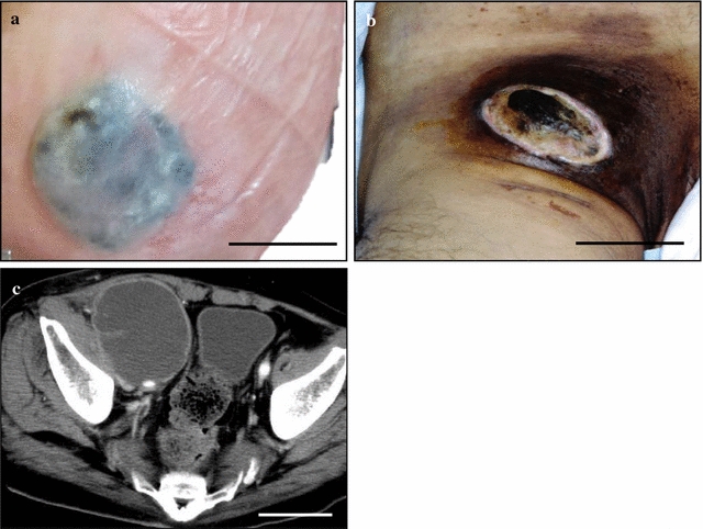
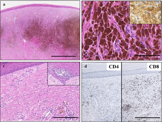
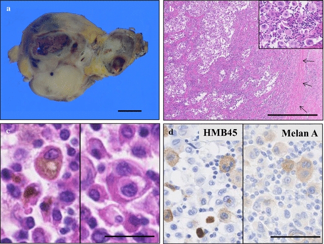
Case presentation: A gradually growing and verrucous hypopigmented macule had been noticed in the right sole of a 65-year-old Japanese male since 2 years before, and it turned to be a solitary bluish to black patch with surrounding depigmentation and was recently decreased in size. In parallel, the patient had a rapidly growing black-pigmented mass lesion at the right inguen. The cutaneous specimen from the sole showed an aggregation of many melanophages predominantly in the middle to deep layer of dermis, associated with surrounding fibrosis, reactive vascular proliferation and CD8-positive T-lymphocytic infiltrate, covered by attenuated epidermis with absence of rete ridge. However, no remnant MM cells were completely seen in the step-serial sections. We first interpreted it as blue nevus. By contrast, the inguinal mass revealed a diffuse proliferation of highly atypical mono- to multi-nucleated large cells having abundant eosinophilic cytoplasm in the enlarged lymph node tissue. Immunohistochemical findings demonstrated that these atypical cells were specifically positive for HMB45 and Melan A. Therefore, we finally made a diagnosis of complete regression of primary cutaneous MM associated with distant lymph node metastasis of MM.
Conclusion: Careful, not only general/cutaneous but histopathological, examinations should be necessary and adjunctive aids for reaching the correct diagnosis of complete regression of cutaneous MM.
Figures
Similar articles
-
Malignant blue nevus: case report of a Japanese man with a distant cutaneous metastasis.Am J Dermatopathol. 2007 Feb;29(1):88-91. doi: 10.1097/01.dad.0000187932.87103.4a. Am J Dermatopathol. 2007. PMID: 17284970
-
Cellular blue nevus (CBN) lymph node metastases of the neck with no primary skin lesion: a case report and review of literature.J Craniomaxillofac Surg. 2010 Dec;38(8):601-4. doi: 10.1016/j.jcms.2010.01.008. Epub 2010 Mar 12. J Craniomaxillofac Surg. 2010. PMID: 20223677 Review.
-
Regressed subungual melanoma simulating cellular blue nevus: managed with sentinel lymph node biopsy.Dermatol Surg. 2006 Apr;32(4):577-80; discussion 580-1. doi: 10.1111/j.1524-4725.2006.32119.x. Dermatol Surg. 2006. PMID: 16681670
-
A pigmented scalp nodule: malignant blue nevus.Cutis. 1996 Jul;58(1):40-2. Cutis. 1996. PMID: 8823547 Review.
-
Benign "metastatic" cellular blue nevus.Ann Plast Surg. 1994 Oct;33(4):426-31. doi: 10.1097/00000637-199410000-00013. Ann Plast Surg. 1994. PMID: 7810962
Cited by
-
Complete regression of primary melanoma associated with nevi involution under BRAF inhibitors: A case report and review of the literature.Oncol Lett. 2019 May;17(5):4176-4182. doi: 10.3892/ol.2018.9738. Epub 2018 Nov 19. Oncol Lett. 2019. PMID: 30944613 Free PMC article.
-
Multiple Primary Cutaneous Melanomas in a Bulgarian Patient: The Possible Role of One Step Melanoma Surgery (OSMS) As the Most Adequate Treatment Approach!Open Access Maced J Med Sci. 2018 Nov 21;6(11):2155-2160. doi: 10.3889/oamjms.2018.487. eCollection 2018 Nov 25. Open Access Maced J Med Sci. 2018. PMID: 30559881 Free PMC article.
-
Primary pulmonary malignant melanoma diagnosed with percutaneous biopsy tissue: A case report.World J Clin Cases. 2020 Dec 26;8(24):6373-6379. doi: 10.12998/wjcc.v8.i24.6373. World J Clin Cases. 2020. PMID: 33392320 Free PMC article.
References
-
- High WA, Stewart D, Wilbers CR, Cockerell CJ, Hoang MP, Fitzpatrick JE. Completely regressed primary cutaneous malignant melanoma with nodal and/or visceral metastases: a report of 5 cases and assessment of the literature and diagnostic criteria. J Am Acad Dermatol. 2005;53:89–100. doi: 10.1016/j.jaad.2005.03.006. - DOI - PubMed
-
- Everson TC, Cole WH. Spontaneous regression of cancer. Philadelphia: WB Saunders; 1966.
MeSH terms
LinkOut - more resources
Full Text Sources
Other Literature Sources
Medical
Research Materials
