Review
. 2016 Aug 28;428(17):3387-98.
doi: 10.1016/j.jmb.2016.07.007. Epub 2016 Jul 22.

Avoidance and Subversion of Eukaryotic Homeostatic Autophagy Mechanisms by Bacterial Pathogens

Affiliations
Review

Avoidance and Subversion of Eukaryotic Homeostatic Autophagy Mechanisms by Bacterial Pathogens

Cheryl Miller et al. J Mol Biol. .

Abstract

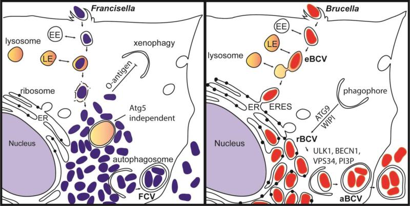
Autophagy is a conserved lysosomal recycling process, which maintains cellular homeostasis during stress and starvation conditions by degrading and recycling proteins, lipids, and carbohydrates, ultimately increasing nutrient availability in eukaryotes. An additional function of autophagy, termed xenophagy, is to detect, capture, and destroy invading microorganisms, such as viruses, bacteria, and protozoa, providing autophagy with a role in innate immunity. Many intracellular pathogens have, however, developed mechanisms to avoid xenophagy and have evolved strategies to take advantage of select autophagic processes to undergo their intracellular life cycle. This review article will discuss the molecular mechanisms used by the intracellular bacterial pathogens Francisella spp. and Brucella spp. to manipulate components of the autophagic pathway, promoting cytosolic growth in the case of Francisella spp. and facilitating cellular egress and cell-to-cell spread in the case of Brucella spp. These examples highlight how successful, highly infectious bacterial pathogens avoid or subvert host autophagy mechanisms normally employed to maintain eukaryotic homeostasis.

Keywords: O-antigen; bacterial secretion system; cellular egress; infection; nutrient acquisition.


Figures

Figure 1
Figure 1. Sequential steps of canonical autophagy
In the presence of amino acids, growth factors and energy, the mTOR complex represses autophagy by inhibiting the kinase activity of ULK1. The PI3K/Akt pathway inhibits autophagy, while AMPK activates autophagy by controlling mTOR activation under nutrient-limiting conditions. Upon autophagy induction (1), the ULK1 complex activates the BECN1/VPS34 complex to initiate (2) phagophore formation and nucleation. BECN1 can be activated directly by the IRE1α/JNK pathway or inhibited by the pharmacological drug 3-methyladenine (3-MA). Phagophore elongation (3) proceeds to engulf and sequester autophagic cargo and the phagophore membrane acquires LC3. Ubiquitin-like conjugation systems mediate the closure of the autophagosome (4). Maturation of the autophagosome (5) occurs via fusion with late endocytic/lysosomal compartments, forming the autolysosome where material is degraded. Autolysosomes are then recycled in a process that allows for lysosome reformation (6).
Figure 2
Figure 2. Autophagy complexes that coordinate autophagosome formation
The ULK1 complex includes ULK1/Atg1, Atg17/RB1CC1/FIP200, Atg29, Atg31, Atg13, Atg11 and Atg101. Atg13 and Atg17/RB1CC1/FIP200 are substrates of ULK1 kinase activity. In addition to autophagy initiation, Atg17, Atg29 and Atg31 are also involved in phagophore closure. The transmembrane protein Atg9 cycling system mediates membrane recruitment during nucleation and elongation and involves interactions between the phosphoinositide PI3P, Atg9, Atg18/WIPI, VMP1 and Atg2 on the phagophore membrane. The Vps34 phosphatidylinositol 3-kinase (PI3P/PtdIns3K/PI3C3) complex includes, either PI3P, Vps15/PIK3R4/p150, BECN1/Atg6 and Atg14L or UVRAG. The Vps34 complex is required for the nucleation of autophagosomal membranes. Two ubiquitin-like conjugating systems, Atg12-Atg5-Atg16L1 and Atg8/LC3, are required for maturation and closure of the autophagosomal membrane. The cysteine protease Atg4 cleaves LC3 to generate LC3-I; Atg3 and Atg7 subsequently conjugate LC3-I to phosphatidylethanolamine (PE), forming the membrane-associated LC3-II. All membranes depicted represent the phagophore membrane.
Figure 3
Figure 3. Intracellular niches of Francisella spp. and Brucella spp
(A) Following internalization, Francisella resides in a vacuole that sequentially acquires markers of early endosomes (EE) and late endosomes (LE). Francisella escapes the original vacuole before lysosomal fusion to reach the cytosol. Once in the cytosol, the F. tularensis capsular and lipopolysaccharide O-antigen is an essential component of xenophagy avoidance. Francisella replicates rapidly in the cytosol of host cells and is found adjacent to autophagosomes. F. tularensis acquires amino acids generated via autophagy in an Atg5 independent manner. After extensive replication in murine macrophages, some bacteria are found in Francisella-containing vacuoles (FCVs). (B) Brucella exploits functions of the host secretory pathway to grow within membrane bound compartments called Brucella-containing vacuoles (BCV) throughout its intracellular life cycle. Brucella first traffics along the endocytic pathway in an endosomal BCV (eBCV), which acquires early endosomal (EE) markers then late endosomal (LE) markers, and partially fuses with lysosomes. Brucella then accesses the secretory pathway via interactions with endoplasmic reticulum (ER) exit sites (ERES) to access the ER. B. abortus requires the autophagy proteins ATG9 and WIPI, for biogenesis of the replicative BCV (rBCV) derived from ER membranes. After extensive replication, some rBCV are engulfed by autophagosome-like structures to become autophagic BCVs (aBCVs), which are involved in bacterial release and cell-to-cell spread. B. abortus selectively subverts several autophagy initiation complexes containing ULK1, BECN1, VPS34 and PI3P to generate an autophagic vacuole important for egress and cell-to-cell spread.

References

    1. Feng Y, He D, Yao Z, Klionsky DJ. The machinery of macroautophagy. Cell Research. 2013;24:24–41. doi:10.1038/cr.2013.168. - PMC - PubMed
    1. Mizushima N. Autophagy: process and function. Genes Dev. 2007;21:2861–2873. doi:10.1101/gad.1599207. - PubMed
    1. Yang Z, Huang J, Geng J, Nair U, Klionsky DJ. Atg22 Recycles Amino Acids to Link the Degradative and Recycling Functions of Autophagy. Mol. Biol. Cell. 2006;17:5094–5104. doi:10.1091/mbc.E06-06-0479. - PMC - PubMed
    1. Yu L, McPhee CK, Zheng L, Mardones GA, Rong Y, Peng J, et al. Termination of autophagy and reformation of lysosomes regulated by mTOR. Nature. 2010;465:942–946. doi:10.1038/nature09076. - PMC - PubMed
    1. Yang Z, Klionsky DJ. Eaten alive: a history of macroautophagy. Nature Cell Biology. 2010;12:814–822. doi:10.1038/ncb0910-814. - PMC - PubMed

Publication types

LinkOut - more resources