

Genetically modified laboratory mice with sebaceous glands abnormalities
- PMID: 27457558
- PMCID: PMC11108334
- DOI: 10.1007/s00018-016-2312-0
Genetically modified laboratory mice with sebaceous glands abnormalities
Abstract
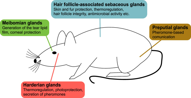
Sebaceous glands (SG) are exocrine glands that release their product by holocrine secretion, meaning that the whole cell becomes a secretion following disruption of the membrane. SG may be found in association with a hair follicle, forming the pilosebaceous unit, or as modified SG at different body sites such as the eyelids (Meibomian glands) or the preputial glands. Depending on their location, SG fulfill a number of functions, including protection of the skin and fur, thermoregulation, formation of the tear lipid film, and pheromone-based communication. Accordingly, SG abnormalities are associated with several diseases such as acne, cicatricial alopecia, and dry eye disease. An increasing number of genetically modified laboratory mouse lines develop SG abnormalities, and their study may provide important clues regarding the molecular pathways regulating SG development, physiology, and pathology. Here, we summarize in tabulated form the available mouse lines with SG abnormalities and, focusing on selected examples, discuss the insights they provide into SG biology and pathology. We hope this survey will become a helpful information source for researchers with a primary interest in SG but also as for researchers from unrelated fields that are unexpectedly confronted with a SG phenotype in newly generated mouse lines.
Keywords: Mouse models; Sebaceous gland; Skin.
Conflict of interest statement
There are no conflicts of interest to declare.
Figures
References
-
- Sundberg JP. Handbook of mouse mutations with skin and hair abnormalities. Boca Raton: CRC Press; 1994.
Publication types
MeSH terms
LinkOut - more resources
Full Text Sources
Other Literature Sources
