. 2016 Sep 12;36(19):2514-23.
doi: 10.1128/MCB.00142-16. Print 2016 Oct 1.

An Arginine Finger Regulates the Sequential Action of Asymmetrical Hexameric ATPase in the Double-Stranded DNA Translocation Motor

Affiliations

An Arginine Finger Regulates the Sequential Action of Asymmetrical Hexameric ATPase in the Double-Stranded DNA Translocation Motor

Zhengyi Zhao et al. Mol Cell Biol. .

Abstract

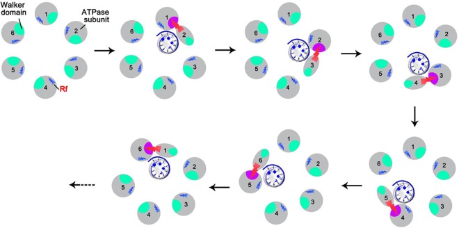
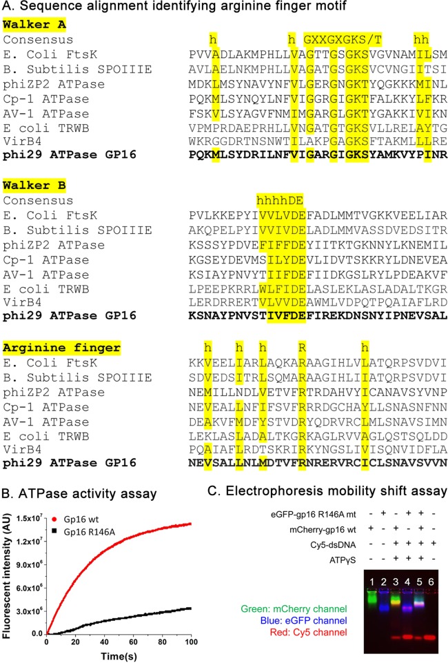
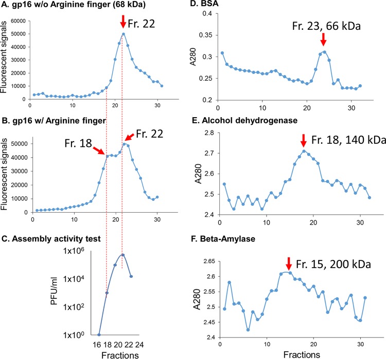
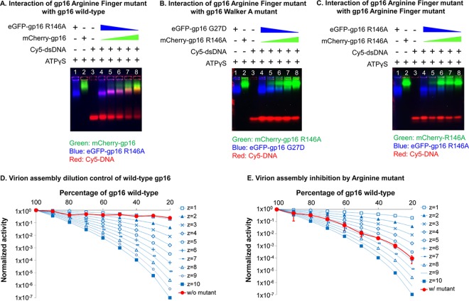
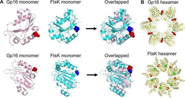
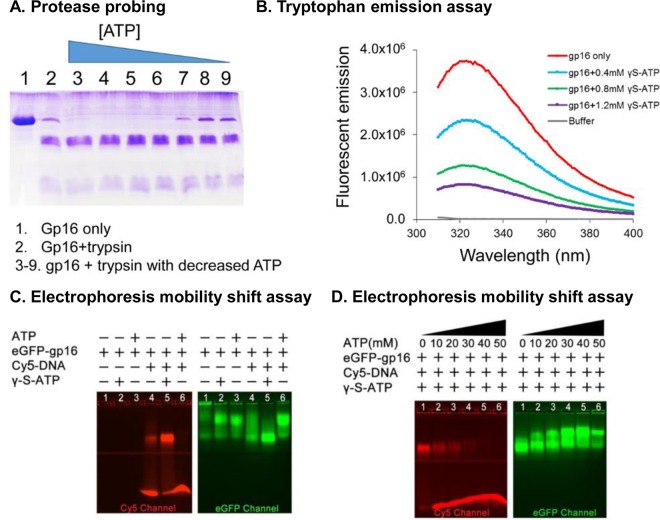
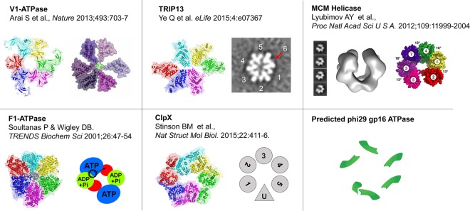
Biological motors are ubiquitous in living systems. Currently, how the motor components coordinate the unidirectional motion is elusive in most cases. Here, we report that the sequential action of the ATPase ring in the DNA packaging motor of bacteriophage ϕ29 is regulated by an arginine finger that extends from one ATPase subunit to the adjacent unit to promote noncovalent dimer formation. Mutation of the arginine finger resulted in the interruption of ATPase oligomerization, ATP binding/hydrolysis, and DNA translocation. Dimer formation reappeared when arginine mutants were mixed with other ATPase subunits that can offer the arginine to promote their interaction. Ultracentrifugation and virion assembly assays indicated that the ATPase was presenting as monomers and dimer mixtures. The isolated dimer alone was inactive in DNA translocation, but the addition of monomer could restore the activity, suggesting that the hexameric ATPase ring contained both dimer and monomers. Moreover, ATP binding or hydrolysis resulted in conformation and entropy changes of the ATPase with high or low DNA affinity. Taking these observations together, we concluded that the arginine finger regulates sequential action of the motor ATPase subunit by promoting the formation of the dimer inside the hexamer. The finding of asymmetrical hexameric organization is supported by structural evidence of many other ATPase systems showing the presence of one noncovalent dimer and four monomer subunits. All of these provide clues for why the asymmetrical hexameric ATPase gp16 of ϕ29 was previously reported as a pentameric configuration by cryo-electron microscopy (cryo-EM) since the contact by the arginine finger renders two adjacent ATPase subunits closer than other subunits. Thus, the asymmetrical hexamer would appear as a pentamer by cryo-EM, a technology that acquires the average of many images.


Figures

FIG 1
FIG 1
The proposed mechanism of ATPase coordination with a series of conformational changes during DNA binding and ATP hydrolysis that are regulated by the arginine finger (Rf, red).
FIG 2
FIG 2
Identification and characterization of the arginine finger in the ϕ29 gp16 ATPase. (A) Sequence alignment among gp16 ATPase and other ATPases in the same family, indicating the location of the Walker A, Walker B, and arginine finger (R) motifs of gp16 ATPase, which are well aligned with previously established domains (11, 27, 34, 39–41). h, hydrophobic residue. (B and C) ATP binding and hydrolysis activity assay of the gp16 arginine mutant. After the R146 residue is mutated, gp16 ATPase loses its ATP hydrolysis activity (B) and DNA binding activity, as shown by EMSA (C). wt, wild type.
FIG 3
FIG 3
Ultracentrifugation assay showing the presence of both dimers and monomers in gp16 ATPase rings. (A and B) One peak of eGFP-gp16 R146A (A) and two peaks of eGFP-gp16 wild type (B) were shown after parallel ultracentrifugation in a 15% to 35% glycerol gradient, indicating that both monomers and dimers were formed in the gp16 wild type, while dimer formation is interrupted by the mutation of the arginine finger. (C) The isolated gp16 dimers did not show any viral assembly activity, supporting the previous finding that addition of fresh gp16 monomers is required for reinitiating the DNA packaging intermediates. (D to F) Ultracentrifugation fractions (Fr) of protein markers, including BSA (66 kDa), alcohol dehydrogenase (140 kDa), and beta-amylase (200 kDa), are shown, with their peak locations around fractions 23, 18, and 15, respectively, to mark the separation of the monomer and dimer of gp16 ATPase. w/, with; w/o, without.
FIG 4
FIG 4
Intersubunit interaction of gp16 arginine mutant with other gp16s. (A to C) EMSAs showing the interaction of the gp16 arginine finger mutant with wild-type gp16 (A), the gp16 Walker A mutant (B), and the arginine finger mutant (C). Interactions between the gp16 arginine finger mutant and wild-type gp16 or the gp16 Walker A mutant are demonstrated by the band shift of both ATPase and DNA in the gel, while no obvious band shifts were observed when the arginine finger mutant ATPases were mixed together. DNA was labeled with Cy5, and different ATPases were labeled with different fluorescent protein tags for observation in the gel. (D and E) Binomial distribution assay to show the blockage of the ATPase arginine finger mutant on motor packaging activity. Different ratios of buffer (D) or eGFP-gp16 arginine finger mutants (E) were mixed with wild-type gp16 ATPase for the in vitro virion assembly activity assay. The experimental curve is plotted with theoretical predictions made according to the equation of Fang et al. (59) The experimental curve matches with the theoretical prediction with z = 6, indicating that six subunits are present in the ATPase ring, and one arginine finger mutant is enough to block the activity of the motor (see Materials and Methods).
FIG 5
FIG 5
Prediction and comparison of gp16 structure. (A) Structural comparison between the crystal structure of FtsK monomer (PDB accession number 2IUU; cyan) and the gp16 ATPase model (pink). The arginine finger is highlighted as a sphere. (B) Comparison of the predicted gp16 hexamer and FtsK hexamer. The ATPase gp16 hexamer structure was constructed using the predicted monomer structure shown in panel A and the P. aeruginosa FtsK (PDB accession number 2IUU) as templates (34). VMD was used to render the image of the structure (35). The ATP domains are highlighted as spheres: residue 27 (green, the conserved Walker ATP domain) and residue 146 (red, the arginine finger). The interaction of the arginine finger with the upstream adjacent subunit is evidenced by the proximity of the red and green spheres in both the constructed structure of the gp16 hexamer and the FtsK hexamer crystal structure.
FIG 6
FIG 6
Demonstration of two separate steps of gp16 conformational changes and entropic landscape alteration after ATP binding and ATP hydrolysis, respectively. (A) Trypsin probing showed that the ATPase-digested band is decreased with a reduced amount of ATP added into gp16 ATPase samples, suggesting that the gp16 ATPase is less constrained after binding to ATP. (B) Intrinsic tryptophan fluorescence assay showing the signal changes of ATPase upon the addition of different concentrations of ATP. (C) EMSA showing that gp16 ATPase bound to ATP and undergoes a conformational change that has a high affinity for DNA and that ATP hydrolysis triggers a second conformational change of gp16 ATPase with a low affinity with DNA. (D) Increasing DNA is released from gp16 ATPase/DNA/ATP complex upon the addition of increased amount of ATP that can be hydrolyzed by the gp16 ATPase.
FIG 7
FIG 7
Asymmetrical structure of various ATPase hexamer models. Structure illustrations of V1-ATPase (adapted from reference with permission of the publisher), TRIP13 (adapted and modified from reference with permission of the publisher), ClpX (adapted and modified from reference with permission of the publisher), MCM helicase (adapted from reference with permission of the publisher), and F1-ATPase (80) are shown as representatives of asymmetrical hexamers. PDB accession numbers are as follows: V1-ATPase, 3VR5; TRIP13, 4XGU; F1-ATPase, 1BMF; ClpX, 4I81. The EM reconstruction of the MCM helicase is deposited in the EMDataBank under accession number EMD-5429.

References

    1. Zhang X, Wigley DB. 2008. The “glutamate switch” provides a link between ATPase activity and ligand binding in AAA+ proteins. Nat Struct Mol Biol 15:1223–1227. doi: 10.1038/nsmb.1501. - DOI - PMC - PubMed
    1. Guo P, Noji H, Yengo CM, Zhao Z, Grainge I. 2016. Biological nanomotors with a revolution, linear, or rotation motion mechanism. Microbiol Mol Biol Rev 80:161–186. doi: 10.1128/MMBR.00056-15. - DOI - PMC - PubMed
    1. Snider J, Thibault G, Houry WA. 2008. The AAA+ superfamily of functionally diverse proteins. Genome Biol 9:216. doi: 10.1186/gb-2008-9-4-216. - DOI - PMC - PubMed
    1. Ammelburg M, Frickey T, Lupas AN. 2006. Classification of AAA+ proteins. J Struct Biol 156:2–11. doi: 10.1016/j.jsb.2006.05.002. - DOI - PubMed
    1. Chen C, Guo P. 1997. Sequential action of six virus-encoded DNA-packaging RNAs during phage phi29 genomic DNA translocation. J Virol 71:3864–3871. - PMC - PubMed

Publication types

MeSH terms

LinkOut - more resources