. 2016 Nov 1;594(21):6319-6331.
doi: 10.1113/JP272504. Epub 2016 Sep 11.

Extracellular K+ rapidly controls NaCl cotransporter phosphorylation in the native distal convoluted tubule by Cl- -dependent and independent mechanisms

Affiliations

Extracellular K+ rapidly controls NaCl cotransporter phosphorylation in the native distal convoluted tubule by Cl- -dependent and independent mechanisms

David Penton et al. J Physiol. .

Abstract

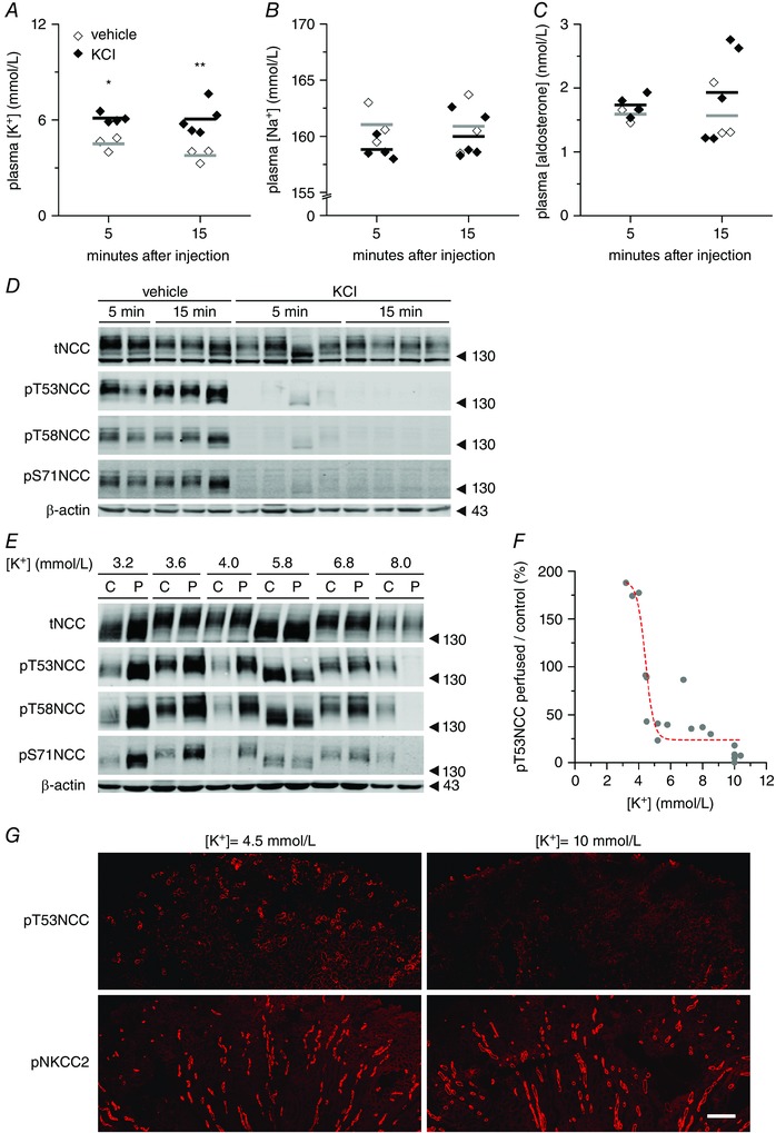
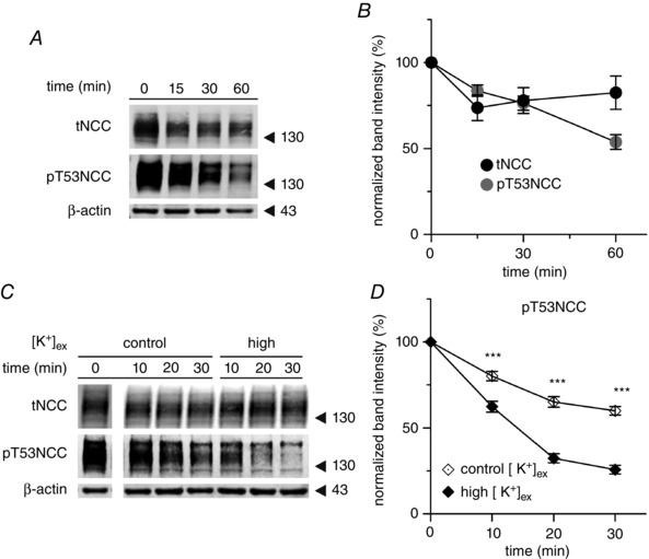
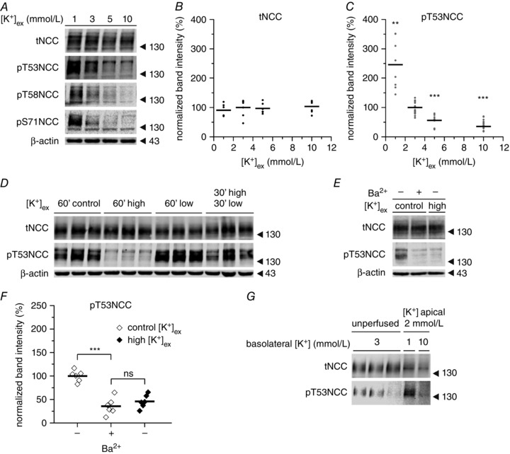
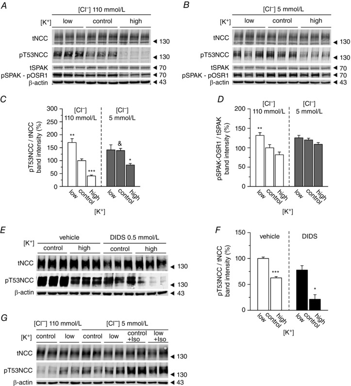
Key points: High dietary potassium (K+ ) intake dephosphorylates and inactivates the NaCl cotransporter (NCC) in the renal distal convoluted tubule (DCT). Using several ex vivo models, we show that physiological changes in extracellular K+ , similar to those occurring after a K+ rich diet, are sufficient to promote a very rapid dephosphorylation of NCC in native DCT cells. Although the increase of NCC phosphorylation upon decreased extracellular K+ appears to depend on cellular Cl- fluxes, the rapid NCC dephosphorylation in response to increased extracellular K+ is not Cl- -dependent. The Cl- -dependent pathway involves the SPAK/OSR1 kinases, whereas the Cl- independent pathway may include additional signalling cascades.

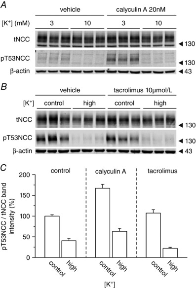
Abstract: A high dietary potassium (K+ ) intake causes a rapid dephosphorylation, and hence inactivation, of the thiazide-sensitive NaCl cotransporter (NCC) in the renal distal convoluted tubule (DCT). Based on experiments in heterologous expression systems, it was proposed that changes in extracellular K+ concentration ([K+ ]ex ) modulate NCC phosphorylation via a Cl- -dependent modulation of the with no lysine (K) kinases (WNK)-STE20/SPS-1-44 related proline-alanine-rich protein kinase (SPAK)/oxidative stress-related kinase (OSR1) kinase pathway. We used the isolated perfused mouse kidney technique and ex vivo preparations of mouse kidney slices to test the physiological relevance of this model on native DCT. We demonstrate that NCC phosphorylation inversely correlates with [K+ ]ex , with the most prominent effects occurring around physiological plasma [K+ ]. Cellular Cl- conductances and the kinases SPAK/OSR1 are involved in the phosphorylation of NCC under low [K+ ]ex . However, NCC dephosphorylation triggered by high [K+ ]ex is neither blocked by removing extracellular Cl- , nor by the Cl- channel blocker 4,4'-diisothiocyano-2,2'-stilbenedisulphonic acid. The response to [K+ ]ex on a low extracellular chloride concentration is also independent of significant changes in SPAK/OSR1 phosphorylation. Thus, in the native DCT, [K+ ]ex directly and rapidly controls NCC phosphorylation by Cl- -dependent and independent pathways that involve the kinases SPAK/OSR1 and a yet unidentified additional signalling mechanism.

Keywords: potassium; signal transduction; sodium transport.


Figures

Figure 1
Figure 1. Plasma [K+] directly and rapidly controls NCC phosphorylation in the intact kidney in vivo and ex vivo
AC, changes in plasma K+, Na+ and aldosterone concentrations, respectively, 5 or 15 min after i.v. injection of 100 μl of vehicle (three mice) or 80 mmol l–1 KCl in mice (four or five mice per group). Dots represent independent measurements and bars represent the mean of each group. * P < 0.05, ** P < 0.01 compared to vehicle injection, using an unpaired Student's t test. D, representative immunoblot (from mice in AC) showing the changes in NCC phosphorylation in response to plasma [K+] elevations. E, representative immunoblot of isolated perfused mouse kidneys (‘P’) showing the modulation of NCC expression and phosphorylation at different phospho‐sites upon changes in [K+]ex (‘C’: non‐perfused contralateral controls) F, summary of 18 experiments showing the dependence of NCC phosphorylation on the [K+]ex. Each dot represents the ratio of NCC phosphorylation between the perfused kidney and the non‐perfused contralateral kidney from one mouse. Dashed red line represents a sigmoidal fitting (for details, see Supporting information, Table S2). G, immunofluorescence staining of pT53NCC (upper images) and the closely‐related cotransporter pNKCC2 phosphorylated in T96 and T101 (lower images) in kidneys perfused with 4.5 or 10 mmol l–1 [K+]ex. Scale bar = 250 μm.
Figure 2
Figure 2. Ex vivo kidney slice preparation recapitulates the findings in intact mice and isolated perfused kidney
A, representative experiment showing the expression and phosphorylation of NCC at T53 in kidney slices incubated for 1 h in control buffer (3 mmol l–1 [K+]ex). B, summary of the expression and phosphorylation of NCC at T53 in kidney slices incubated under control conditions for 1 h (n = 19 slices, 10 mice). C, representative western blot showing the expression and phosphorylation of NCC at position T53 under control (3 mmol l–1) or high (10 mmol l–1) [K+]ex incubation conditions. Samples at t 0 (after 30 min of equilibration) were run in the same gel as the rest for every given antibody but not in sequential position. D, summary of phosphorylation of NCC at position T53 in kidney slices incubated under control (3 mmol l–1) or high (10 mmol l–1) [K+]ex conditions (n = 8–17 slices, three to seven mice). *** P < 0.001; unpaired Student's t test compared to control incubation). The mean ± SEM is shown in (B) and (D).
Figure 3
Figure 3. Regulation of NCC phosphorylation by maneuvers affecting DCT membrane voltage
A, representative experiment showing the dose–response modulation of NCC phosphorylation by [K+]ex at different phosphorylation sites in kidney slices. B, expression of tNCC upon incubation with different [K+]ex (n = 6–7 slices per treatment, two mice). C, changes in NCC phosphorylation at position T53 upon incubation with different [K+]ex (n = 8–17 slices per treatment, three to seven mice). ** P < 0.01, *** P < 0.001; unpaired Student's t test compared to control [K+]ex (3 mmol l–1). D, representative experiment showing the reversibility of NCC dephosphorylation triggered by high [K+]ex. Control, high and low stand for 3, 10 and 1 mmol l–1 [K+]ex respectively. This experiment was repeated twice (two mice) with similar results. E and F, effect of BaCl2 (5 mmol l–1) on NCC phosphorylation (n = 6 slices per treatment, two mice). G, changes in NCC phosphorylation in hand isolated DCTs microperfused with 2 mmol l–1 [K+]ex from the luminal side and either 1 or 10 mmol l–1 [K+]ex from the basolateral side for 10 min. Each band represents a pool of 10 DCTs. *** P < 0.001, ns, non‐significant using an unpaired Student's t test. In (B), (C) and (F), points represent individual experiments (slices) and bars represent the mean of the given treatment.
Figure 4
Figure 4. Effect of [K+]ex on NCC and SPAK/OSR1 phosphorylation in native DCT cells is affected by changes in Cl conductances
A, representative immunoblot showing the changes in tNCC and pT53NCC, as well as SPAK and pSPAK‐pOSR1, in kidney slices treated with low (1 mmol l–1), control (3 mmol l–1) or high (10 mmol l–1) [K+]ex under control (110 mmol l–1) [Cl]ex conditions. B, same experiment as in A but under 5 mmol l–1 [Cl]ex. C, summary of the effect of [K+]ex on NCC phosphorylation at position T53 under control or low [Cl]ex conditions (n = 9–18 slices per treatment, three to six mice). D, summary of the effect of [K+]ex on SPAK/OSR1 phosphorylation at position T53 under control or low [Cl]ex conditions (n = 6 slices per treatment, two mice). C and D, * P < 0.05, ** P < 0.01 and *** P < 0.001, compared to the control condition ([K+]ex 3 mmol l–1) of each [Cl]ex; &P < 0.05 compared to the control condition of 110 mmol l–1 [Cl]ex using ANOVA with Bonferroni's multiple comparison post hoc test. E, representative experiment showing the changes in NCC expression and phosphorylation at T53 in the presence or absence of the [Cl] channel blocker DIDS upon treatment with control (3 mmol l–1) or high (10 mmol l–1) [K+]ex. F, densitometric analysis of (E) (n = 3 slices per treatment, one mouse). * P < 0.05 compared to the control for each condition using an unpaired Student's t test. G, stimulation of NCC phosphorylation by isoproterenol (100 mmol l–1) under low (5 mmol l–1) [Cl]ex.
Figure 5
Figure 5. Inhibition of PP1, PP2A and PP3 does not impair the dephosphorylation of NCC upon high [K+]ex
Representative western blot showing the changes in NCC expression and phosphorylation at position T53 in slices treated with control (3 mmol l–1) or high (10 mmol l–1) [K+]ex under control (110 mmol l–1) [Cl]ex conditions. A, effect of inhibition of PP1 and PP2A with calyculin A. B, effect of inhibition of PP3 (calcineurine) with tacrolimus. C, summary of the densitometric analysis of 6–12 slices per condition, two to four mice per experimental group.

Comment in

References

    1. Alessi DR, Zhang J, Khanna A, Hochdörfer T, Shang Y & Kahle KT (2014). The WNK‐SPAK/OSR1 pathway: master regulator of cation‐chloride cotransporters. Sci Signal 7, re3. - PubMed
    1. Bandulik S, Schmidt K, Bockenhauer D, Zdebik Aa, Humberg E, Kleta R, Warth R & Reichold M (2011). The salt‐wasting phenotype of EAST syndrome, a disease with multifaceted symptoms linked to the KCNJ10 K+ channel. Pflügers Arch 461, 423–435. - PubMed
    1. Bazúa‐Valenti S, Chávez‐Canales M, Rojas‐Vega L, González‐Rodríguez X, Vázquez N, Rodríguez‐Gama A, Argaiz ER, Melo Z, Plata C, Ellison DH, García‐Valdés J, Hadchouel J & Gamba G (2015). The effect of WNK4 on the Na+‐Cl– cotransporter is modulated by intracellular chloride. J Am Soc Nephrol 26, 1781–1786. - PMC - PubMed
    1. Borschewski A, Himmerkus N, Boldt C, Blankenstein KI, McCormick Ja, Lazelle R, Willnow TE, Jankowski V, Plain A, Bleich M, Ellison DH, Bachmann S & Mutig K (2016). Calcineurin and sorting‐related receptor with A‐type repeats interact to regulate the renal Na+‐K+‐2Cl− cotransporter. J Am Soc Nephrol 27, 107–119. - PMC - PubMed
    1. Buendia JR, Bradlee ML, Daniels SR, Singer MR & Moore LL (2015). Longitudinal effects of dietary sodium and potassium on blood pressure in adolescent girls. JAMA Pediatr 02118, 1–9. - PubMed

MeSH terms

LinkOut - more resources