. 2016 Sep 9;198(19):2732-42.
doi: 10.1128/JB.00425-16. Print 2016 Oct 1.

The SaeRS Two-Component System Is a Direct and Dominant Transcriptional Activator of Toxic Shock Syndrome Toxin 1 in Staphylococcus aureus

Affiliations

The SaeRS Two-Component System Is a Direct and Dominant Transcriptional Activator of Toxic Shock Syndrome Toxin 1 in Staphylococcus aureus

Miren L Baroja et al. J Bacteriol. .

Abstract

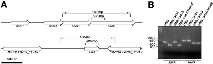
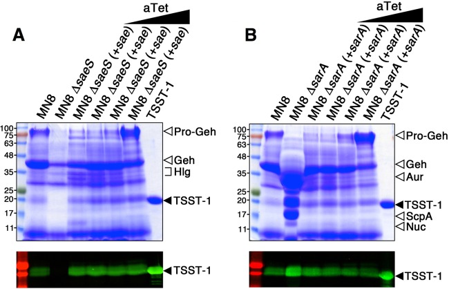
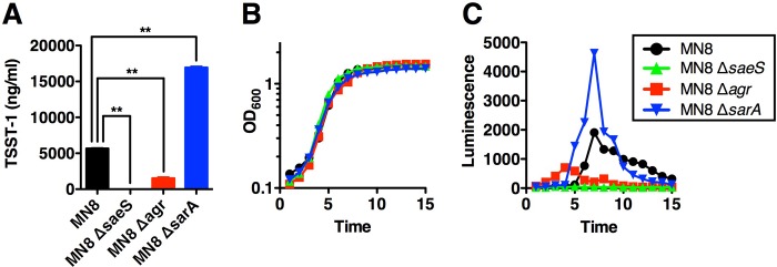
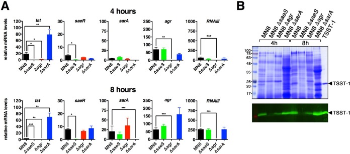
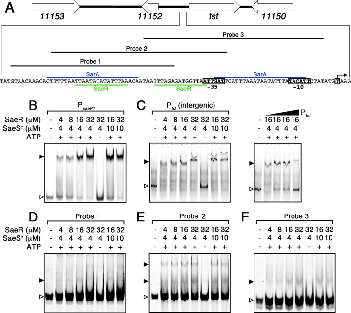
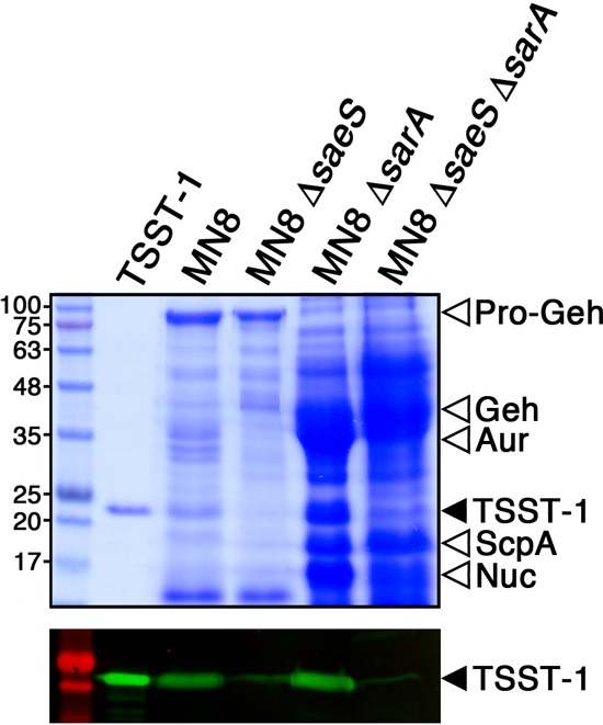
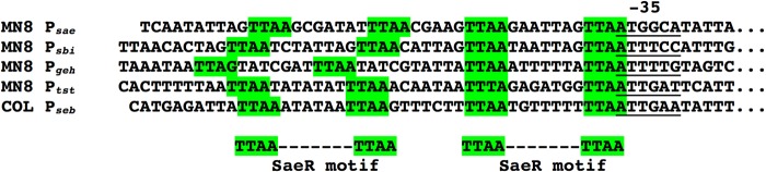
Toxic shock syndrome toxin 1 (TSST-1) is a Staphylococcus aureus superantigen that has been implicated in both menstrual and nonmenstrual toxic shock syndrome (TSS). Despite the important role of TSST-1 in severe human disease, a comprehensive understanding of staphylococcal regulatory factors that control TSST-1 expression remains incomplete. The S. aureus exotoxin expression (Sae) operon contains a well-characterized two-component system that regulates a number of important exotoxins in S. aureus, although regulation of TSST-1 by the Sae system has not been investigated. We generated a defined deletion mutant of the Sae histidine kinase sensor (saeS) in the prototypic menstrual TSS strain S. aureus MN8. Mutation of saeS resulted in a complete loss of TSST-1 expression. Using both luciferase reporter experiments and quantitative real-time PCR, we demonstrate that the Sae system is an important transcriptional activator of TSST-1 expression. Recombinant SaeR was able to bind directly to the tst promoter to a region containing two SaeR consensus binding sites. Although the stand-alone SarA transcriptional regulator has been shown to be both a positive and a negative regulator of TSST-1, deletion of sarA in S. aureus MN8 resulted in a dramatic overexpression of TSST-1. As expected, mutation of agr also reduced TSST-1 expression, but this phenotype appeared to be independent of Sae. A double mutation of saeS and sarA resulted in the loss of TSST-1 expression. This work indicates that the Sae system is a dominant and direct transcriptional activator that is required for expression of TSST-1.

Importance: The TSST-1 superantigen is an exotoxin, produced by some strains of S. aureus, that has a clear role in both menstrual and nonmenstrual TSS. Although the well-characterized agr quorum sensing system is a known positive regulator of TSST-1, the molecular mechanisms that directly control TSST-1 expression are only partially understood. Our studies demonstrate that the Sae two-component regulatory system is a positive transcriptional regulator that binds directly to the TSST-1 promoter, and furthermore, our data suggest that Sae is required for expression of TSST-1. This work highlights how major regulatory circuits can converge to fine-tune exotoxin expression and suggests that the Sae regulatory system may be an important target for antivirulence strategies.


Figures

FIG 1
FIG 1
Schematic and PCR analysis of the saeS and sarA in-frame deletions in S. aureus MN8. (A) Scale schematic of the 381-bp deletion in saeS (top) and the 358-bp deletion in sarA (bottom) and location of the PCR products used for analysis and sequencing of the corresponding deletions. (B) DNA agarose gel analysis using PCR products indicated in panel A for the individual and double MN8 sarA and saeS deletions as indicated.
FIG 2
FIG 2
Sae is a positive regulator and SarA is a negative regulator of TSST-1 expression in S. aureus MN8. Shown are exoprotein profiles (top panels) and Western blot analysis (bottom panels) of TSST-1 for wild-type S. aureus MN8 and the corresponding (A) saeS and (B) sarA deletion mutant and complemented strains. Concentrated supernatants from the indicated strains grown in BHI medium for 18 h were loaded onto 12% SDS-PAGE gels. Increasing concentrations (0, 5, 50, and 500 ng/ml) of anhydrotetracycline (aTet) were used to induce the promoter in the complemented strains. Molecular mass markers were loaded on the left and labeled in kilodaltons, and purified recombinant TSST-1 was loaded on the right and is indicated by the solid arrowheads.
FIG 3
FIG 3
TSST-1 quantitation, growth curve analysis, and TSST-1 promoter activity of wild-type S. aureus MN8 and the corresponding saeS, sarA, and agr mutants. (A) S. aureus MN8 strains were grown for 18 h in BHI medium, and TSST-1 secretion was quantified by ELISA. The data represent the means ± standard errors of the means (SEM) from three biological replicates (**, P < 0.05 by one-way ANOVA with Tukey's multiple comparison test). (B and C) The indicated S. aureus strains were grown at 37°C in a Biotek Synergy H4 multimode plate reader, and OD600 and luminescence readings were taken every hour for 15 h. Results are expressed as OD600 units (B) and luminescence in relative light units (defined as counts per minute) (C). The data represent the means from three independent experiments, each done with quadruplicate technical replicates.
FIG 4
FIG 4
qRT-PCR analysis of tst transcripts and relevant regulators in wild-type S. aureus MN8 and the saeS, sarA, and agr mutants. (A) cDNA was prepared at the indicated time points, and copies were normalized to the housekeeping rpoB gene. Data represent the means ± SEM from four biological replicates (*, P < 0.05, **, P < 0.01, and ***, P < 0.001, by one-way ANOVA with Tukey's multiple comparison test). (B) Exoprotein profiles (top panel) and anti-TSST-1 Western blot (bottom panel) analysis of the indicated S. aureus strains at the 4- and 8-h time points. Molecular mass markers were loaded on the left (labeled in kilodaltons), and purified recombinant TSST-1 was loaded on the right (indicated by solid arrowheads).
FIG 5
FIG 5
DNA binding analysis of phosphorylated SaeR (P∼SaeR) to the PsaeP1 and Ptst promoters. (A) Schematic map and nucleotide sequence of Ptst in S. aureus MN8 (locus tag prefix HMPREF0769). The characterized transcriptional start site and −10 and −35 regions are labeled with boxes, and the characterized SarA binding sites is highlighted in blue (13). The proposed SaeR binding sequences are highlighted in green. Probes 1, 2, and 3 correspond to the EMSAs shown in panels D, E, and F, respectively. (C to F) DNA binding analysis of P∼SaeR to PsaeP1 (B), Ptst (C), probe 1 (D), probe 2 (E), and probe 3 (F). Panel C (right panel) also shows a competitive binding analysis using 0-, 5-, 10-, and 50-fold excess unlabeled Ptst (gradient denoted by the triangle). Unbound DNA probes are indicated by open arrowheads, and DNA shifts are indicated by solid arrowheads. Protein concentrations, and the presence of ATP in the reaction buffer, are indicated for each lane.
FIG 6
FIG 6
The Sae two-component regulator is necessary for TSST-1 expression in the absence of repression by SarA. Shown are exoprotein profiles (top panel) and Western blot (bottom panel) analysis of TSST-1 for wild-type S. aureus MN8 and the corresponding mutants. Concentrated supernatants from the indicated strains grown in BHI medium were loaded onto 12% SDS-PAGE gels. Molecular mass markers in kilodaltons are shown on the left. Purified recombinant TSST-1 is indicated by the solid arrowhead. The locations of pro-Geh, mature Geh, Aur, ScpA, and nuclease (Nuc) are indicated by open arrowheads.
FIG 7
FIG 7
Alignment of promoter element nucleotide sequences of SaeR binding motifs located upstream of PsaeP1, Psbi, Pgeh, Ptst, and Pseb. The −35 promoter binding regions are underlined, and green highlights outline the primary and secondary SaeR binding motifs.

References

    1. Lowy FD. 1998. Staphylococcus aureus infections. N Engl J Med 339:520–532. doi:10.1056/NEJM199808203390806. - DOI - PubMed
    1. Dinges MM, Orwin PM, Schlievert PM. 2000. Exotoxins of Staphylococcus aureus. Clin Microbiol Rev 13:16–34. doi:10.1128/CMR.13.1.16-34.2000. - DOI - PMC - PubMed
    1. McCormick JK, Yarwood JM, Schlievert PM. 2001. Toxic shock syndrome and bacterial superantigens: an update. Annu Rev Microbiol 55:77–104. doi:10.1146/annurev.micro.55.1.77. - DOI - PubMed
    1. Centers for Disease Control and Prevention. 2011. Toxic shock syndrome; 2011 case definition. Centers for Disease Control and Prevention, Atlanta, GA.
    1. DeVries AS, Lesher L, Schlievert PM, Rogers T, Villaume LG, Danila R, Lynfield R. 2011. Staphylococcal toxic shock syndrome 2000–2006: epidemiology, clinical features, and molecular characteristics. PLoS One 6:e22997. doi:10.1371/journal.pone.0022997. - DOI - PMC - PubMed

MeSH terms

LinkOut - more resources