. 2016 Aug 9;113(32):9057-62.
doi: 10.1073/pnas.1606667113. Epub 2016 Jul 25.

Chromosomal landscape of UV damage formation and repair at single-nucleotide resolution

Affiliations

Chromosomal landscape of UV damage formation and repair at single-nucleotide resolution

Peng Mao et al. Proc Natl Acad Sci U S A. .

Abstract

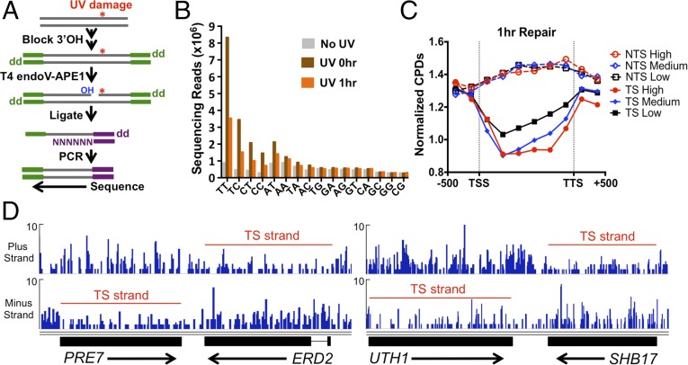
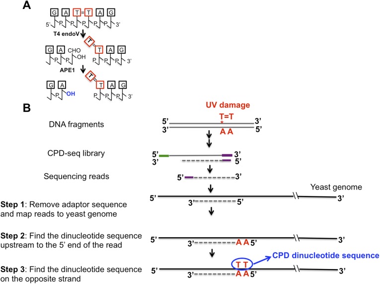
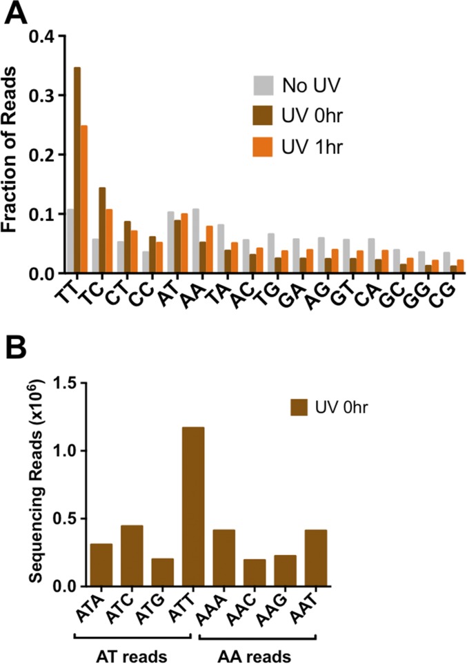
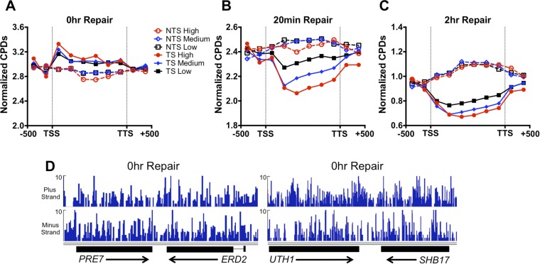
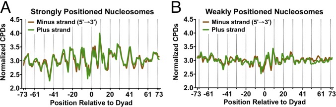
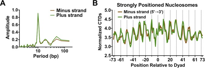
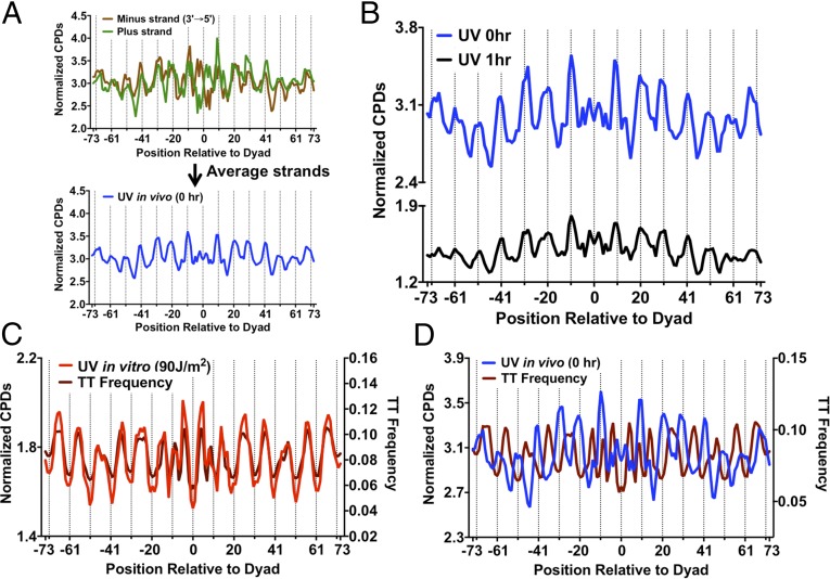
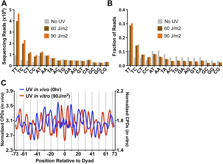
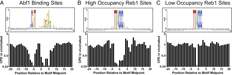
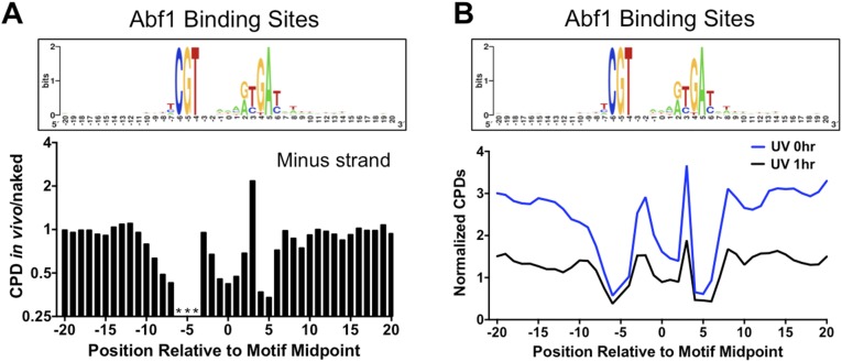
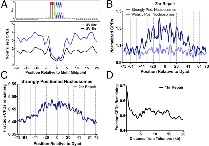
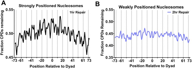
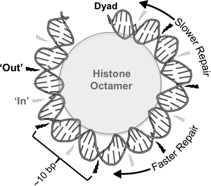
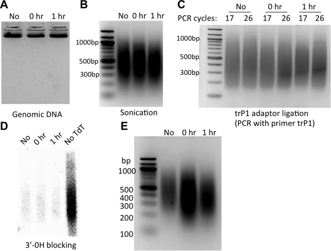
UV-induced DNA lesions are important contributors to mutagenesis and cancer, but it is not fully understood how the chromosomal landscape influences UV lesion formation and repair. Genome-wide profiling of repair activity in UV irradiated cells has revealed significant variations in repair kinetics across the genome, not only among large chromatin domains, but also at individual transcription factor binding sites. Here we report that there is also a striking but predictable variation in initial UV damage levels across a eukaryotic genome. We used a new high-throughput sequencing method, known as CPD-seq, to precisely map UV-induced cyclobutane pyrimidine dimers (CPDs) at single-nucleotide resolution throughout the yeast genome. This analysis revealed that individual nucleosomes significantly alter CPD formation, protecting nucleosomal DNA with an inward rotational setting, even though such DNA is, on average, more intrinsically prone to form CPD lesions. CPD formation is also inhibited by DNA-bound transcription factors, in effect shielding important DNA elements from UV damage. Analysis of CPD repair revealed that initial differences in CPD damage formation often persist, even at later repair time points. Furthermore, our high-resolution data demonstrate, to our knowledge for the first time, that CPD repair is significantly less efficient at translational positions near the dyad of strongly positioned nucleosomes in the yeast genome. These findings define the global roles of nucleosomes and transcription factors in both UV damage formation and repair, and have important implications for our understanding of UV-induced mutagenesis in human cancers.

Keywords: DNA damage; DNA repair; chromatin; nucleosome; transcription factor.


Conflict of interest statement

The authors declare no conflict of interest.

Figures

Fig. 1.
Fig. 1.
CPD-seq method for mapping UV damage formation and repair in the yeast genome. (A) Experimental strategy for the CPD-seq method. The trP1 adapter is colored green, and the A adapter is in purple. “OH” indicates a free 3′OH; “dd” indicates dideoxy (i.e., 3′H). (B) Analysis of dinucleotide sequences associated with CPD-seq sequencing reads. In UV-treated samples, there is an enrichment of sequencing reads at dipyrimidine sequences. (C) Significantly fewer CPDs remain in the TS following 1 h of repair than the NTS. The number of CPD reads normalized by the total number of nucleotides in dipyrimidine sequences for each DNA strand was calculated for the promoter, coding region, and termination site for high (>10 mRNAs per hour), medium (1–10 mRNAs per hour), low (<1 mRNAs per hour) transcribed genes (20). TSS is the transcription start site; TTS is the transcription termination site. (D) Snapshot of the distribution of CPD reads following 1 h of repair for the PRE7, ERD2, UTH1, and SHB17 genes. The red line indicates the transcribed strand [gene coordinates and image drawn using the Integrative Genomics Viewer (33)]. y axis depicts the number of CPD reads.
Fig. S1.
Fig. S1.
(A) T4 endonuclease V and APE1 cleave at CPD lesions, generating a free 3′OH immediately upstream of the CPD damage site. (B) Bioinformatics procedure to extract CPD dinucleotide sequences. Green, trP1 adaptor; purple, A adaptor.
Fig. S2.
Fig. S2.
(A) Fraction of dinucleotide sequences associated with CPD-seq sequencing reads after normalizing by the total number of reads for each sample. (B) Frequency of trinucleotide sequences associated with AT and AA reads. The high frequency of ATC- and ATT-associated reads suggests that the enrichment of AT reads in the UV-treated samples is likely derived from lesions associated with adjacent TT and TC sequences, potentially because of the weak exonuclease activity of APE1. In contrast, there was no enrichment of AAC- or AAT-associated reads.
Fig. S3.
Fig. S3.
(A) More UV damage is initially present on the TS than the NTS in the UV 0-h sample. (B) TC-NER of TS strand is apparent after 20 min of repair. (C) Preferential repair of TS strand is apparent even after 2 h of repair. (D) No significant difference in CPD reads between the TS and NTS strands of the PRE7, ERD2, UTH1, and SHB17 genes in the UV 0-h sample.
Fig. 2.
Fig. 2.
Strongly positioned nucleosomes in yeast cause an ∼10-bp UV photo-footprint. (A) CPD damage in strongly positioned nucleosomes (nucleosome score > 5) is higher at outward rotational settings (dashed lines). The normalized CPD distribution for the 0-h UV sample was analyzed along the plus and minus strands of ∼10,000 strongly positioned nucleosomes. Both plus and minus strands were oriented in the 5′ to 3′ direction. (B) CPD damage in weakly positioned nucleosomes (nucleosome score < 1) does not show a significant UV photo-footprint.
Fig. S4.
Fig. S4.
(A) Periodogram of nucleosome UV photo-footprint reveals a periodicity of CPD formation of ∼10 bp. (B) CTD show a particularly prominent UV photo-footprint in strongly positioned nucleosomes. The number of sequencing reads associated with TT dinucleotides in the UV 0-h sample was normalized by the number of TT dinucleotides at positions relative to the nucleosome dyad.
Fig. 3.
Fig. 3.
The nucleosome UV photo-footprint protects T-rich DNA sequences from UV damage. (A) Method for averaging normalized CPD counts between plus and minus DNA strands in strongly positioned nucleosomes. The DNA strands were aligned in their normal antiparallel orientation to remove strand-specific sequence biases, and the normalized CPD data were calculated as a weighted average. (B) The nucleosome UV photo-footprint persists at 1-h repair. DNA regions that fell within the transcribed strand of a gene were excluded from this analysis. (C) In vitro CPD formation in the absence of nucleosomes is strongly correlated with the TT frequency in DNA. Both in vitro CPD formation and TT frequency peak at inward rotational positions. (D) In vivo CPD formation in the presence of strongly positioned nucleosomes is anticorrelated with TT frequency in nucleosomal DNA.
Fig. S5.
Fig. S5.
(A) Counts of dinucleotide sequences associated with CPD-seq reads for naked DNA irradiated in vitro with 60 or 90 J/m2 UVC light. The “No UV” data are the same as in Fig. 1B. (B) Fraction of dinucleotide sequences associated with CPD-seq sequencing reads after normalizing by the total number of reads for each sample. (C) When yeast genomic DNA was irradiated in vitro (UV 90J sample), CPD formation occurs more frequently in DNA that adopts an inward rotational setting in vivo. Inward rotational settings occur at the midpoints between the dashed lines. CPD formation in vivo (0-h UV sample) shows the opposite trend.
Fig. 4.
Fig. 4.
The yeast transcription factors Abf1 and Reb1 induce a significant UV photo-footprint at their DNA binding sites. (A) Abf1-bound DNA sites show altered CPD formation. (Upper) The DNA consensus sequence of 661 Abf1 binding sites [generated using WebLogo (34)], including DNA flanking each binding site. (Lower) The scaled ratio of normalized CPDs in the UV 0-h sample (in vivo) relative to the UV 90-J/m2 sample (naked) for the plus strand of the Abf1 binding sites. Asterisks indicate that the indicated position in the motif cannot form CPD lesions because of DNA sequence constraints. (B) Same as A, except the plus strand of high-occupancy Reb1 binding sites (Reb1 occupancy > 10) were analyzed. (C) Same as A, except a plus strand of low-occupancy Reb1 binding sites (Reb1 occupancy < 10) were analyzed.
Fig. S6.
Fig. S6.
(A) Abf1-bound DNA sites show altered CPD formation. (Upper) The DNA consensus sequence of 661 Abf1 binding sites [generated using WebLogo (34)], including DNA flanking each binding site. (Lower) The scaled ratio of normalized CPDs in the UV 0-h sample (in vivo) relative to the UV 90J sample (naked) for the minus strands of Abf1 binding sites. Asterisks indicate that the indicated position in the motif cannot form CPD lesions because of DNA sequence constraints. (B) Comparison of normalized CPDs at Abf1 binding sites at 0-h and 1-h repair time points. The weighted average of normalized CPDs of both strands is depicted.
Fig. 5.
Fig. 5.
Analysis of repair of CPD lesions. (A) Comparison of normalized CPDs at high-occupancy Reb1 binding sites at 0-h and 1-h repair time points. The weighted average of normalized CPDs of both strands is depicted. (B) Plot of normalized CPD reads in strongly positioned (nucleosome score > 5) and weakly positioned (nucleosome score < 5) nucleosomes after 2 h of repair. Dashed lines indicate outward rotational settings (dashed lines). The weighted average of normalized CPDs of both strands is shown. (C) More CPDs remain unrepaired adjacent to the nucleosome dyad in strongly positioned nucleosomes. The fraction of CPDs remaining was calculated by comparing the 2-h repair sample to its matched 0-h control. (D) Telomere regions show slower CPD removal following 1 h of repair. The fraction of CPDs remaining was calculated by comparing the 1-h repair sample to its matched 0-h control.
Fig. S7.
Fig. S7.
(A) Fraction of CPDs remaining following 1 h of repair relative to the dyad of strongly positioned nucleosomes. (B) Positioning relative to the dyad does not affect CPD removal in weakly positioned nucleosomes. The fraction of CPDs remaining was calculated by comparing the 2-h repair sample to its matched 0-h control.
Fig. S8.
Fig. S8.
Model of the how strongly positioned nucleosomes affect UV damage formation and repair. In strongly positioned nucleosomes, CPD damage formation is highest at outward (“Out”) rotational settings (indicated by large black lightning bolt), which gives rise to the ∼10-bp periodicity. Inward (“In”) rotational settings have the lowest CPD damage formation (indicated by small gray lightning bolt), even though such regions are TT rich. NER is inhibited at translational positions near the nucleosome dyad. DNA structure was depicted using Pymol to visualize 1ID3.
Fig. S9.
Fig. S9.
Results at intermediate steps in CPD-seq procedure. Yeast cells were irradiated with 125 J/m2 UVC light. Samples were taken before UV treatment (“No”), immediately after UV (0 h), and after 1 h of repair (1 h). (A) Isolated genomic DNA. (B) Sonicated genomic DNA. (C) PCR confirmation of trP1 adaptor ligation. (D) PCR confirmation of 3′OH blocking by TdT and ddATP (see SI Materials and Methods). (E) Gel verification of CPD-seq libraries.

References

    1. Friedberg EC, et al. DNA Repair and Mutagenesis. 2nd Ed ASM Press; Washington, DC: 2006.
    1. Hanawalt PC, Spivak G. Transcription-coupled DNA repair: Two decades of progress and surprises. Nat Rev Mol Cell Biol. 2008;9(12):958–970. - PubMed
    1. Marteijn JA, Lans H, Vermeulen W, Hoeijmakers JH. Understanding nucleotide excision repair and its roles in cancer and ageing. Nat Rev Mol Cell Biol. 2014;15(7):465–481. - PubMed
    1. Pleasance ED, et al. A comprehensive catalogue of somatic mutations from a human cancer genome. Nature. 2010;463(7278):191–196. - PMC - PubMed
    1. Lawrence MS, et al. Mutational heterogeneity in cancer and the search for new cancer-associated genes. Nature. 2013;499(7457):214–218. - PMC - PubMed

Publication types