. 2016 Sep 23;60(10):6013-22.
doi: 10.1128/AAC.01067-16. Print 2016 Oct.

Crystal Structure of the Metallo-β-Lactamase GOB in the Periplasmic Dizinc Form Reveals an Unusual Metal Site

Affiliations

Crystal Structure of the Metallo-β-Lactamase GOB in the Periplasmic Dizinc Form Reveals an Unusual Metal Site

Jorgelina Morán-Barrio et al. Antimicrob Agents Chemother. .

Abstract

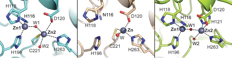
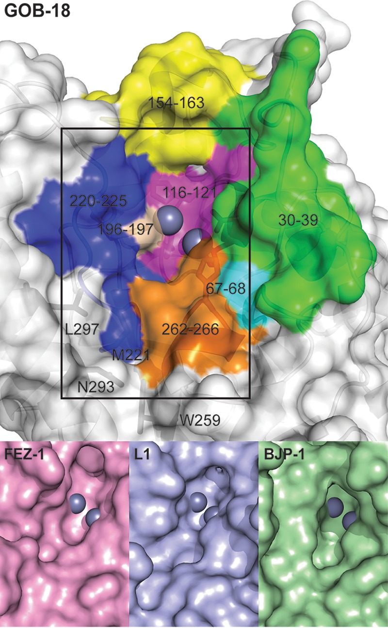
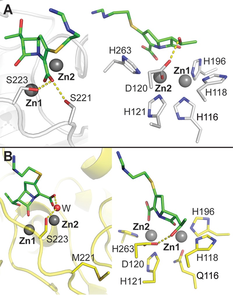
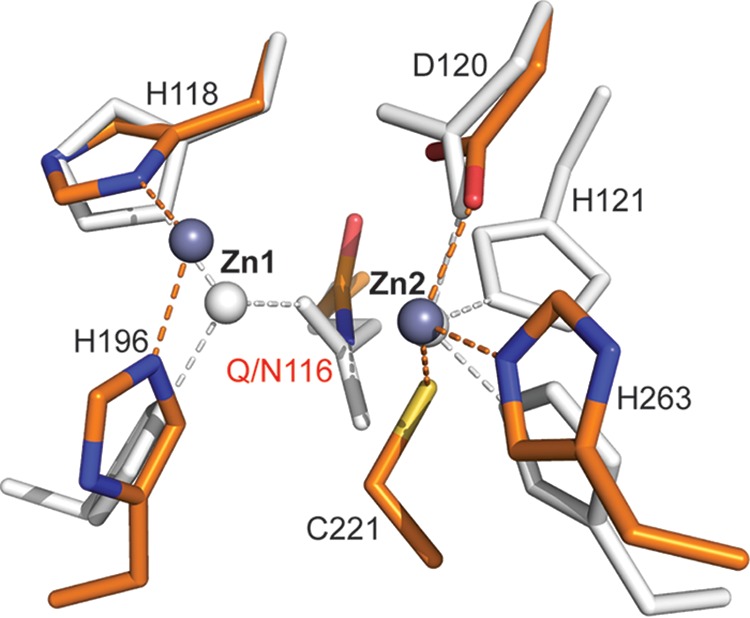
Metallo-beta-lactamases (MBLs) are broad-spectrum, Zn(II)-dependent lactamases able to confer resistance to virtually every β-lactam antibiotic currently available. The large diversity of active-site structures and metal content among MBLs from different sources has limited the design of a pan-MBL inhibitor. GOB-18 is a divergent MBL from subclass B3 that is expressed by the opportunistic Gram-negative pathogen Elizabethkingia meningoseptica This MBL is atypical, since several residues conserved in B3 enzymes (such as a metal ligand His) are substituted in GOB enzymes. Here, we report the crystal structure of the periplasmic di-Zn(II) form of GOB-18. This enzyme displays a unique active-site structure, with residue Gln116 coordinating the Zn1 ion through its terminal amide moiety, replacing a ubiquitous His residue. This situation contrasts with that of B2 MBLs, where an equivalent His116Asn substitution leads to a di-Zn(II) inactive species. Instead, both the mono- and di-Zn(II) forms of GOB-18 are active against penicillins, cephalosporins, and carbapenems. In silico docking and molecular dynamics simulations indicate that residue Met221 is not involved in substrate binding, in contrast to Ser221, which otherwise is conserved in most B3 enzymes. These distinctive features are conserved in recently reported GOB orthologues in environmental bacteria. These findings provide valuable information for inhibitor design and also posit that GOB enzymes have alternative functions.


Figures

FIG 1
FIG 1
Metallo-beta-lactamase zinc-binding sites. B. cereus BcII (B1; PDB entry 1BC2) (left), S. fonticola SfhI (B2; PDB entry 3SD9) (center), and S. maltophilia L1 (B3; PDB entry 1SML) (right) are shown. Zinc atoms are shown as gray spheres, and water molecules (W) are shown as small red spheres. Coordination bonds are indicated with dashed lines.
FIG 2
FIG 2
Purification and characterization of periplasmic GOB-18. (A) SDS-PAGE analysis of fractions along different steps of the purification process. Lanes: 1, total cell extract (T); 2, periplasmic fraction corresponding to the sample loaded on the CM-Sephadex column (L); 3, CM-Sephadex flowthrough (FCM); 4, CM-Sephadex elution, corresponding to the sample loaded on the Mono S column (E); 5, Mono S flowthrough (FS); 6 to 17, Mono S elution fractions (f1 to f12). Molecular mass markers (in kilodaltons) are indicated at the right. The vertical gray line between lanes 3 and 4 indicates the junction of two different gels prepared and run together, since the Mini Protean III system (Bio-Rad) holds up to 15 lanes. (B) Circular dichroism spectra of periplasmic GOB-18 (black line), remetallated Zn(II)-GOB-18 (gray), and apo-GOB-18 (light gray). ΔA/cb is the molar elipticity, where c is the protein concentration in mg/ml and b is the path length in cm.
FIG 3
FIG 3
Overall structure of periplasmic GOB-18. (A) Cartoon representation of GOB-18 (chain A) with helices colored in red, strands in yellow, and nonstructured loops in green. The inset highlights selected residues (in stick representation). Dashed lines represent atomic interactions. (B) Comparison of GOB-18 with the B3 MBLs FEZ-1 (PDB entry 1K07), BJP-1 (PDB entry 3LVZ), and L1 (PDB entry 1SML). The segments of GOB-18 exhibiting the highest RMSD values are highlighted in different colors. Zn atoms are shown as gray spheres. N-term, N terminus.
FIG 4
FIG 4
Active site of periplasmic GOB-18. (A, upper) Comparison of the active site in the two monomers present in the crystal structure of GOB-18. Chain A is colored by atom in yellow (C atoms), blue (N atoms), red (oxygen atoms), and dark gray (zinc atoms). Chain B is shown in light gray throughout. Protein residues are depicted as sticks, zinc atoms are shown as big spheres, and water molecules are small spheres. The 2mFo-DFc sigmaA-weighted electron density, contoured at 1.7 σ and represented as a gray mesh, corresponds to chain A. Dashed lines represent atomic interactions, and the related distances are 2.1 Å in all cases, except for the Zn1-Gln98 and Zn1-axial water interactions, where distances are 2.0 Å and 2.2 Å, respectively. (Lower) Comparison of the active site of GOB-18 (chain A, yellow) with those of the B3 MBLs FEZ-1 (PDB entry 1K07; pink), BJP-1 (PDB entry 3LVZ; green), and L1 (PDB entry 1SML; blue). (B) Residues in the second coordination sphere in the active site of GOB-18 (chain A) are shown in stick representation with C atoms colored in cyan.
FIG 5
FIG 5
Catalytic groove in GOB-18. Surface representation of GOB-18 (chain A), with different colors highlighting the structural elements that define the catalytic groove. Zinc atoms are shown as gray spheres. For comparison, the region delimited by the rectangle is compared to the equivalent regions of the B3 MBLs FEZ-1 (PDB entry 1K07), L1 (PDB entry 1SML), and BJP-1 (PDB entry 3LVZ) shown at the bottom.
FIG 6
FIG 6
Molecular dynamics simulations calculated using in silico docked imipenem in complex with L1 and GOB-18. (A) Snapshot of the L1-imipenem molecular dynamics simulation showing the interaction between imipenem and residues Asp120, Ser221, and Ser223. (B) Snapshot of the GOB-18-imipenem molecular dynamics simulation showing the interaction between imipenem and residues Asp120 and Ser223. Imipenem is shown in stick representation with green carbons, L1 and GOB-18 are displayed in gray and yellow, respectively, and zinc atoms and a water molecule are depicted as gray and red spheres, respectively.
FIG 7
FIG 7
Comparison of the active sites of di-Zn(II) GOB-18 and di-Zn(II) CphA. GOB-18 (chain A) is shown in light gray, and CphA (PDB entry 3F9O) is colored in orange (C atoms), blue (N atoms), red (oxygen atoms), and dark gray (zinc atoms). Protein residues are shown as sticks, zinc atoms are depicted as spheres, and dashed lines highlight atomic interactions.

References

    1. Meini MR, Llarrull LI, Vila AJ. 2015. Overcoming differences: the catalytic mechanism of metallo-beta-lactamases. FEBS Lett 589:3419–3432. doi: 10.1016/j.febslet.2015.08.015. - DOI - PMC - PubMed
    1. Meini MR, Llarrull LI, Vila AJ. 2014. Evolution of metallo-beta-lactamases: trends revealed by natural diversity and in vitro evolution. Antibiotics (Basel) 3:285–316. doi: 10.3390/antibiotics3030285. - DOI - PMC - PubMed
    1. Palzkill T. 2013. Metallo-beta-lactamase structure and function. Ann N Y Acad Sci 1277:91–104. doi: 10.1111/j.1749-6632.2012.06796.x. - DOI - PMC - PubMed
    1. Llarrull LI, Testero SA, Fisher JF, Mobashery S. 2010. The future of the beta-lactams. Curr Opin Microbiol 13:551–557. doi: 10.1016/j.mib.2010.09.008. - DOI - PMC - PubMed
    1. Fisher JF, Meroueh SO, Mobashery S. 2005. Bacterial resistance to beta-lactam antibiotics: compelling opportunism, compelling opportunity. Chem Rev 105:395–424. doi: 10.1021/cr030102i. - DOI - PubMed

MeSH terms