. 2016 Dec;5(12):1695-1706.
doi: 10.5966/sctm.2015-0397. Epub 2016 Jul 25.

Hair Follicle and Sebaceous Gland De Novo Regeneration With Cultured Epidermal Stem Cells and Skin-Derived Precursors

Affiliations

Hair Follicle and Sebaceous Gland De Novo Regeneration With Cultured Epidermal Stem Cells and Skin-Derived Precursors

Xiaoxiao Wang et al. Stem Cells Transl Med. 2016 Dec.

Abstract

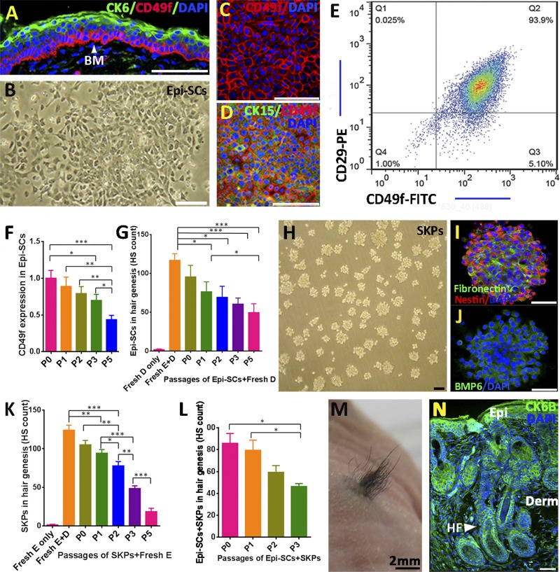
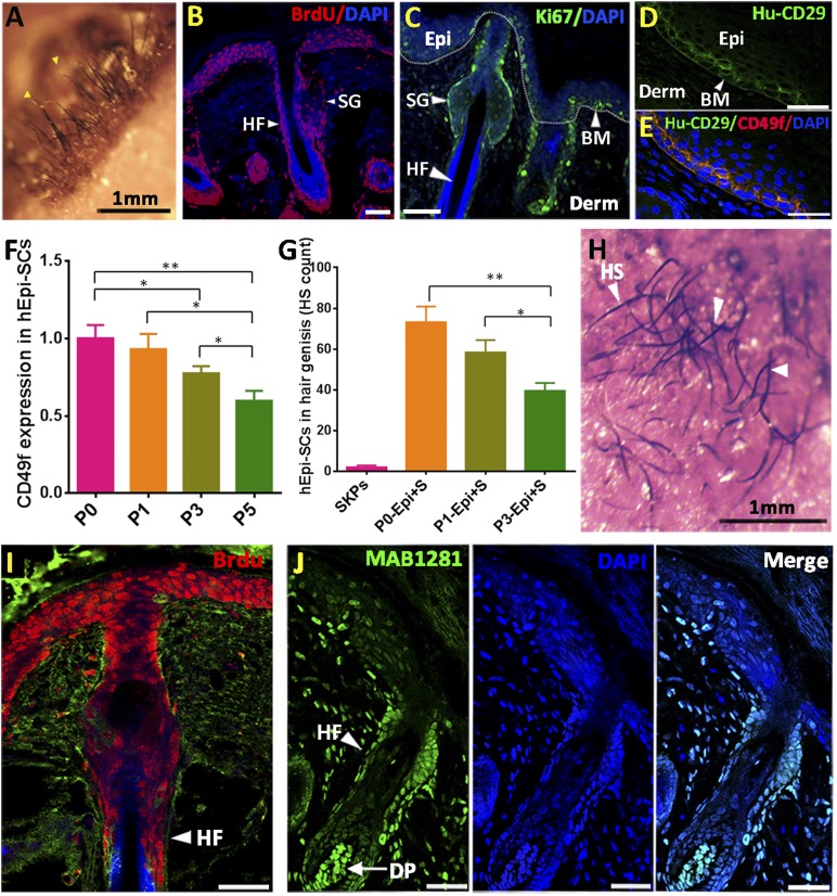
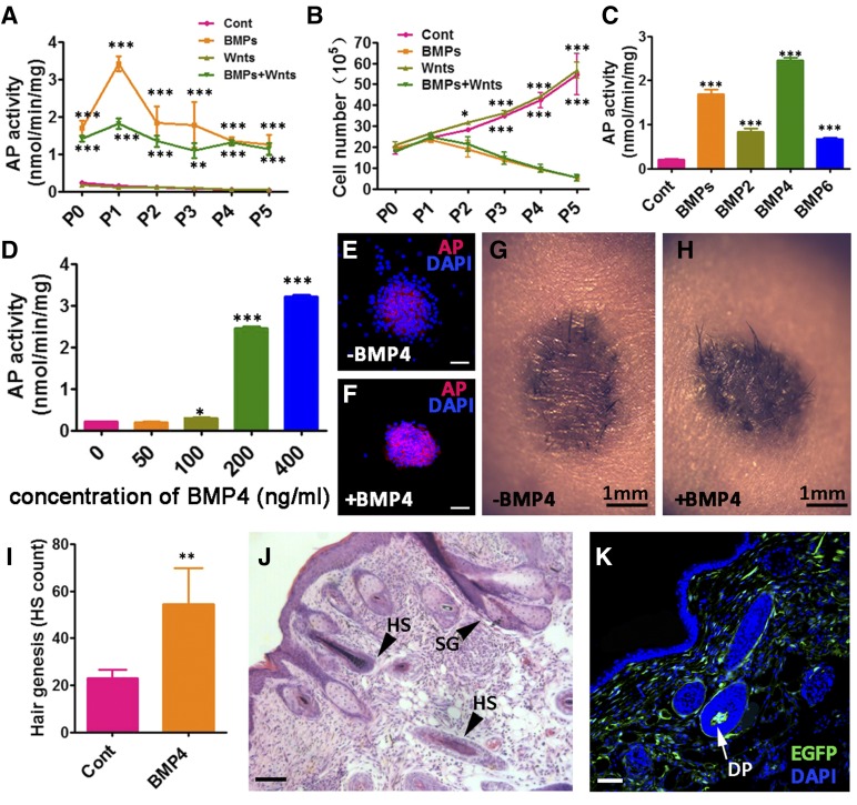
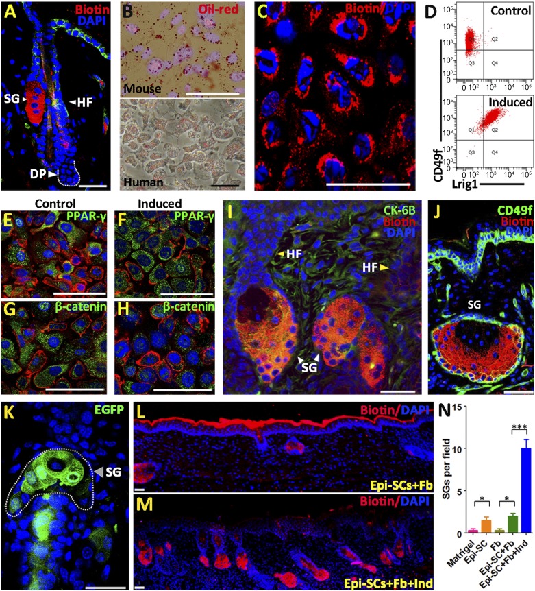
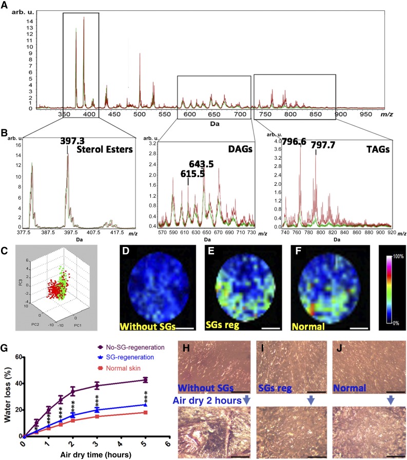
: Stem cell-based organ regeneration is purported to enable the replacement of impaired organs in the foreseeable future. Here, we demonstrated that a combination of cultured epidermal stem cells (Epi-SCs) derived from the epidermis and skin-derived precursors (SKPs) was capable of reconstituting functional hair follicles and sebaceous glands (SG). When Epi-SCs and SKPs were mixed in a hydrogel and implanted into an excisional wound in nude mice, the Epi-SCs formed de novo epidermis along with hair follicles, and SKPs contributed to dermal papilla in the neogenic hair follicles. Notably, a combination of culture-expanded Epi-SCs and SKPs derived from the adult human scalp were sufficient to generate hair follicles and hair. Bone morphogenetic protein 4, but not Wnts, sustained the expression of alkaline phosphatase in SKPs in vitro and the hair follicle-inductive property in vivo when SKPs were engrafted with neonatal epidermal cells into excisional wounds. In addition, Epi-SCs were capable of differentiating into sebocytes and formed de novo SGs, which excreted lipids as do normal SGs. Thus our results indicate that cultured Epi-SCs and SKPs are sufficient to generate de novo hair follicles and SGs, implying great potential to develop novel bioengineered skin substitutes with appendage genesis capacity.

Significance: In postpartum humans, skin appendages lost in injury are not regenerated, despite the considerable achievement made in skin bioengineering. In this study, transplantation of a combination of culture-expanded epidermal stem cells and skin-derived progenitors from mice and adult humans led to de novo regeneration of functional hair follicles and sebaceous glands. The data provide transferable knowledge for the development of novel bioengineered skin substitutes with epidermal appendage regeneration capacity.

Keywords: BMP4; Epidermal stem cells; Hair follicle regeneration; Sebaceous glands; Skin-derived precursors.


Figures

Figure 1.
Figure 1.
Hair neogenesis with cultured epidermal stem cells (Epi-SCs) and skin-derived precursors (SKPs). (A): Putative epidermal stem cells residing in the basal layer of neonatal mouse epidermis expressed CD49f (red) in immunofluorescence stain, and mature keratinocytes in the top layers of the epidermis expressed cytokeratin (CK)6 (green). Nuclei were stained with 4′,6-diamidino-2-phenylindole. (B–E): Cultured Epi-SCs derived from neonatal mice (B) were positive for CD49f (C) and CK15 (D) in immunofluorescence stain; fluorescence-activated cell sorting analysis of the Epi-SCs indicated high levels of surface CD29 and CD49f (E). (F): The expression level of CD49f decreased progressively upon successive passages (P) in culture as determined by immunofluorescence analysis (in relation to the fluorescence intensity of P0 cells). Triple wells were used for each of the above experiments, and each experiment was repeated three times with similar results (∗, p < .05; ∗∗, p < .01; ∗∗∗, p < .001). (G): Hair genesis of cultured Epi-SCs in different passages. Cultured Epi-SCs derived from neonatal mice in different passages (P0 to P5) were implanted into excisional wounds in nude mice in combination with freshly isolated neonatal dermal cells (fresh D) in Matrigel; dermal cells alone or freshly isolated neonatal epidermal cells plus dermal cells (fresh E+D) were used as controls. Hair shafts generated 20 days posttransplant were counted (n = 6; ∗, p < .05; ∗∗∗, p < .001). (H–J): SKPs derived from neonatal mice in spheroid culture (H) expressed nestin, fibronectin (I), and BMP6 (J) in immunofluorescence analysis. (K): Hair genesis of SKPs in different passages. SKPs in P0 to P5 were implanted into excisional wounds in nude mice in combination with freshly isolated neonatal mouse epidermal cells (fresh E), and freshly isolated neonatal mouse epidermal cells alone or in combination with freshly isolated neonatal mouse dermal cells (fresh E+D) were used as controls. Twenty days posttransplant, hairs generated were counted (n = 6; ∗, p < .05; ∗∗, p < .01; ∗∗∗, p < .001). (L-N): Cultured Epi-SCs and SKPs in hair genesis. Combinations of cultured neonatal mouse Epi-SCs (P0 to P3) and SKPs (P0 to P3) were engrafted into excisional wounds in nude mice, and the number of hairs generated were counted 20 days posttransplant (n = 3, ∗, p < .05). (L). A representative image of hairs generated 20 days after a transplantation of P1 Epi-SCs and SKPs (M). Immunofluorescence analysis of the skin tissue with hair genesis showed densely populated hair follicles and sebaceous glands (N). Scale bars = 50 μm. Abbreviations: BM, basement membrane; BMP6, bone morphogenetic protein 6; CK, cytokeratin; DAPI, 4′,6-diamidino-2-phenylindole; Derm, dermis; Epi, epidermis; Epi-SC, epidermal stem cells; FITC, fluorescein isothiocyanate; fresh D, freshly isolated neonatal dermal cells; fresh D+E, freshly isolated neonatal epidermal cells plus dermal cells; HF, hair follicle; HS, hair shafts; P, passage; PE, phycoerythrin.
Figure 2.
Figure 2.
Cultured human epidermal stem cells (Epi-SCs) and skin-derived precursors (SKPs) form de novo hair follicles. (A-E): Hair follicle reconstitution. A mixture of cultured human Epi-SCs (hEpi-SCs) with cultured neonatal mouse SKPs (in passage 1) in Matrigel was implanted into excisional wounds in nude mice. A representative image at 15 days posttransplantation shows neogenic hairs (A). Histological analysis of the wound at 15 days showed that hEpi-SCs that were prelabeled with bromodeoxyuridine (BrdU) formed epidermis and hair follicles (B). Ki67+ cells were found in the hair follicle and epidermis of the newly formed skin (C). Immunofluorescence staining with an antibody specifically against human CD29 (Hu-CD29) detected positive cells in the basal layer epidermis, suggesting that the transplanted Epi-SCs reconstituted the stem cell pool in the epidermis (D, E). (F, G): The expression level of CD49f in hEpi-SCs decreased progressively upon culture passages as assessed by immunofluorescence analysis (F), which correlated with a reduction in hair genesis ability of the cells in hair follicle reconstitution analysis (G) (n = 6; ∗, p < .05; ∗∗, p < .01). (H–J): A mixture of cultured human scalp-derived Epi-SCs, which were prelabeled with BrdU and SKPs in Matrigel, was implanted into excisional wounds in nude mice. Black hairs grew out 15 days posttransplant (H). Immunofluorescence analysis of the skin tissue showed that BrdU-positive hEpi-SCs contributed to the epidermis and hair follicles in the newly formed skin (I); staining with MAB1281 confirmed that hair follicles, including the DP and numerous dermal cells in the regenerated skin, were of human origin (green) (J). Scale bars = 50 μm. Abbreviations: BM, basement membrane; BrdU, bromodeoxyuridine; DAPI, 4′,6-diamidino-2-phenylindole; Derm, dermis; DP, dermal papilla; Epi, epidermis; Epi+S, epidermis + SKPs; hEpi-SCs, human epidermal stem cells; HF, hair follicle; HS, hair shaft; Hu-CD 29, human CD 29; P, passage; SG, sebaceous gland; SKPs, skin-derived precursors.
Figure 3.
Figure 3.
Effects of bone morphogenetic proteins (BMPs) and Wnts on the inductive ability of skin-derived precursors (SKPs) in hair genesis. (A, B): The effect of BMPs and Wnts on AP activity (A) and the proliferation (B) of SKPs. Freshly isolated SKPs (passage [P]0) were seeded in regular culture medium without (Cont) or with supplementation of BMPs, including BMP2, BMP4, and BMP6 (200 ng/ml each); Wnts, including Wnt3a, Wnt5a, and Wnt10b (200 ng/ml each); or a combination of the above BMPs and Wnts (BMPs+Wnts) and incubated for up to 5 passages (P5). AP activity (A) and the number (B) of SKPs in each passage were assessed. (C): The effect of different BMPs on AP activity of SKPs. SKPs were cultured in regular medium without (Cont) or with supplementation of BMP2, BMP4, and BMP6 at 200 ng/ml each or a combination of the three BMPs (200 ng/ml each) for 3 days, and the AP activity of SKPs were measured. (D): The effect of different concentrations of BMP4 on AP activity of SKPs. SKPs were cultured in regular medium with supplementation of different concentrations of BMP4 (0∼400 ng/ml) for 3 days, and the AP activity of SKPs was measured. Triple wells were used for each of the above experiments, and each experiment was repeated three times with similar results. ∗, p < .05; ∗∗, p < .01; ∗∗∗, p < .001. (E, F): Immunofluorescence analysis of AP in SKPs. SKPs in p3 were cultured without (E) or with supplementation of BMP4 at 200 ng/ml (F) for 3 days. The expression of AP (red) was detected under confocal microscope. Nuclei are highlighted with 4′,6-diamidino-2-phenylindole (blue). (G–I): The effect of BMP4 on the inductive ability of SKPs in the neogenesis of hair follicles. A combination of freshly isolated neonatal mouse epidermal cells (105) and enhanced green fluorescent protein-expressing SKPs in p5 (2 × 10⁵) that were pretreated without (G) or with (H) 200 ng/ml BMP4 for 3 days was engrafted into an excisional wound in Matrigel in nude mice. After 20 days, hairs grown from the wounds were photographed, and representative images are shown (G, H). The number of hair shafts per wound were counted (I) (n = 3; ∗∗, p < .01). (J, K): Histological analysis of the wound tissue: hematoxylin and eosin stain showed hair follicles with hair shaft and sebaceous glands (J); immunofluorescence analysis of the tissue sections showed that SKPs contributed to the dermal papilla in the hair follicle and numerous cells (green) in the dermis (K). Scale bars = 50 μm. Abbreviations: AP, alkaline phosphatase; BMP, bone morphogenetic protein; Cont, without supplementation of bone morphogenetic proteins; DP, dermal papilla; DAPI, 4′,6-diamidino-2-phenylindole; EGFP, enhanced green fluorescent protein; HS, hair shaft; P, passage; SG, sebaceous glands.
Figure 4.
Figure 4.
Epidermal stem cells (Epi-SCs) differentiate into sebocytes and form sebaceous glands (SGs). (A): Abundant biotin was detected in sebocytes of the SG in mouse skin after immunofluorescence stain. (B–H): Sebocyte differentiation. Epi-SCs derived from neonatal mouse epidermis were cultured in an induction medium containing dexamethasone, insulin, rosiglitazone, and XAV939 for 3 days; lipid droplets (red) appeared in the cells as shown after oil red stain (B). Meanwhile, high levels of biotin were detected in the cells (C). Fluorescence-activated cell sorting analysis indicated an increase in Lrig1 expression in the cells (D). Immunofluorescence stain showed an increase in peroxisome proliferator-activated receptor γ expression (E, F) and a decrease in β-catenin expression (G, H). (I–N): Formation of SGs by Epi-SCs. Epi-SCs derived from the epidermis of neonatal mice were mixed with neonatal mouse skin-derived precursors (SKPs) or neonatal dermal fibroblasts (Fb) and implanted into excisional wounds in nude mice. Fourteen days later, wound tissue sections were stained for biotin and cytokeratin (CK)6. SGs were formed in wounds received implantation of Epi-SCs and SKPs, which were in association with hair follicles (I). Independent SGs in disassociation with hair follicles were found in wounds receiving implantation of Epi-SCs and dermal fibroblasts (J). Cultured mouse Epi-SCs were labeled with enhanced green fluorescent protein (EGFP)-lentiviruses and then implanted into excisional wounds in nude mice with SKPs. De novo SGs expressing EGFPs were found (K). (L–N): Neonatal mouse Epi-SCs and neonatal dermal fibroblasts were implanted into excisional wounds in nude mice in the absence (L) or presence (M) of the induction cocktail (dexamethasone, insulin, rosiglitazone, and XAV939), which significantly increased the number of SGs in the wound (N). The SG number is quantified via counting of the SG structure in the tissue section of the cells’ implanted area (within the wound area); the SG number is the average of more than nine sections of three mice in one group. ∗, p < .05; ∗∗∗, p < .001. Scale bars = 50 μm. Abbreviations: DAPI, 4′,6-diamidino-2-phenylindole; DP, dermal papilla; Epi, epidermis; EGFP, enhanced green fluorescent protein; Fb, fibroblasts; HF, hair follicle; Ind, induction; PPARγ, peroxisome proliferator-activated receptor γ; SC, stem cell; SG, sebaceous gland.
Figure 5.
Figure 5.
Regenerated sebaceous glands secrete lipids. (A, B): MALDI imaging mass spectrometry analysis of excretions of the normal skin indicated that the peak at m/z 397.3 corresponded to cholesterol esters (A) and that 615.5 and 769.6 corresponded to diacylglycerols (C37H68O5) and triacylglycerols (TAG) (C47H86O6), respectively (B). (C): Principal component analysis showed a high similarity in lipid composition between samples collected from the surface of normal mouse skin and the surface of healed skin wounds, which received transplantation of Epi-SCs and SKPs. (D–F): Visualization of m/z 797.7 (TAG, C49H90O6) on the normal skin or the healed skin wounds without or with sebaceous gland regeneration. (G–J): Water loss assay. Tissues of normal mouse skin or healed skin wound with or without SG regeneration were subjected to transepidermal water loss analysis. Weights of the tissues before and after air drying for different times were measured (n = 4; ∗, p < .05; ∗∗∗, p < .001) (G), and their surface appearances were photographed under a dissecting microscope (H–J). Scale bars = 0.5 mm. Abbreviations: arb. u., arbitrary unit; DAG, diacylglycerols; PC, principal component, reg, regeneration; SG, sebaceous gland; TAG, triacylglycerols.

Similar articles

Cited by

References

    1. Langer R, Vacanti JP. Tissue engineering. Science. 1993;260:920–926. - PubMed
    1. Fuchs E. Scratching the surface of skin development. Nature. 2007;445:834–842. - PMC - PubMed
    1. Martin P. Wound healing—Aiming for perfect skin regeneration. Science. 1997;276:75–81. - PubMed
    1. Boyce ST, Kagan RJ, Yakuboff KP, et al. Cultured skin substitutes reduce donor skin harvesting for closure of excised, full-thickness burns. Ann Surg. 2002;235:269–279. - PMC - PubMed
    1. Blanpain C, Fuchs E. Epidermal homeostasis: A balancing act of stem cells in the skin. Nat Rev Mol Cell Biol. 2009;10:207–217. - PMC - PubMed

Publication types

MeSH terms