Over-Expression of Meteorin Drives Gliogenesis Following Striatal Injury
- PMID: 27458346
- PMCID: PMC4932119
- DOI: 10.3389/fncel.2016.00177
Over-Expression of Meteorin Drives Gliogenesis Following Striatal Injury
Abstract
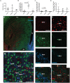
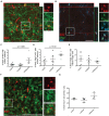
A number of studies have shown that damage to brain structures adjacent to neurogenic regions can result in migration of new neurons from neurogenic zones into the damaged tissue. The number of differentiated neurons that survive is low, however, and this has led to the idea that the introduction of extrinsic signaling factors, particularly neurotrophic proteins, may augment the neurogenic response to a level that would be therapeutically relevant. Here we report on the impact of the relatively newly described neurotrophic factor, Meteorin, when over-expressed in the striatum following excitotoxic injury. Birth-dating studies using bromo-deoxy-uridine (BrdU) showed that Meteorin did not enhance injury-induced striatal neurogenesis but significantly increased the proportion of new cells with astroglial and oligodendroglial features. As a basis for comparison we found under the same conditions, glial derived neurotrophic factor significantly enhanced neurogenesis but did not effect gliogenesis. The results highlight the specificity of action of different neurotrophic factors in modulating the proliferative response to injury. Meteorin may be an interesting candidate in pathological settings involving damage to white matter, for example after stroke or neonatal brain injury.
Keywords: GDNF; brain repair; forebrain injury; neurogenesis; oligodendrogenesis; striatum.
Figures


Similar articles
-
Intracerebral infusion of glial cell line-derived neurotrophic factor promotes striatal neurogenesis after stroke in adult rats.Stroke. 2006 Sep;37(9):2361-7. doi: 10.1161/01.STR.0000236025.44089.e1. Epub 2006 Jul 27. Stroke. 2006. PMID: 16873711
-
Lentiviral delivery of meteorin protects striatal neurons against excitotoxicity and reverses motor deficits in the quinolinic acid rat model.Neurobiol Dis. 2011 Jan;41(1):160-8. doi: 10.1016/j.nbd.2010.09.003. Epub 2010 Sep 16. Neurobiol Dis. 2011. PMID: 20840868
-
Treatment with UDP-glucose, GDNF, and memantine promotes SVZ and white matter self-repair by endogenous glial progenitor cells in neonatal rats with ischemic PVL.Neuroscience. 2015 Jan 22;284:444-458. doi: 10.1016/j.neuroscience.2014.10.012. Epub 2014 Oct 16. Neuroscience. 2015. PMID: 25453769
-
Neuroprotection by neurotrophins and GDNF family members in the excitotoxic model of Huntington's disease.Brain Res Bull. 2002 Apr;57(6):817-22. doi: 10.1016/s0361-9230(01)00775-4. Brain Res Bull. 2002. PMID: 12031278 Review.
-
Seizure-induced neurogenesis: are more new neurons good for an adult brain?Prog Brain Res. 2002;135:121-31. doi: 10.1016/S0079-6123(02)35012-X. Prog Brain Res. 2002. PMID: 12143334 Review.
Cited by
-
Meteorin Is a Novel Therapeutic Target for Wet Age-Related Macular Degeneration.J Clin Med. 2021 Jul 2;10(13):2973. doi: 10.3390/jcm10132973. J Clin Med. 2021. PMID: 34279457 Free PMC article.
-
Antihyperalgesic effects of Meteorin in the rat chronic constriction injury model: a replication study.Pain. 2019 Aug;160(8):1847-1855. doi: 10.1097/j.pain.0000000000001569. Pain. 2019. PMID: 31335652 Free PMC article.
-
Impacts of systemic milieu on cerebrovascular and brain aging: insights from heterochronic parabiosis, blood exchange, and plasma transfer experiments.Geroscience. 2025 May 23. doi: 10.1007/s11357-025-01657-y. Online ahead of print. Geroscience. 2025. PMID: 40407975 Review.
-
Immune genes involved in synaptic plasticity during early postnatal brain development contribute to post-stroke damage in the aging male rat brain.Biogerontology. 2025 Feb 18;26(2):60. doi: 10.1007/s10522-025-10203-4. Biogerontology. 2025. PMID: 39966204 Free PMC article.
-
Circular METRN RNA hsa_circ_0037251 Promotes Glioma Progression by Sponging miR-1229-3p and Regulating mTOR Expression.Sci Rep. 2019 Dec 24;9(1):19791. doi: 10.1038/s41598-019-56417-8. Sci Rep. 2019. PMID: 31875034 Free PMC article.
References
-
- Arvidsson A., Kokaia Z., Airaksinen M. S., Saarma M., Lindvall O. (2001). Stroke induces widespread changes of gene expression for glial cell line-derived neurotrophic factor family receptors in the adult rat brain. Neuroscience 106 27–41. - PubMed
-
- Brandt M. D., Jessberger S., Steiner B., Kronenberg G., Reuter K., Bick-Sander A., et al. (2003). Transient calretinin expression defines early postmitotic step of neuronal differentiation in adult hippocampal neurogenesis of mice. Mol. Cell. Neurosci. 24 603–613. 10.1016/S1044-7431(03)00207-0 - DOI - PubMed
LinkOut - more resources
Full Text Sources
Other Literature Sources
Research Materials

