

Extracellular Vesicle-Associated Transitory Cell Wall Components and Their Impact on the Interaction of Fungi with Host Cells
- PMID: 27458437
- PMCID: PMC4937017
- DOI: 10.3389/fmicb.2016.01034
Extracellular Vesicle-Associated Transitory Cell Wall Components and Their Impact on the Interaction of Fungi with Host Cells
Abstract
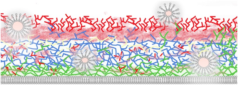
Classic cell wall components of fungi comprise the polysaccharides glucans and chitin, in association with glycoproteins and pigments. During the last decade, however, system biology approaches clearly demonstrated that the composition of fungal cell walls include atypical molecules historically associated with intracellular or membrane locations. Elucidation of mechanisms by which many fungal molecules are exported to the extracellular space suggested that these atypical components are transitorily located to the cell wall. The presence of extracellular vesicles (EVs) at the fungal cell wall and in culture supernatants of distinct pathogenic species suggested a highly functional mechanism of molecular export in these organisms. Thus, the passage of EVs through fungal cell walls suggests remarkable molecular diversity and, consequently, a potentially variable influence on the host antifungal response. On the basis of information derived from the proteomic characterization of fungal EVs from the yeasts Cryptoccocus neoformans and Candida albicans and the dimorphic fungi Histoplasma capsulatum and Paracoccidioides brasiliensis, our manuscript is focused on the clear view that the fungal cell wall is much more complex than previously thought.
Keywords: cell wall remodeling; extracellular vesicles; fungal cell wall; host cell; proteomics.
Figures
References
-
- Albuquerque P. C., Nakayasu E. S., Rodrigues M. L., Frases S., Casadevall A., Zancope-Oliveira R. M., et al. (2008). Vesicular transport in Histoplasma capsulatum: an effective mechanism for trans-cell wall transfer of proteins and lipids in ascomycetes. Cell. Microbiol. 10 1695–1710. 10.1111/j.1462-5822.2008.01160.x - DOI - PMC - PubMed
Publication types
Grants and funding
LinkOut - more resources
Full Text Sources
Other Literature Sources