Case Reports
. 2016 May 19:3:40.
doi: 10.3389/fvets.2016.00040. eCollection 2016.

Canine Butterfly Glioblastomas: A Neuroradiological Review

Affiliations
Case Reports

Canine Butterfly Glioblastomas: A Neuroradiological Review

John H Rossmeisl et al. Front Vet Sci. .

Abstract

In humans, high-grade gliomas may infiltrate across the corpus callosum resulting in bihemispheric lesions that may have symmetrical, winged-like appearances. This particular tumor manifestation has been coined a "butterfly" glioma (BG). While canine and human gliomas share many neuroradiological and pathological features, the BG morphology has not been previously reported in dogs. Here, we describe the magnetic resonance imaging (MRI) characteristics of BG in three dogs and review the potential differential diagnoses based on neuroimaging findings. All dogs presented for generalized seizures and interictal neurological deficits referable to multifocal or diffuse forebrain disease. MRI examinations revealed asymmetrical (2/3) or symmetrical (1/3), bihemispheric intra-axial mass lesions that predominantly affected the frontoparietal lobes that were associated with extensive perilesional edema, and involvement of the corpus callosum. The masses displayed heterogeneous T1, T2, and fluid-attenuated inversion recovery signal intensities, variable contrast enhancement (2/3), and mass effect. All tumors demonstrated classical histopathological features of glioblastoma multiforme (GBM), including glial cell pseudopalisading, serpentine necrosis, microvascular proliferation as well as invasion of the corpus callosum by neoplastic astrocytes. Although rare, GBM should be considered a differential diagnosis in dogs with an MRI evidence of asymmetric or symmetric bilateral, intra-axial cerebral mass lesions with signal characteristics compatible with glioma.

Keywords: astrocytoma; brain tumor; canine; glioblastoma; magnetic resonance imaging.


Figures
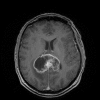
Figure 1
Figure 1
Coronal, post-contrast T1 image of a posterior human butterfly GBM.
Figure 2
Figure 2
MRI and pathological features of asymmetrical butterfly glioblastoma, Case 1. Transverse T2 (A,D), T1 (B,E), FLAIR (G), and post-contrast T1 (C,F,H) MR images from the rostral aspect of the intra-axial mass at the level of the cruciate gyrus (A–C), the midportion of the lesion (D–F), and caudal aspects of the mass at the level of the mesencephalic aqueduct (G,H). The mass is T1 hypo- to isointense, T2/FLAIR hyperintense and demonstrates marked contrast enhancement. Mass effect, manifest as left lateral ventricular compression and a falx shift to the right, and T2/FLAIR hyperintensity in the perilesional white matter are evident. The extensive corpus callosal and bihemispheric involvement of the contrast-enhancing lesion burden is depicted in the T1 post-contrast dorsal planar (I), transverse (C,F,H), and parasagittal images (L). Microscopic features of GBM (J) include serpentine necrosis, glial pseudopalisading, and microvascular proliferation, H&E stain, bar = 125 μm. Diffuse neoplastic infiltration of the periventricular white matter [(K); LV, right lateral ventricle; bar = 3.5 mm] of the right cerebrum, H&E stain, bar = 40 μm, inset.
Figure 3
Figure 3
Asymmetrical butterfly glioblastoma, Case 2. Transverse T2 (A,E,I), T1 (B,F,J), and post-contrast T1 (C,G,K) MR images from the rostral aspect of the left cerebral lesion (A–C), the midportion of the lesions (E–G), and caudal portions of the mass lesions (I–K). The masses are heterogeneously T1 hypo- to isointense, T2 hyperintense, and the right cerebral lesion demonstrates heterogeneous contrast enhancement. In the rostral aspect of the lesion, T2 hyperintensity extending from the left cerebral mass lesion across midline (A) via the corpus callosum is evident, which was demonstrated to be caused by neoplastic invasion of the corpus callosum at necropsy [(D), H&E, bar = 500 μm]. Morphology of GBM [(D), inset] within right cerebral mass illustrating serpentine necrosis and hemorrhage, H&E, bar = 75 μm. Bilateral mass effect, manifest as lateral ventricular compression and a falx shift to the right is present. Gross brain specimen (H) at the level of the optic chiasm demonstrating mass lesion in right cerebrum (white arrow) and malacia of the subcortical white matter on the left (black arrows). At the caudal level, the left and right intra-axial mass lesions are connected via T2 hyperintensity (I) extending across corpus callosum. Photomicrograph (L) of multifocal infiltration of neoplastic astrocytes within the body of the corpus callosum, H&E stain, bar = 3 mm, LV, left lateral ventricle.
Figure 4
Figure 4
Symmetrical butterfly glioblastoma, Case 3. Transverse T1 (A) and FLAIR (B) MR images at the level of the cruciate gyrus, demonstrating an intra-axial, bilaterally symmetric mass lesion present in the frontoparietal regions. Schematic, MR intensity-segmented transverse (C) and dorsal planar (G) representations of symmetrical bihemispheric appearance of butterfly GBM (stippled gray), perilesional edema (white), brain parenchyma (light gray), and ventricles (black). Histopathological features of GBM (D) include marked hypercellularity, cellular pleomorphism, and microvascular proliferation. Oligodendroglial components of the tumor are present throughout the section (H&E stain, bar = 200 μm). In the dorsal planar PD-T2 (E) and FLAIR (F) images, the mass attenuates the rostral aspects of the lateral ventricles and is associated with extensive symmetrical perilesional hyperintensity within the surrounding white matter, consistent with edema. The mass demonstrates heterogeneous signal intensity in all sequences. (H) 50% of neoplastic astrocytes demonstrate intense GFAP staining, while neoplastic oligodendrocytes lack GFAP immunoreactivity (GFAP stain, horseradish peroxidase with 3,3′-diaminobenzidine substrate, bar = 50 μm).

References

    1. Rees JH, Smirniotopoulos JG, Jones RV, Wong K. Glioblastoma multiforme: radiologic-pathologic correlation. Radiographics (1996) 16:1413–38.10.1148/radiographics.16.6.8946545 - DOI - PubMed
    1. Dziurzynski K, Blas-Boria D, Suki D, Cahill DP, Prabhu SS, Puduvalli V, et al. Butterfly glioblastomas: a retrospective review and qualitative assessment of outcomes. J Neurooncol (2012) 109:555–63.10.1007/s11060-012-0926-0 - DOI - PMC - PubMed
    1. Song RB, Vite CH, Bradley CE, Cross RJ. Postmortem evaluation of 435 cases of intracranial neoplasia in dogs and relationship of neoplasm with breed, age, and body weight. J Vet Intern Med (2013) 27:1143–52.10.1111/jvim.12136 - DOI - PubMed
    1. Bentley RT, Ober CP, Anderson KL, Feeney DA, Naughton JF, Ohlfest JR, et al. Canine intracranial gliomas: relationship between magnetic resonance imaging criteria and tumor type and grade. Vet J (2013) 198:463–71.10.1016/j.tvjl.2013.08.015 - DOI - PMC - PubMed
    1. Young BD, Levine JM, Porter BF, Chen-Allen AV, Rossmeisl JH, Platt SR, et al. Magnetic resonance imaging features of intracranial astrocytomas and oligodendrogliomas in dogs. Vet Radiol Ultrasound (2011) 52:132–41.10.1111/j.1740-8261.2010.01758.x - DOI - PubMed

Publication types

LinkOut - more resources