. 2016 Sep 8;537(7619):197-201.
doi: 10.1038/nature19316. Epub 2016 Jul 26.

Cryo-EM structure of the spliceosome immediately after branching

Affiliations

Cryo-EM structure of the spliceosome immediately after branching

Wojciech P Galej et al. Nature. .

Abstract

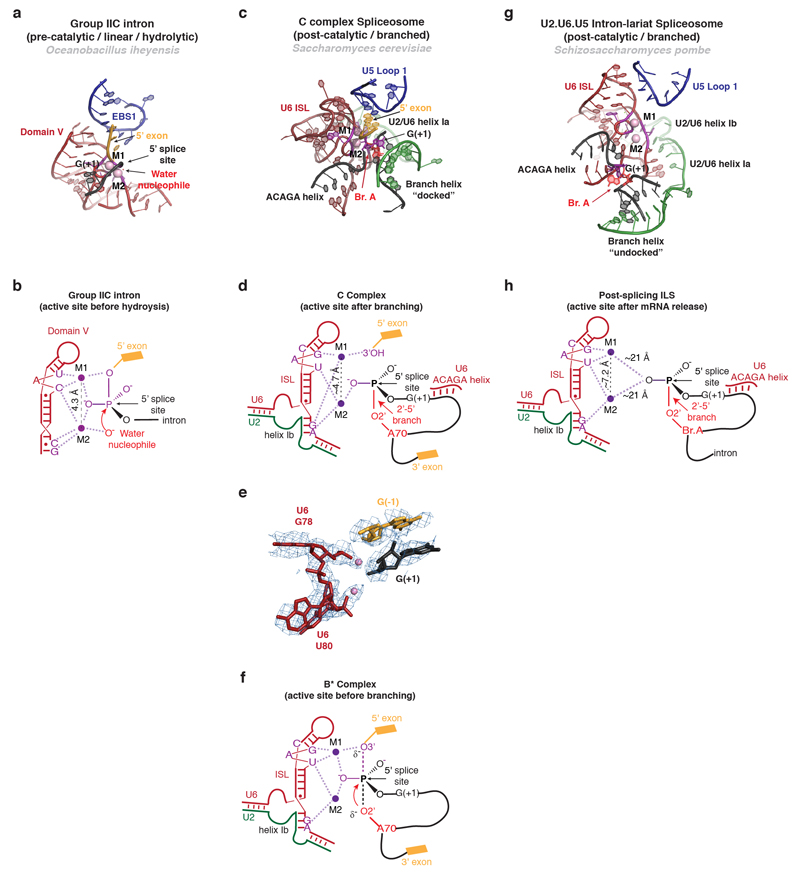
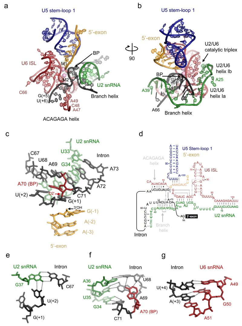
Precursor mRNA (pre-mRNA) splicing proceeds by two consecutive transesterification reactions via a lariat-intron intermediate. Here we present the 3.8 Å cryo-electron microscopy structure of the spliceosome immediately after lariat formation. The 5'-splice site is cleaved but remains close to the catalytic Mg2+ site in the U2/U6 small nuclear RNA (snRNA) triplex, and the 5'-phosphate of the intron nucleotide G(+1) is linked to the branch adenosine 2'OH. The 5'-exon is held between the Prp8 amino-terminal and linker domains, and base-pairs with U5 snRNA loop 1. Non-Watson-Crick interactions between the branch helix and 5'-splice site dock the branch adenosine into the active site, while intron nucleotides +3 to +6 base-pair with the U6 snRNA ACAGAGA sequence. Isy1 and the step-one factors Yju2 and Cwc25 stabilize docking of the branch helix. The intron downstream of the branch site emerges between the Prp8 reverse transcriptase and linker domains and extends towards the Prp16 helicase, suggesting a plausible mechanism of remodelling before exon ligation.


Conflict of interest statement

The authors declare no competing financial interests.

Figures

Extended Data Figure 1
Extended Data Figure 1. Biochemical characterisation of the complex and initial cryo-EM analysis.
a, SDS-PAGE analysis of the purified sample. Protein identities were confirmed by mass spectrometry analysis. Protein labels are coloured according to sub-complex identity (dark blue, U5 snRNP; light blue, helicase module; orange, NTC; yellow, NTR; green, U2 snRNP; purple, splicing factors; grey, not found in density) b, analysis of the fluorescently labelled substrate in the sample by denaturing PAGE, showing conversion of linear pre-mRNA (time point 0’) into branched lariat-intron intermediate (time point 30’), which is a predominant species in the purified sample (C complex). The two hairpins on the right depict the 2xMS2 stem-loops attached to the 5’end of the UBC4 pre-mRNA substrate for affinity purification. c, a typical cryo-EM micrograph collected on an FEI Titan Krios microscope operated at 300 kV and detected with a Gatan K2 Summit camera. d, reference-free 2D classification results. e, detail of a single class average with major domains labelled.
Extended Data Figure 2
Extended Data Figure 2. Overview of the data processing scheme used in this study.
Iterative 2D classification, template selection and automated particle picking resulted in 248K particles which were classified in 3D with a scaled and low-pass filtered model of ILS (EMDB-6413) as a reference. The best class was refined to 3.8 Å resolution overall. Focused classification allowed us to obtain two other maps with improved quality of the peripheral regions (Prp19 and helicase modules, EMD-4056 and EMD-4057). Classification of the core complex with fine angular sampling and local searches revealed a subtle movement of the U2 snRNP which correlates with the appearance of the extra density, interpreted as a WD40 domain which belongs to Prp17 or Prp19.
Extended Data Figure 3
Extended Data Figure 3. Global and local resolution analysis.
a, two orthogonal sections through the map showing variation in the local resolution as estimated by Resmap. b, an overall map of the core complex c, Gold-standard FSC plots for three maps used in this study. d, map of the core complex with a helicase module. e, a map of the core complex with Prp19 module.
Extended Data Figure 4
Extended Data Figure 4. Examples of cryo-EM density at the core of the complex with atomic models built in.
a, U5 snRNA loop 1 with 5’-exon bound. b, the active site with exon, intron, U2 and U6 snRNAs. c, two helices of the Prp8 Reverse Transcriptase Thumb/X domain, showing a clear helical pitch and excellent densities for the side chains. d, Fourier Shell Correlation between model and the map and cross-validation of the model fitting. (The original atom positions have been randomly displaced up to 0.5Å and refined with restraints against the half1 map only. FSC was calculated for two half maps. Excellent correlation up to the high resolution between the model and the half2 map (which was not used in refinement) cross-validates the model for overfitting.
Extended Data Figure 5
Extended Data Figure 5. Metal binding by the catalytic core of C complex.
a,b, Structure (a) and schematic representation (b) of the active site of a group IIC intron trapped in the pre-catalytic state in the presence of Ca2+ (PDB 4FAQ, ref. 75). The 5’ splice site scissile phosphate is aligned with the two metals bound at the core in a catalytic configuration, as shown in b. Note that, in this pre-catalytic structure, the group II domain VI is not present and therefore the structure does not contain the bulged adenosine nucleophile required for the branching reaction. As a result, the nucleophile is a water molecule, rather than the 2’-OH of the branch site adenosine found in spliceosomal introns. c-d, Structure of the RNA at the active site of spliceosomal C complex, showing the overall architecture (c), schematic of metal binding (d), and comparison of the model with the EM density (e). Note conservation of the metal binding residues compared to the group II intron (c.f. ref. 36) and proximity of the cleaved G(-1)-G(+1) bond to putative M1. f, Proposed interactions between U6 snRNA and the two catalytic Mg2+ during the transition state for branching, as inferred from biochemistry. g, h, Structure (g) and schematic (h) of the RNA core of the U2.U6.U5 ILS complex in a post-catalytic configuration (PDB 3JB9, ref. 26), likely following release of the mRNA. The two Mg2+ are shown as modelled in the coordinates deposited by the authors of the ILS structure (PDB 3JB9, ref. 26). In the ILS structure M1 and M2 are further apart (7.2 A) than in most other structures of RNAs that coordinate catalytic metals (usually 3.9-5 A); nonetheless the ligands modeled for M1 and M2 are consistent with the ligands identified biochemically for the two catalytic Mg2+ necessary for splicing (compare PDB 3JB9 and 4R0D with the data in refs. 34 and 36). Note that the branch helix is undocked from the U6 snRNA metal binding site and G(+1) is far away from the two Mg2+ at the core. The substrate and snRNAs are colour-coded while residues that position the catalytic metals are shown in magenta.
Extended Data Figure 6
Extended Data Figure 6. Examples of the structures of isolated components.
De novo built proteins are shown in cartoon form, along with a secondary structure diagram for the novel zinc finger fold of Yju2. Proteins that were modelled into low-resolution regions by rigid-body docking of crystal structures or homology models (Prp19 module, Brr2, Prp16, Prp8Jab1/MPN) are shown in their cryo-EM densities.
Extended Data Figure 7
Extended Data Figure 7. Conformational changes between U4/U6.U5 tri-snRNP, Complex C and Intron-Lariat Spliceosome.
a, rearrangement of the RNaseH-like domain with respect to the main body of Prp8 in all three complexes. b, α-finger (1575-1598) contacting the key RNA and proteins in a context-dependent manner. c, Prp8 N-terminal domain movements along with Prp8 residues 1406-1436 transiently docking on top of the 5’-exon and Cwc21 in complex C, stabilising the 5’-exon and interdomain contacts in Prp8. d, conformational rearrangements between complex C and S.pombe ILS showing a coupled movement of the U2 snRNP, Syf1 and Prp19.
Extended Data Figure 8
Extended Data Figure 8. Implications for deposition of the Exon-Junction Complex.
In higher eukaryotes exon-junction complexes (EJCs) are deposited 20 – 24 nt upstream of splice junctions, and form a binding platform for factors involved in nuclear export, translation, alternative splicing and nonsense-mediated mRNA decay. The core EJC components eIF4AIII, MAGOH and Y14 are found in human B and C complexes. Cwc22 is required for eIF4AIII recruitment to spliceosomes– and holds it in an open, inactive conformation. a, Crystal structure of the eIF4AIII:Cwc22 complex docked onto the spliceosomal C complex via superposition on Cwc22. b, Crystal structure of the core EJC, superimposed on the previous model via the second RecA domain of eIF4AIII. c, The 5’-exon exiting the channel at the interface between the Prp8 Large and N-terminal domains is positioned perfectly for the deposition of the EJC, explaining how the Cwc22 MIF4G domain is involved in determining the distance of EJC deposition from the splice junction.
Figure 1
Figure 1. Subunit architecture of the spliceosomal complex C.
a-c, three orthogonal views of the complex coloured according to the subunit identity. d, a list of all 44 modelled subunits of the complex grouped into functional sub-complexes.
Figure 2
Figure 2. Overview of the core structure.
a, Prp8 and its central role in organizing the entire assembly (SII denotes U2/U6 stem II). b, RNA only in the same orientation as in a (ISL, U6 snRNA Internal Stem-Loop; 5’SL, U6 snRNA 5’ Stem-Loop; SL1, U5 snRNA Stem-Loop 1; VSL, U5 snRNA Variable Stem-Loop; S3, U5 snRNA Stem III). c, Ecm2, Cwc2 and Bud31 binding to the 5’-end of the U6 snRNA. d, top view of the complex. e, RNA only in the same orientation as in d. f, Secondary structure diagram for the 3'-end of U2 snRNA.
Figure 3
Figure 3. Structure of the RNA catalytic core.
a, key RNA elements at the active site. ISL denotes Internal Stem-Loop. b, orthogonal view illustrating the branch helix and helices Ia and Ib of U2/U6 snRNA duplex. c, the branch helix and 5’-exon with the 2’-5’ phosphodiester linkage (red arrow). d, intricate RNA interactions at the active site (dotted lines indicate base triples; dot and star indicate G-U wobble and other non-canonical base-pairs). e, base-triple interaction between the branch helix and 5’-splice site. f, a network of interactions in the branch helix. g, Hoogsteen base-pair between intron A(+3) and G50 of U6 snRNA.
Figure 4
Figure 4. Proteins at the active site.
a, 5’exon channel formed between the Large and N-terminal domains of Prp8, Cwc21 and Cwc22. b, 5’exon:U5 loop 1 interaction surrounded by Prp8. Th/X denotes Thumb/domain X of Prp8 (residues 1300-1375). c, interactions between the 5’-exon, the N-terminal (purple) and Large (blue) domains of Prp8, and Yju2 (green). Interactions involving protein main and side chains are shown by solid and dotted lines. d, components surrounding U6 Internal Stem-Loop. e, Prp8 and Cef1 (myb1 domain) stabilise the catalytic triplex. HB denotes helix bundle of the RT domain (residues 750-870). f, structure of the catalytic triplex.
Figure 5
Figure 5. Step 1 factors and branch site positioning
a, interaction between the RNA catalytic core and Prp8. b, positioning of the branch helix by step 1 factors. c, corresponding view in S.pombe post splicing ILS complex, showing dramatic repositioning of the branch helix and its further stabilisation by debranching co-factor Cwf19. d, a close-up view of step 1 factors interacting with the branch helix.
Figure 6
Figure 6. The role of helicases in active site remodelling.
a, the intron sequence downstream from the branch site exits the spliceosome via a channel in Prp8 and extends towards Prp16. Translocation of Prp16 towards the branch helix would destabilise step 1 factors and displace the branch helix from its pocket. b, schematic illustrating how step 1 or step 2 specific factors can determine the specificity of the helicase recruited to the spliceosome at particular stages of splicing.

Comment in

References

    1. Will CL, Lührmann R. Spliceosome structure and function. Cold Spring Harb Perspect Biol. 2011;3:a003707. - PMC - PubMed
    1. Burge CB, Tuschl T, Sharp PA. In: The RNA World II. Gesteland RF, Cech TR, Atkins JF, editors. Cold Spring Harbor Laboratory Press; 1999. pp. 525–560.
    1. Lambowitz AM, Zimmerly S. Group II introns: mobile ribozymes that invade DNA. Cold Spring Harb Perspect Biol. 2011;3:a003616. - PMC - PubMed
    1. Chan S-P, Kao D-I, Tsai W-Y, Cheng S-C. The Prp19p-associated complex in spliceosome activation. Science. 2003;302:279–282. - PubMed
    1. Ohi MD, Gould KL. Characterization of interactions among the Cef1p-Prp19p-associated splicing complex. RNA. 2002;8:798–815. - PMC - PubMed

MeSH terms

Substances