. 2016 Sep:45:88-97.
doi: 10.1016/j.neurobiolaging.2016.05.015. Epub 2016 May 21.

Age-related changes in tonic activation of presynaptic versus extrasynaptic γ-amniobutyric acid type B receptors in rat medial prefrontal cortex

Affiliations

Age-related changes in tonic activation of presynaptic versus extrasynaptic γ-amniobutyric acid type B receptors in rat medial prefrontal cortex

Haley E Carpenter et al. Neurobiol Aging. 2016 Sep.

Abstract

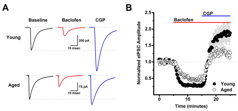
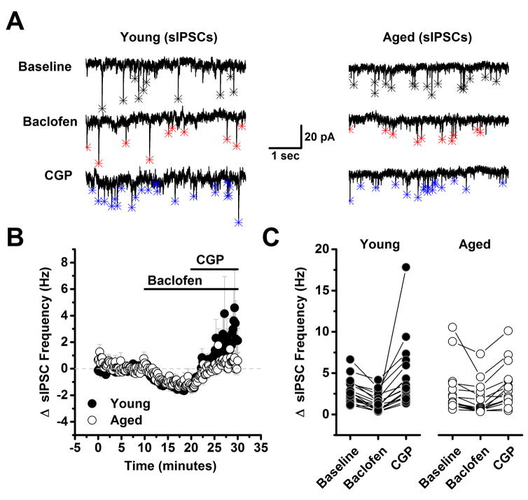
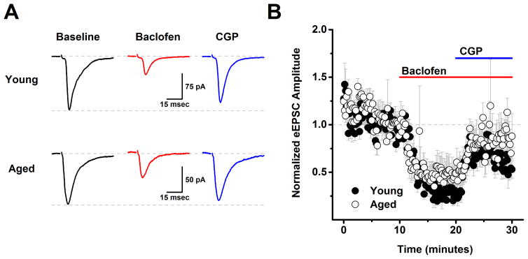
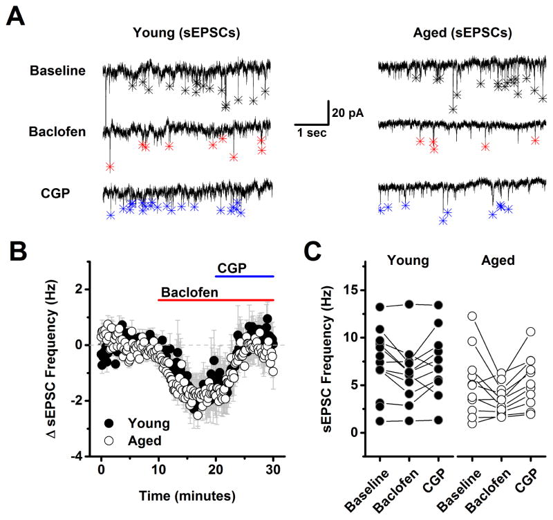
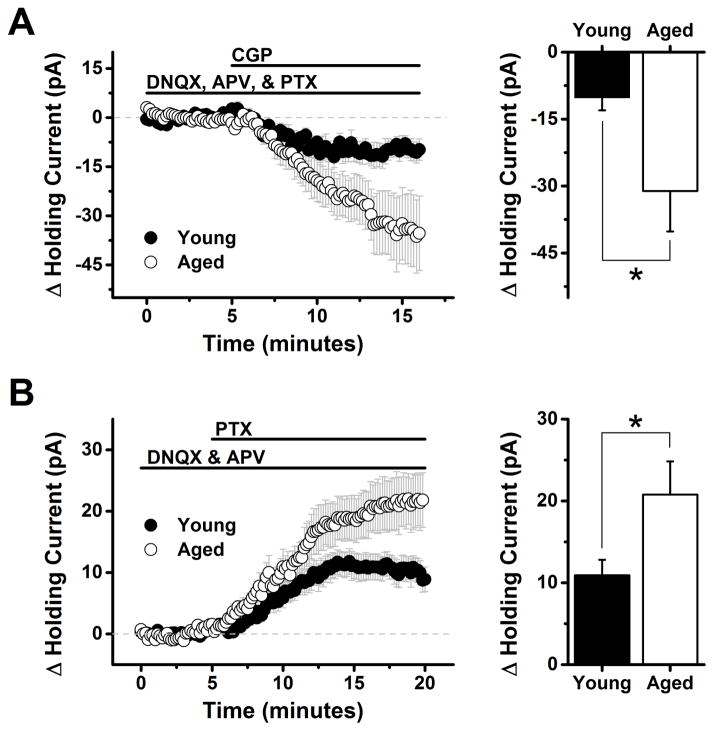
The present study examined the effect of age on both glutamatergic and γ-aminobutyric acid mediated (GABAergic) signaling in the rodent medial prefrontal cortex (mPFC), with an emphasis on revealing novel changes contributing to increased inhibition in age. Whole-cell patch clamp recordings were obtained from layer 2/3 mPFC pyramidal neurons in acute cortical slices prepared from either young (4 months) or aged (20-24 months) male F344 rats. Results indicated that GABAB receptors on GABAergic, but not on glutamatergic, inputs to layer 2/3 pyramidal cells are tonically activated by ambient GABA in young animals and further demonstrated that this form of tonic inhibition is significantly attenuated in aged mPFC. Moreover, concurrent with loss of tonic presynaptic GABAB autoreceptor activation, layer 2/3 pyramidal cells in aged mPFC are subjected to increased tonic activation of extrasynaptic GABAA and GABAB receptors. These data demonstrate a shift in the site of GABAB receptor-mediated inhibitory tone in the aged mPFC that clearly promotes increased inhibition of pyramidal cells in aged animals, and that may plausibly contribute to impaired executive function.

Keywords: Age-related cognitive decline; GABA(A); GABA(B); L2/3 pyramidal neurons; Prefrontal cortex; Working memory.


Conflict of interest statement

statement The authors have no conflicts of interest, and all animal procedures were performed in compliance with the Institutional Animal Care and Use Committee (IACUC) of the University of Florida.

Figures

Figure 1
Figure 1. Aging reduces tonic activation of presynaptic GABAB autoreceptors on inhibitory inputs to layer 2/3 pyramidal neurons
A) Raw data traces representative of the population mean are illustrated from a young neuron (top) and an aged neuron (bottom) in baseline conditions, after application of 10 μM baclofen, and in the presence of both baclofen and 10 μM CGP. Each trace is an average of 15–30 consecutive sweeps (generated at 0.1 Hz) in the stated conditions. Note that while baclofen reduced evoked IPSC amplitude in both cells, amplitude in the presence of baclofen and CGP exceeded baseline amplitude much more strongly in the neuron extracted from a young animal. The top dashed line in each set of traces illustrates a common baseline, while the bottom dashed line represents the evoked IPSC amplitude in baseline conditions. B) Population data illustrate that the same effects are apparent across cells when evoked IPSC amplitude is normalized to the baseline mean (young: n=39, 8, aged: n=23, 7, in neurons, animals respectively). See text for specific values. DNQX (10 μM) and APV (40 μM) were present in the bath solution throughout the experiment.
Figure 2
Figure 2. Spontaneous inhibitory postsynaptic currents are less reliable reporters of presynaptic GABAB autoreceptor activation than are evoked inhibitory postsynaptic currents
A) Representative traces from a young neuron (left) and aged neuron (right) in baseline conditions, after application of 10 μM baclofen, and in the presence of both baclofen and 10 μM CGP. Asterisks in each trace indicate the peak of detected events. Baclofen clearly reduced spontaneous IPSC frequency in both neurons (middle traces, red asterisks). Notably, event frequency also exceeded baseline after application of 10 μM CGP in young, but not in the aged neuron (bottom traces, blue asterisks). B) Illustrates the average change in spontaneous IPSC frequency across all cells tested as in panel A. Although the mean spontaneous IPSC frequency in the presence of both baclofen and CGP more strongly trended above baseline in neurons from young animals, overall the effect was not statistically significant (young: n=19, 9, aged: n=15, 8, in neurons, animals respectively, p=0.09, see text of the results section for more details). C) Raw spontaneous IPSC frequency is plotted in all three conditions for every neuron tested in both the young and aged population. DNQX (10 μM) and APV (40 μM) were present in the bath solution throughout the experiment.
Figure 3
Figure 3. Analysis of evoked EPSCs indicates that GABAB heteroreceptors are expressed on excitatory inputs to layer 2/3 pyramidal neurons, but are not subject to tonic activation in either young or aged neurons
A) Raw data traces representative of the population mean are illustrated from a young neuron (top) and an aged neuron (bottom) in baseline conditions, after application of 10 μM baclofen, and in the presence of both baclofen and 10 μM CGP. Each trace is an average of 15–30 consecutive sweeps (generated at 0.1 Hz) in the stated conditions. Baclofen reduced evoked IPSC amplitude in both cells, but amplitude in the presence of baclofen and CGP did not exceed baseline values in either cell. The top dashed line in each set of traces illustrates a common baseline, while the bottom dashed line represents the evoked IPSC amplitude in baseline conditions. B) Population data illustrate that the same effects are apparent across cells when evoked EPSC amplitude is normalized to the baseline mean (young: n=5, 2, aged: n= 5, 2, in neurons, animals respectively). See text for specific values. PTX (50 μM) was present in the bath solution throughout the experiment.
Figure 4
Figure 4. Frequency of spontaneous excitatory postsynaptic currents is also sensitive to baclofen but not subject to tonic GABAB receptor mediated inhibition
A) Representative traces from a young neuron (left) and aged neuron (right) in baseline conditions, after application of 10 μM baclofen, and in the presence of both baclofen and 10 μM CGP. Asterisks in each trace indicate the peak of detected events. Baclofen clearly reduced spontaneous EPSC frequency in both neurons (middle traces, red asterisks). Event frequency returned to, but did not notably exceed baseline, after application of 10 μM CGP in both the young and the aged neuron (bottom traces, blue asterisks). B) Population data illustrating the average change in spontaneous EPSC frequency across all cells tested as in panel A. As per the representative cells, baclofen clearly inhabited spontaneous EPSC frequency, and subsequent application of CGP reversed this effect, however no difference was noted between ages, and no evidence of tonic GABAB heteroreceptors activation was apparent in either population (young: n=11, 4, aged: n=11, 5, in neurons, animals respectively). See text of the results section for more details. C) Raw spontaneous EPSC frequency is plotted in all three conditions for every neuron tested in both the young and aged population. PTX (100 μM) was present in the bath solution throughout the experiment.
Figure 5
Figure 5. Extrasynaptic high affinity GABAA and GABAB receptors are expressed by L2/3 pyramidal neurons, and are subject to greater tonic activation in aged animals
A) Left panel: Bath application of 10 μM CGP (a GABAB receptor antagonist) causes a clear shift in holding current in L2/3 pyramidal cells voltage clamped at −70 mV with a K-gluconate based internal solution (see Methods). Antagonists for ionotropic glutamate (DNQX, 10 μM, APV 40 μM) and GABAA receptors (PTX, 100 μM) were present throughout the experiment. The polarity of the holding current shift is consistent with the hypothesis that CGP is blocking an inhibitory current mediated by tonic activation of extrasynaptic GABAB receptors. Right panel: When quantified between 12 and 16 minutes into the experiment (7 to 11 minutes after application of CGP) this effect was significantly greater in neurons from aged animals than in neurons from young animals (−10.2 ± 2.9 pA in young neurons vs. −31.1 ± 9.06 pA in aged neurons, n=8, 15 respectively, p=0.04, see test of results section for further details). Note: although we refer to GABAB receptors on layer 2/3 pyramidal cells that are tonically activated by ambient GABA as ‘extrasynaptic’ here, and throughout the manuscript, it is plausible that synaptically located GABAB receptors also have sufficiently high affinity for GABA to contribute to these tonic currents, particularly if GABA uptake near synapses is not prohibitive. B) Left panel: illustrates a similar experiment conducted in conditions designed to isolate and amplify current through extrasynaptic GABAA receptors. Specifically, L2/3 pyramidal neurons were voltage clamped at −70 mV in the presence of ionotropic glutamate receptor antagonists (DNQX, 10 μM, APV 40 μM) using a high chloride CsMeSO3 based internal solution. Application of the GABAA receptor antagonist PTX (100 μM) revealed a clear shift in holding current in both young and aged neurons. The polarity of the holding current shift is consistent with the hypothesis that PTX is blocking an excitatory current mediated by tonic activation of extrasynaptic GABAA receptors. The current is excitatory due to the strong outward driving force on chloride at −70 mV that is created when using an internal solution with a high chloride concentration. Right panel: When quantified between 15 and 20 minutes into the experiment (4 to 10 minutes after application of PTX) this effect was significantly greater in neurons from aged animals than in neurons from young animals (10.9 ± 1.91 pA in young neurons vs. 20.8 ± 4.07 pA in aged neurons, n=18 cells (7 animals), 17 cells (7 animals) respectively, p<0.001, see test of results section for further details).

References

    1. Alexander GE, Ryan L, Bowers D, Foster TC, Bizon JL, Geldmacher DS, Glisky EL. Characterizing cognitive aging in humans with links to animal models. Front Aging Neurosci. 2012;4:21. doi: 10.3389/fnagi.2012.00021. - DOI - PMC - PubMed
    1. Arnsten AF, Wang MJ, Paspalas CD. Neuromodulation of thought: flexibilities and vulnerabilities in prefrontal cortical network synapses. Neuron. 2012;76(1):223–39. doi: 10.1016/j.neuron.2012.08.038. - DOI - PMC - PubMed
    1. Bakker A, Krauss GL, Albert MS, Speck CL, Jones LR, Stark CE, Yassa MA, Bassett SS, Shelton AL, Gallagher M. Reduction of hippocampal hyperactivity improves cognition in amnestic mild cognitive impairment. Neuron. 2012;74(3):467–74. doi: 10.1016/j.neuron.2012.03.023. - DOI - PMC - PubMed
    1. Banuelos C, Beas BS, McQuail JA, Gilbert RJ, Frazier CJ, Setlow B, Bizon JL. Prefrontal cortical GABAergic dysfunction contributes to age-related working memory impairment. J Neurosci. 2014;34(10):3457–66. doi: 10.1523/JNEUROSCI.5192-13.2014. - DOI - PMC - PubMed
    1. Beas BS, McQuail JA, Banuelos C, Setlow B, Bizon JL. Prefrontal cortical GABAergic signaling and impaired behavioral flexibility in aged F344 rats. Neuroscience. 2016 doi: 10.1016/j.neuroscience.2016.02.014. - DOI - PMC - PubMed

Publication types