

Epithelioid inflammatory myofibroblastic sarcoma: a clinicopathological, immunohistochemical and molecular cytogenetic analysis of five additional cases and review of the literature
- PMID: 27460384
- PMCID: PMC4962498
- DOI: 10.1186/s13000-016-0517-z
Epithelioid inflammatory myofibroblastic sarcoma: a clinicopathological, immunohistochemical and molecular cytogenetic analysis of five additional cases and review of the literature
Abstract
Background: To explore the clinical characteristics and pathological features of epithelioid inflammatory myofibroblastic sarcoma (EIMS) with emphasis on the diagnostic spectrum.
Methods: The clinical data and histological features in 5 additional cases of EIMS were retrospectively reviewed. Immunohistochemical study and interphase fluorescence in situ hybridization (FISH) analysis were carried out.
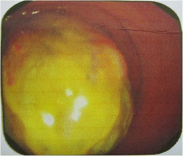
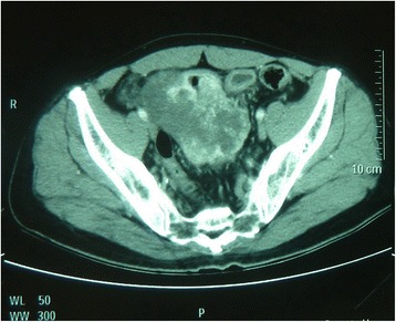
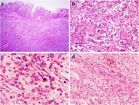
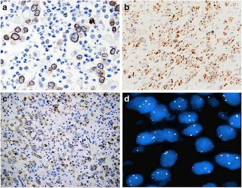
Results: There were 2 males and 3 females with age at presentation ranging from 15 to 58 years (mean, 37 years). All 5 tumors were intra-abdominal with 2 arising in the mesentery and 1 each in the omentum, rectum and transverse colon. The tumor size ranged from 5 to 20 cm in maximum diameter (mean, 10.7 cm). Histologically, all 5 tumors were composed predominantly of large epithelioid cells possessing vesicular nuclei, prominent nucleoli, and amphophilic cytoplasm. Mitotic figures were easily identified (mean, 20/10HPF). Tumor cells were arranged in clusters or sheets embedded in a myxoid stroma containing prominent neutrophils. A minor component of spindle cells was present in focal areas. By immunohistochemistry, all 5 cases were positive for anaplastic lymphoma kinase (ALK) with a nuclear membrane pattern in 4 and cytoplasmic staining with perinuclear accentuation in 1. Besides ALK, tumor cells stained variably for desmin (4/5), alpha smooth muscle actin (2/5), muscle-specific actin (1/2) and pan-cytokeratin (1/4). FISH analysis demonstrated the presence of ALK rearrangement in all 5 cases. Of 5 patients, 3 developed local recurrence, 1 died of disease 8 months after surgery.
Conclusion: EIMS represents a highly aggressive variant of inflammatory myofibroblastic tumor characterized by epithelioid morphology, prominent neutrophilic infiltrate, and nuclear membrane staining of ALK with ALK rearrangement. As patients with ALK-rearrangement tumors may benefit from targeted therapy, accurate diagnosis of EIMS is very important. Familiar with the characteristic features of EIMS will help pathologists avoid misdiagnosing the tumor as other malignancies.
Keywords: Fluorescence in situ hybridization; Gastrointestinal tract; Inflammatory myofibroblastic tumor; RANBP2-ALK; Soft tissue tumor.
Figures
Similar articles
-
Epithelioid inflammatory myofibroblastic sarcoma: An aggressive intra-abdominal variant of inflammatory myofibroblastic tumor with nuclear membrane or perinuclear ALK.Am J Surg Pathol. 2011 Jan;35(1):135-44. doi: 10.1097/PAS.0b013e318200cfd5. Am J Surg Pathol. 2011. PMID: 21164297
-
Cytopathologic features of epithelioid inflammatory myofibroblastic sarcoma with correlation of histopathology, immunohistochemistry, and molecular cytogenetic analysis.Cancer Cytopathol. 2015 Aug;123(8):495-504. doi: 10.1002/cncy.21558. Epub 2015 Jul 2. Cancer Cytopathol. 2015. PMID: 26139079
-
Pulmonary epithelioid inflammatory myofibroblastic sarcoma with multiple bone metastases: case report and review of literature.Diagn Pathol. 2015 Jul 16;10:106. doi: 10.1186/s13000-015-0358-1. Diagn Pathol. 2015. PMID: 26178751 Free PMC article. Review.
-
[Clinicopathological features of inflammatory myofibroblastic tumor].Zhonghua Bing Li Xue Za Zhi. 2021 Mar 8;50(3):194-200. doi: 10.3760/cma.j.cn112151-20200806-00627. Zhonghua Bing Li Xue Za Zhi. 2021. PMID: 33677881 Chinese.
-
Epithelioid inflammatory myofibroblastic sarcoma in abdominal cavity: a case report and review of literature.Int J Clin Exp Pathol. 2015 Apr 1;8(4):4213-9. eCollection 2015. Int J Clin Exp Pathol. 2015. PMID: 26097614 Free PMC article. Review.
Cited by
-
Epithelioid Inflammatory Myofibroblastic Sarcoma Presenting as Gastrointestinal Bleed: Case Report and Literature Review.JPGN Rep. 2020 Dec 17;2(1):e019. doi: 10.1097/PG9.0000000000000019. eCollection 2021 Feb. JPGN Rep. 2020. PMID: 37206935 Free PMC article. Review.
-
Epithelioid inflammatory myofibroblastic sarcoma: a case report and brief literature review.Front Oncol. 2023 Sep 29;13:1212529. doi: 10.3389/fonc.2023.1212529. eCollection 2023. Front Oncol. 2023. PMID: 37841422 Free PMC article.
-
Primary epithelioid inflammatory myofibroblastic sarcoma of the brain with EML4::ALK fusion mimicking intra-axial glioma: a case report and brief literature review.J Pathol Transl Med. 2024 May;58(3):141-145. doi: 10.4132/jptm.2024.04.12. Epub 2024 May 14. J Pathol Transl Med. 2024. PMID: 38766740 Free PMC article.
-
Thoracic epithelioid inflammatory myofibroblastic sarcoma: a rare and aggressive disease with case report and literature review.Discov Oncol. 2024 Sep 27;15(1):484. doi: 10.1007/s12672-024-01375-5. Discov Oncol. 2024. PMID: 39331206 Free PMC article.
-
New Perspectives in the Treatment of Inflammatory Myofibroblastic Tumor with ALK Translocation: Case Report.Case Rep Oncol. 2024 Jul 20;17(1):763-772. doi: 10.1159/000539739. eCollection 2024 Jan-Dec. Case Rep Oncol. 2024. PMID: 39144250 Free PMC article.
References
-
- Fletcher CDM, Bridge JA, Hogendoorn PCW, Mertens F. World Health Organization Classification of Tumors of Soft Tissue and Bone. 4. Lyon, France: IARC Press; 2013. pp. 83–84.
-
- Griffin CA, Hawkins AL, Dvorak C, Henkle C, Ellingham T, Perlman EJ. Recurrent involvement of 2p23 in inflammatory myofibroblastic tumors. Cancer Res. 1999;59:2776–80. - PubMed
-
- Gleason BC, Hornick JL. Inflammatory myofibroblastic tumors: where are we now? J ClinPathol. 2008;61:428–37. - PubMed
-
- Mariño-Enríquez A, Wang WL, Roy A, Lopez-Terrada D, Lazar AJ, Fletcher CD, et al. Epithelioid inflammatory myofibroblastic sarcoma:An aggressive intra-abdominal variant of inflammatory myofibroblastic tumor with nuclear membrane or perinuclear ALK. Am J Surg Pathol. 2011;35:135–44. doi: 10.1097/PAS.0b013e318200cfd5. - DOI - PubMed
Publication types
MeSH terms
Substances
LinkOut - more resources
Full Text Sources
Other Literature Sources
Medical
Research Materials
Miscellaneous
