Cellular Cations Control Conformational Switching of Inositol Pyrophosphate Analogues
- PMID: 27460418
- PMCID: PMC5076471
- DOI: 10.1002/chem.201601754
Cellular Cations Control Conformational Switching of Inositol Pyrophosphate Analogues
Abstract
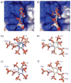
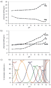
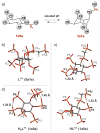
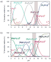
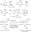
The inositol pyrophosphate messengers (PP-InsPs) are emerging as an important class of cellular regulators. These molecules have been linked to numerous biological processes, including insulin secretion and cancer cell migration, but how they trigger such a wide range of cellular responses has remained unanswered in many cases. Here, we show that the PP-InsPs exhibit complex speciation behaviour and propose that a unique conformational switching mechanism could contribute to their multifunctional effects. We synthesised non-hydrolysable bisphosphonate analogues and crystallised the analogues in complex with mammalian PPIP5K2 kinase. Subsequently, the bisphosphonate analogues were used to investigate the protonation sequence, metal-coordination properties, and conformation in solution. Remarkably, the presence of potassium and magnesium ions enabled the analogues to adopt two different conformations near physiological pH. Understanding how the intrinsic chemical properties of the PP-InsPs can contribute to their complex signalling outputs will be essential to elucidate their regulatory functions.
Keywords: biological activity; conformation analysis; phosphorylation; protonation; signal transduction.
© 2016 WILEY-VCH Verlag GmbH & Co. KGaA, Weinheim.
Figures



Similar articles
-
One Scaffold, Two Conformations: The Ring-Flip of the Messenger InsP8 Occurs under Cytosolic Conditions.Biomolecules. 2023 Apr 4;13(4):645. doi: 10.3390/biom13040645. Biomolecules. 2023. PMID: 37189392 Free PMC article.
-
Dissecting the activation of insulin degrading enzyme by inositol pyrophosphates and their bisphosphonate analogs.Chem Sci. 2021 Jul 8;12(32):10696-10702. doi: 10.1039/d1sc02975d. eCollection 2021 Aug 18. Chem Sci. 2021. PMID: 34476054 Free PMC article.
-
Inositol polyphosphates intersect with signaling and metabolic networks via two distinct mechanisms.Proc Natl Acad Sci U S A. 2016 Nov 1;113(44):E6757-E6765. doi: 10.1073/pnas.1606853113. Epub 2016 Oct 19. Proc Natl Acad Sci U S A. 2016. PMID: 27791083 Free PMC article.
-
Inositol Pyrophosphate Pathways and Mechanisms: What Can We Learn from Plants?Molecules. 2020 Jun 17;25(12):2789. doi: 10.3390/molecules25122789. Molecules. 2020. PMID: 32560343 Free PMC article. Review.
-
Versatile signaling mechanisms of inositol pyrophosphates.Curr Opin Chem Biol. 2022 Oct;70:102177. doi: 10.1016/j.cbpa.2022.102177. Epub 2022 Jun 30. Curr Opin Chem Biol. 2022. PMID: 35780751 Review.
Cited by
-
Metabolism and Functions of Inositol Pyrophosphates: Insights Gained from the Application of Synthetic Analogues.Molecules. 2020 Oct 2;25(19):4515. doi: 10.3390/molecules25194515. Molecules. 2020. PMID: 33023101 Free PMC article. Review.
-
Synthesis of Unsymmetrical Difluoromethylene Bisphosphonates.Org Lett. 2024 Jan 26;26(3):739-744. doi: 10.1021/acs.orglett.3c04211. Epub 2024 Jan 12. Org Lett. 2024. PMID: 38215221 Free PMC article.
-
Features and regulation of non-enzymatic post-translational modifications.Nat Chem Biol. 2018 Feb 14;14(3):244-252. doi: 10.1038/nchembio.2575. Nat Chem Biol. 2018. PMID: 29443975 Review.
-
One Scaffold, Two Conformations: The Ring-Flip of the Messenger InsP8 Occurs under Cytosolic Conditions.Biomolecules. 2023 Apr 4;13(4):645. doi: 10.3390/biom13040645. Biomolecules. 2023. PMID: 37189392 Free PMC article.
-
Dissecting the activation of insulin degrading enzyme by inositol pyrophosphates and their bisphosphonate analogs.Chem Sci. 2021 Jul 8;12(32):10696-10702. doi: 10.1039/d1sc02975d. eCollection 2021 Aug 18. Chem Sci. 2021. PMID: 34476054 Free PMC article.
References
-
- Shears SB. Adv Biol Regul. 2015;57:203–216. - PMC - PubMed
- Wilson MS, Livermore TM, Saiardi A. Biochem J. 2013;452:369–379. - PubMed
- Wundenberg T, Mayr GW. Biol Chem. 2012;393:979–998. - PubMed
- Chakraborty A, Kim S, Snyder SH. Science Signal. 2011;4:re1–11. - PMC - PubMed
- Barker CJ, Illies C, Gaboardi GC, Berggren PO. Cell Mol Life Sci. 2009;66:3851–3871. - PMC - PubMed
- Monserrate JP, York JD. Curr Opin Cell Biol. 2010;22:365–373. - PubMed
-
- Saiardi A, Erdjument-Bromage H, Snowman AM, Tempst P, Snyder SH. Curr Biol. 1999;9:1323–1326. - PubMed
- Saiardi A, Nagata E, Luo HR, Snowman AM, Snyder SH. J Biol Chem. 2001;276:39179–39185. - PubMed
- Schell MJ, Letcher AJ, Brearley CA, Biber J, Murer H, Irvine RF. FEBS Lett. 1999;461:169–172. - PubMed
- Voglmaier SM, Bembenek ME, Kaplin AI, Dorman G, Olszewski JD, Prestwich GD, Snyder SH. Proc Natl Acad Sci USA. 1996;93:4305–4310. - PMC - PubMed
-
- Mulugu S, Bai W, Fridy PC, Bastidas RJ, Otto JC, Dollins DE, Haystead TA, Ribeiro AA, York JD. Science. 2007;316:106–109. - PubMed
- Choi JH, Williams J, Cho J, Falck JR, Shears SB. J Biol Chem. 2007;282:30763–30775. - PMC - PubMed
- Lin H, Fridy PC, Ribeiro AA, Choi JH, Barma DK, Vogel G, Falck JR, Shears SB, York JD, Mayr GW. J Biol Chem. 2008;284:1863–1872. - PMC - PubMed
Grants and funding
LinkOut - more resources
Full Text Sources
Other Literature Sources
Miscellaneous

