. 2016 Sep;38(3):713-20.
doi: 10.3892/ijmm.2016.2688. Epub 2016 Jul 22.

Effects of an intravitreal injection of interleukin-35-expressing plasmid on pro-inflammatory and anti-inflammatory cytokines

Affiliations

Effects of an intravitreal injection of interleukin-35-expressing plasmid on pro-inflammatory and anti-inflammatory cytokines

Chao Hou et al. Int J Mol Med. 2016 Sep.

Abstract

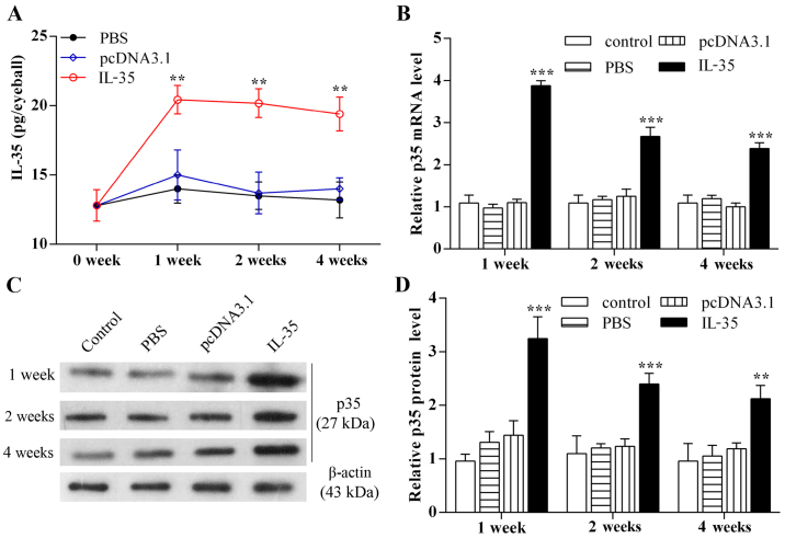
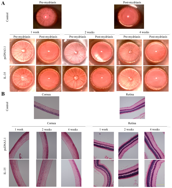
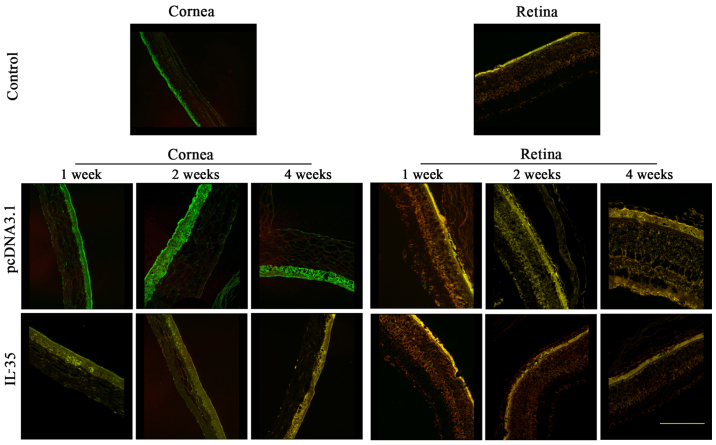
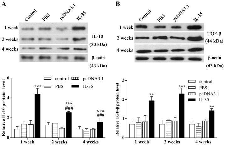
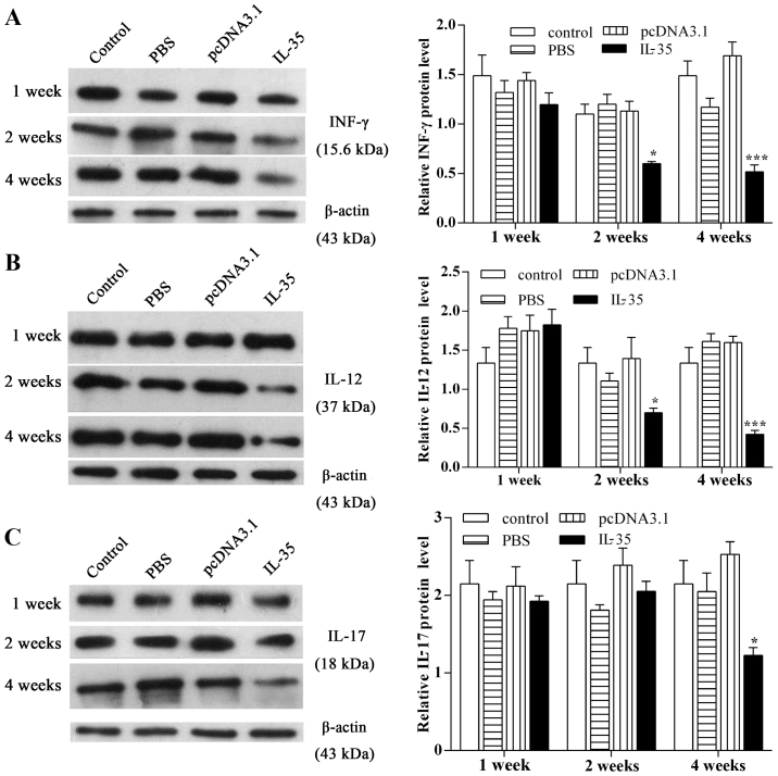
In order to explore the potential effects of interleukin (IL)-35 on IL-10, transforming growth factor-β (TGF-β), interferon-γ (INF)-γ, IL-12 and IL-17, a pcDNA3.1‑IL-35 plasmid was injected into the vitreous cavity of BALB/c mice. Enzyme-linked immunosorbent assay, western blot analysis and quantitative PCR analysis were performed to confirm the successful expression of IL-35. Slit-lamp biomicroscopy, hematoxylin and eosin staining and immunofluorescence were employed to detect the status of eyes, and western blot analysis was performed to examine the expression of corneal graft rejection-related cytokines. There were no abnormalities in the eyes pre-mydriasis or post-mydriasis and no injuries to the cornea or retina following the injection of IL-35-expressing plasmid. An immunofluorescence assay detected the positive expression of IL-35 in corneal epithelial cells from IL-35‑injected mice and negative staining in the control group. Further study revealed that IL-35 enhanced the expression of IL-10 and TGF-β which reached their highest levels at 1 and 2 weeks after injection, respectively (p<0.01). Moreover, the expression of INF-γ and IL-12 was decreased significantly at 2 weeks after the injection of IL-35-expressing plasmid (p<0.05), and the expression of IL-17 was suppressed notably at 4 weeks after the injection (p<0.05). The intravitreal injection of IL-35-expressing plasmid in mice downregulates the expression of pro-inflammatory cytokines and upregulates the expression of anti-inflammatory cytokines. Thus, IL-35 may further be assessed as a potential target for the treatment of corneal graft rejection.


Figures

Figure 1
Figure 1
Intravitreal injection of pcDNA3.1-interleukin (IL)-35 plasmid increases the intraocular level of IL-35. The plasmid pcDNA3.1-IL-35 encoding IL-35 sequences was injected into the vitreous cavity of mouse eyes. Mice were raised for 1, 2 and 4 weeks, respectively, prior to sacrifice. Thereafter, the mouse eyes were enucleated and lysed. (A) Enzyme linked immunosorbent assay (ELISA) was employed to examine the secretion level of IL-35 with five replicates at each testing point. (B) RT-qPCR analysis of p35 mRNA levels. β-actin was used as an internal control. Six to eight mice were randomly selected in each group and experiments were performed in triplicate for statistical significance. (C) Representative images of western blot analysis of p35 protein levels. β-actin served as the internal control. (D) The relative expression of p35 protein normalized to β-actin. The results are expressed as the means ± standard deviation (SD). **p<0.01, ***p<0.001 vs. pcDNA3.1.
Figure 2
Figure 2
Intravitreal injection of pcDNA3.1-interleukin (IL)-35 plasmid is safe in ocular tissues. (A) Ocular status of mouse eyes pre-mydriasis and post-mydriasis at the indicated time periods were observed by slit-lamp biomicroscopy. Representative examples of images are shown. (B) Representative photomicrographs of corneal and retinal tissues with or without IL-35 plasmid intravitreal injection. Nuclei labeled with hematoxylin were observed to be blue and cytoplasm stained with eosin was red. Scale bar, 50 µm. The above two experiments were repeated three times.
Figure 3
Figure 3
Interleukin (IL)-35 is uniformly distributed in corneal epithelial cells. An immunofluorescence double assay was performed to examine the distribution of IL-35 in the cornea and retina. The representative merged images were selected from three independent experiments. p35 was visualized using FITC-labeled goat anti-rabbit IgG (H+L) as green and EBI3 was stained with Cy3-labeled goat anti-mouse IgG (H+L) as red. Scale bar, 50 µm.
Figure 4
Figure 4
Elevated interleukin (IL)-35 levels enhance the expression of intraocular IL-10 and transforming growth factor-β (TGF-β). Western blot analysis was performed to evaluate the protein expression of (A) IL-10 and (B) TGF-β at different time-points following an intravitreal injection of pcDNA3.1-IL-35 plasmid. Representative blots are shown and corresponding densitometric analysis is presented as the means ± standard deviation (SD) from three independent experiments. β-actin was used as the internal control. **p<0.01, ***p<0.001 vs. control; ###p<0.001 vs. 1 week of IL-35.
Figure 5
Figure 5
Elevated interleukin (IL)-35 levels suppress the expression of interferon-γ (INF-γ), IL-12 and IL-17. The protein expression of (A) INF-γ, (B) IL-12 and (C) IL-17 were detected by western blot analysis and normalized to β-actin. Representative examples of images are shown and the data are presented as the means ± standard deviation (SD), and the error bars represent the SD of three independent experiments. *P<0.05, ***P<0.001 vs. control.

Similar articles

Cited by

References

    1. Qin Q, Shi Y, Zhao Q, Luo D, Chen Y, Wu J, Zhao M. Effects of CD25siRNA gene transfer on high-risk rat corneal graft rejection. Graefes Arch Clin Exp Ophthalmol. 2015;253:1765–1776. doi: 10.1007/s00417-015-3067-2. - DOI - PubMed
    1. Pan Q, Xu Q, Boylan NJ, Lamb NW, Emmert DG, Yang JC, Tang L, Heflin T, Alwadani S, Eberhart CG, et al. Corticosteroid-loaded biodegradable nanoparticles for prevention of corneal allograft rejection in rats. J Control Release. 2015;201:32–40. doi: 10.1016/j.jconrel.2015.01.009. - DOI - PMC - PubMed
    1. Chen Y, Liao C, Gao M, Belin MW, Wang M, Yu H, Yu J. Efficacy and safety of corneal transplantation using corneas from foreign donors versus domestic donors: a prospective, randomized, controlled trial. J Ophthalmol. 2015;2015:178289. doi: 10.1155/2015/178289. - DOI - PMC - PubMed
    1. Paunicka KJ, Mellon J, Robertson D, Petroll M, Brown JR, Niederkorn JY. Severing corneal nerves in one eye induces sympathetic loss of immune privilege and promotes rejection of future corneal allografts placed in either eye. Am J Transplant. 2015;15:1490–1501. doi: 10.1111/ajt.13240. - DOI - PMC - PubMed
    1. Zhang H, Wang L, Zhang L. Cyclosporine nanomicelle eye drop: a novel medication for corneal graft transplantation treatment. Biol Pharm Bull. 2015;38:893–900. doi: 10.1248/bpb.b15-00111. - DOI - PubMed

MeSH terms