

How Do Meningeal Lymphatic Vessels Drain the CNS?
- PMID: 27460561
- PMCID: PMC5002390
- DOI: 10.1016/j.tins.2016.07.001
How Do Meningeal Lymphatic Vessels Drain the CNS?
Abstract
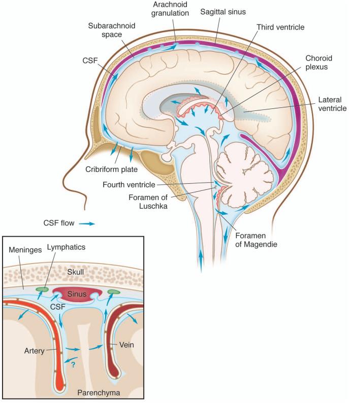
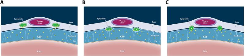
The many interactions between the nervous and the immune systems, which are active in both physiological and pathological states, have recently become more clearly delineated with the discovery of a meningeal lymphatic system capable of carrying fluid, immune cells, and macromolecules from the central nervous system (CNS) to the draining deep cervical lymph nodes. However, the exact localization of the meningeal lymphatic vasculature and the path of drainage from the cerebrospinal fluid (CSF) to the lymphatics remain poorly understood. Here, we discuss the potential differences between peripheral and CNS lymphatic vessels and examine the purported mechanisms of CNS lymphatic drainage, along with how these may fit into established patterns of CSF flow.
Copyright © 2016 Elsevier Ltd. All rights reserved.
Figures
References
-
- Cserr HF, et al. Drainage of brain extracellular fluid into blood and deep cervical lymph and its immunological significance. Brain Pathol. Zurich Switz. 1992;2:269–276. - PubMed
-
- Steinman L. Elaborate interactions between the immune and nervous systems. Nat. Immunol. 2004;5:575–581. - PubMed
-
- Schwartz M, Raposo C. Protective Autoimmunity: A Unifying Model for the Immune Network Involved in CNS Repair. Neurosci. Rev. J. Bringing Neurobiol. Neurol. Psychiatry. 2014;20:343–358. - PubMed
Publication types
MeSH terms
Grants and funding
LinkOut - more resources
Full Text Sources
Other Literature Sources
