. 2016 Jul 27:6:30575.
doi: 10.1038/srep30575.

Protective effects of miR-29a on diabetic glomerular dysfunction by modulation of DKK1/Wnt/β-catenin signaling

Affiliations

Protective effects of miR-29a on diabetic glomerular dysfunction by modulation of DKK1/Wnt/β-catenin signaling

Yung-Chien Hsu et al. Sci Rep. .

Abstract

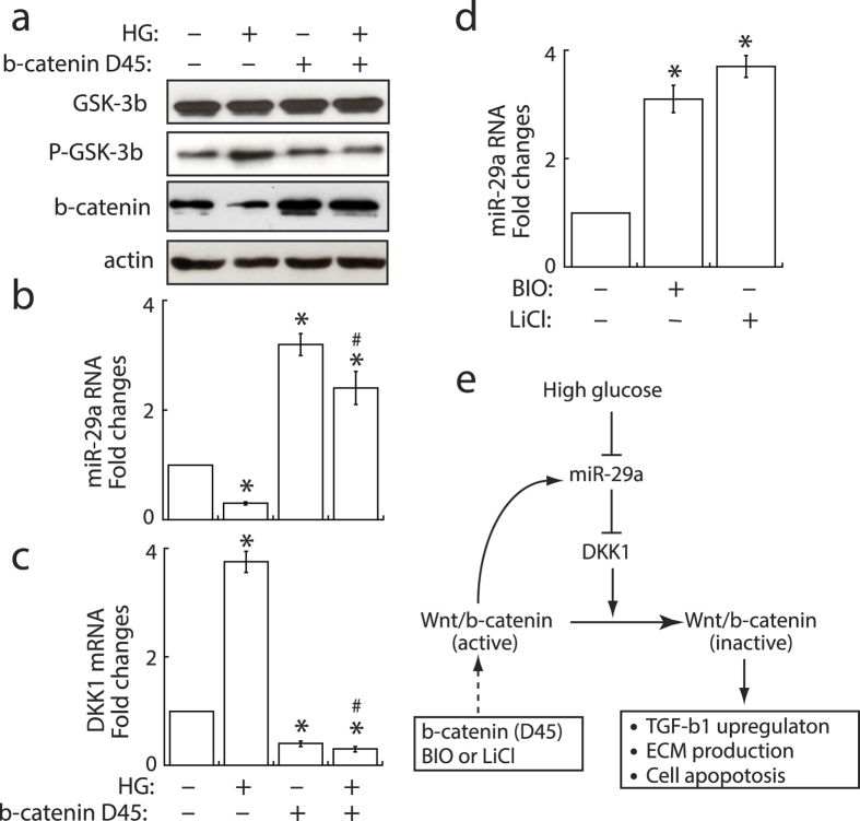
Dysregulation of specific microRNAs or Wnt/β-catenin signaling pathway is critically implicated in the pathogenesis of various renal diseases. However, the relationship between microRNAs and Wnt/β-catenin signaling in diabetes-induced glomerular sclerosis remains unknown. Here, we found that decreased miR-29a expression and attenuated Wnt/β-catenin signaling were concomitantly detected in glomeruli of streptozotocin-induced diabetic mice. Gain of miR-29a function in diabetic mice substantially increased the expression of β-catenin and blocked the expressions of profibrotic gene markers, including DKK1 (a Wnt antagonist), TGF-β1 and fibronectin, in glomerular mesangium. Moreover, in the normal mice treated with miR-29a inhibitor, renal fibrosis was induced with an attenuated Wnt/β-catenin signaling activity. Consistently, the constructed miR-29a transgenic mice that supported sustained Wnt/β-catenin signaling had the ability to block the expressions of profibrotic genes after induction of diabetes. We also demonstrated that miR-29a acts as a positive regulator of Wnt/β-catenin signaling in cultured mesangial cells and functions to protect cell apoptosis and fibrosis. Importantly, we showed that activation of Wnt/β-catenin signaling in cultured mesangial cells by transfecting the β-catenin (Δ45) mutant or by a GSK-3β inhibitor reversely upregulated miR29a. Our findings suggest that the reciprocal relationship between miR-29a and DKK1/Wnt/β-catenin signaling may play an important part in protecting renal fibrogenesis.


Figures

Figure 1
Figure 1. Effects of hyperglycemia on renal function and the expression of miR-29a and profibrotic genes in glomeruli of diabetic kidneys.
Diabetic mice (DM; n = 6) displayed increased blood glucose levels (a), urinary protein excretion (b), and kidney weight (c) as compared to normal controls (NC; n = 6). (d) Isolation of glomerular compartments in renal tissues by laser-captured microdissection (LCM). Expression levels of TGF-β1 (e), fibronectin (f), DKK1 (g), miR-29a (h), miR-29b (i) and miR-29c (j) in glomeruli of normal and diabetic mice (n = 6 each) were evaluated by quantatative RT-PCR. Experimental results are presented as means ± SEM. *Significant differences (P < 0.05) compared with normal controls. (k) Representative in situ hybridization images of miR-29a in glomeruli of normal and diabetic mice. The box region is enlarged and arrows indicate miR-29a-positive cells (brown-colored cells).
Figure 2
Figure 2. Influence of exogenous miR-29a precursor and miR-29a inhibitor on the expression of profibrotic genes and Wnt/β-catenin signaling components in renal glomeruli.
(a) In situ hybridization of miR-29a in renal tissues of mice that were infected with empty control lentivirus (Ctl) or pre-miR-29-expressing lentivirus (Pre-miR-29a). (bf) Changes in expression of glomerular miR-29a, β-catenin, TGF-β1, fibronectin and DKK1 in mice after treatment with STZ (DM; n = 6), pre-miR-29a (NC + miR-29a; n = 6), STZ plus miR-29a precursor (DM + miR-29a; n = 6), a control lentiviral vector (anti-miR-Ctl; n = 6), or miR-29a inhibitor (anti-miR-29a; n = 6). Data are presented as mean ± SEM. NC, normal control; DM, diabetes. Symbol * indicates significant difference vs. NC group and symbol # indicates significant difference vs. DM group (P < 0.05).
Figure 3
Figure 3. Overexpression of miR-29a precursor in diabetic mice alleviates renal fibrosis and alters Wnt/β-catenin signaling activation in glomerular mesangium.
Representative photographs of PAS staining (pink) and immunohistochemical staining (brown) of fibronectin, DKK1 and β-catenin in kidney tissues of normal mice (NC) and diabetic mice (DM) with or without overexpression of miR-29a precursor.
Figure 4
Figure 4. Knockdown of miR-29a in normal mice induces renal fibrosis and downregulates the Wnt/β-catenin signaling.
Representative photographs of PAS staining (pink) and immunohistochemical staining (brown) of fibronectin, DKK1 and β-catenin in kidney tissues of normal mice (NC) and normal mice infected with empty control lentivirus (Anti-miR-Ctl) or with lentivirus expressing miR-29a inhibitor (Anti-miR-29a).
Figure 5
Figure 5. MiR-29a transgenic mice are protected against diabetes-induced renal injury.
(a) In situ hybridization of miR-29a in renal glomeruli of wild-type mice and miR-29a transgenic mice. (bf) Expression of DKK1, TGF-β1, fibronectin, collagen IV and β-catenin mRNAs in renal glomeruli of the wild-type (WT) and miR-29a transgenic mice (Tg) after induction of diabetes (n = 6 for each group). NC, normal control; DM, diabetes. Data are expressed as mean ± SEM. Symbol * indicates significant difference vs. wild-type normal (WT-NC) group and symbol # indicates significant difference vs. wild-type diabetic (WT-DM) group (P < 0.05). (g) Representative photographs of immunohistochemical staining of DKK1 and β-catenin in glomeruli of wild-type mice and miR-29 transgenic mice without or with STZ treatment.
Figure 6
Figure 6. MiR-29a acts a positive regulator of Wnt/β-catenin signaling and functions to prevent apoptosis and fibrogenic activation in cultured renal mesangial cell.
(a) Effects of high glucose, miR-29a precursor and miR-29a inhibitor on cell apoptosis. Renal mesangial cells were treated with high glucose, miR-29a precursor, miR-29a inhibitor, or a combination of high glucose and miR-29a precursor. TUNEL staining was used to detect apoptotic cells (red). Experiments were performed at least three times and representative results are shown. NC, normal glucose control; HG, high glucose. (bd) Changes in the levels of TGF-β1, fibronectin and DKK1 mRNAs in mesangial cells under the above-mentioned conditions. All the experiments were repeated at least three times. Data are indicated as mean ± SEM. Symbol * represents significant difference vs. the normal glucose group, and symbol # represents significant difference vs. the high glucose group (P < 0.05). (e) Western blot analysis of GSK-3β phosphorylation and nuclear β-catenin in mesangial cells. Representative blots from three experiments are shown.
Figure 7
Figure 7. Specific activation of Wnt/β-catanin signaling upregulates miR-29a expression in mesangial cells.
(a) Western blot analysis of GSK-3β phosphorylation and nuclear β-catenin in mesangial cells that were treated with high glucose (HG) or transfected with β-catenin (Δ45) mutant. Experiments were repeated three times and representative blots are shown. (b,c) Stabilization of β-catenin by transfecting the β-catenin (Δ45) mutant significantly enhanced miR-29a, but reduced DKK1, in mesangial cells in either normal or high glucose conditions. (d) Treatment of mesangial cells with a GSK-3β inhibitor, BIO or LiCL, increased miR-29a expression. All quantitative RT-PCR experiments shown above were independently repeated at least three times. Symbol * indicates significant difference vs. the normal glucose group, and symbol # indicates significant difference vs. the high glucose group (P < 0.05). (e) Proposed model for the reciprocal regulation of miR-29a and Wnt/β-catenin signaling.

References

    1. Locatelli F., Pozzoni P. & Del Vecchio L. Renal replacement therapy in patients with diabetes and end-stage renal disease. J Am Soc Nephrol. 15, S25–S29 (2004). - PubMed
    1. Murphy M. et al.. IHG-1 amplifies TGF-β1 signaling and is increased in renal fibrosis. J Am Soc Nephrol. 19, 1672–180 (2008). - PMC - PubMed
    1. Zheng S. et al.. Development of late-stage diabetic nephropathy in OVE26 diabetic mice. Diabetes 53, 3248–3257 (2004). - PubMed
    1. Sasser J. M. et al.. Endothelin A receptor blockade reduces diabetic renal injury via an anti-inflammatory mechanism. J Am Soc Nephrol. 18, 143–154 (2007). - PMC - PubMed
    1. Lauer M. E., Hascall V. C. & Wang A. Heparan sulfate analysis from diabetic rat glomeruli. J Biol Chem. 282, 843–852 (2007). - PubMed

Publication types

MeSH terms

LinkOut - more resources