. 2016 Jul 27:6:30498.
doi: 10.1038/srep30498.

Internalized Tau sensitizes cells to stress by promoting formation and stability of stress granules

Affiliations

Internalized Tau sensitizes cells to stress by promoting formation and stability of stress granules

Cecilia A Brunello et al. Sci Rep. .

Abstract

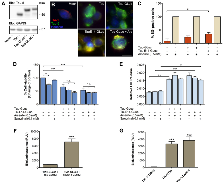
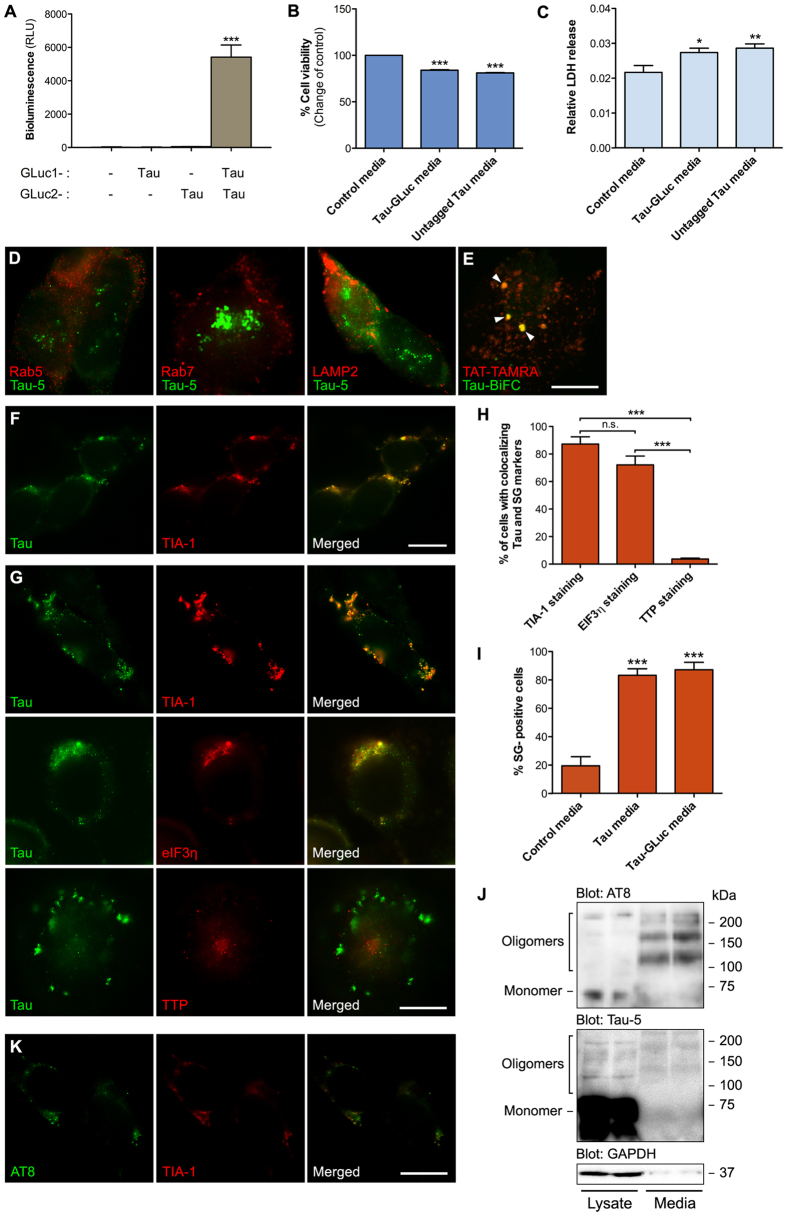
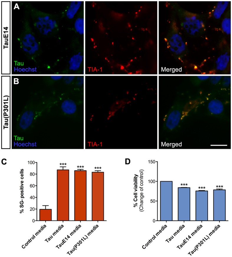
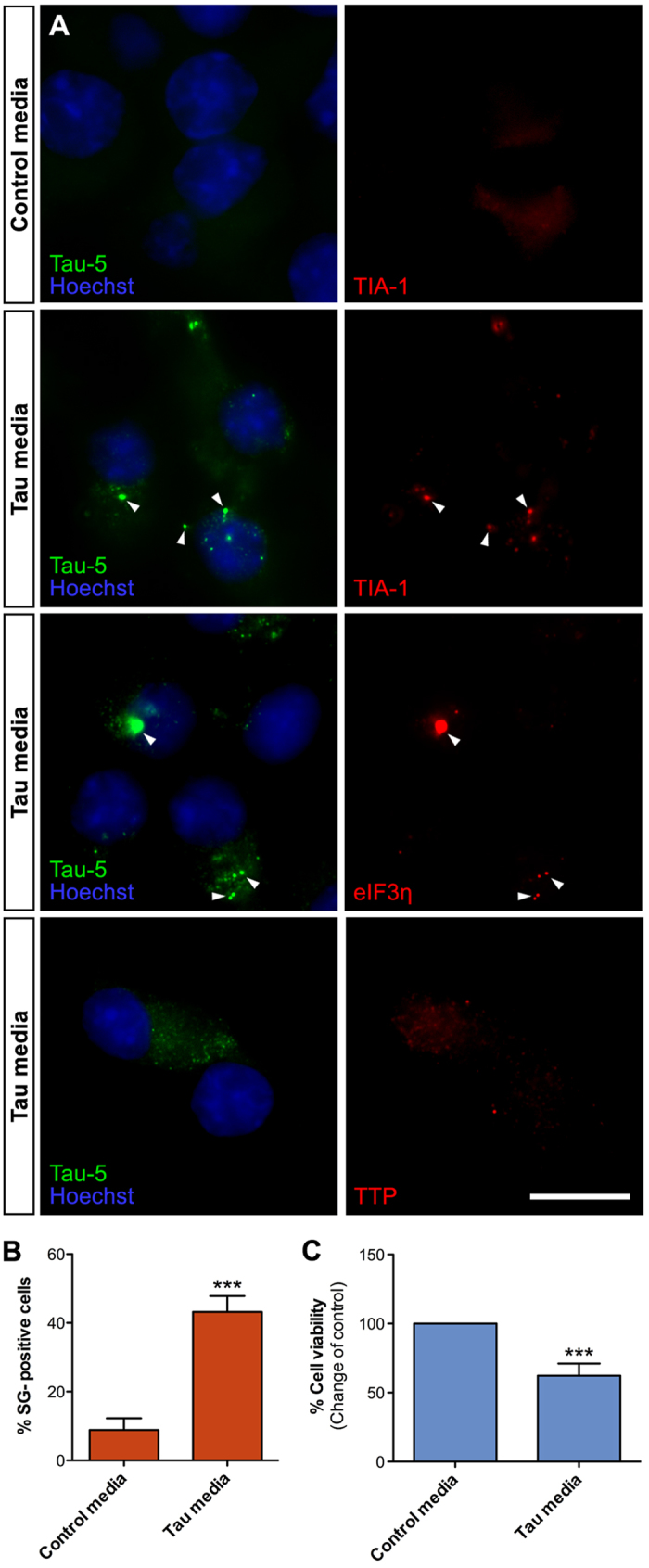
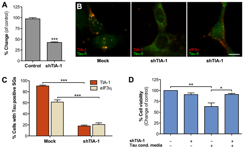
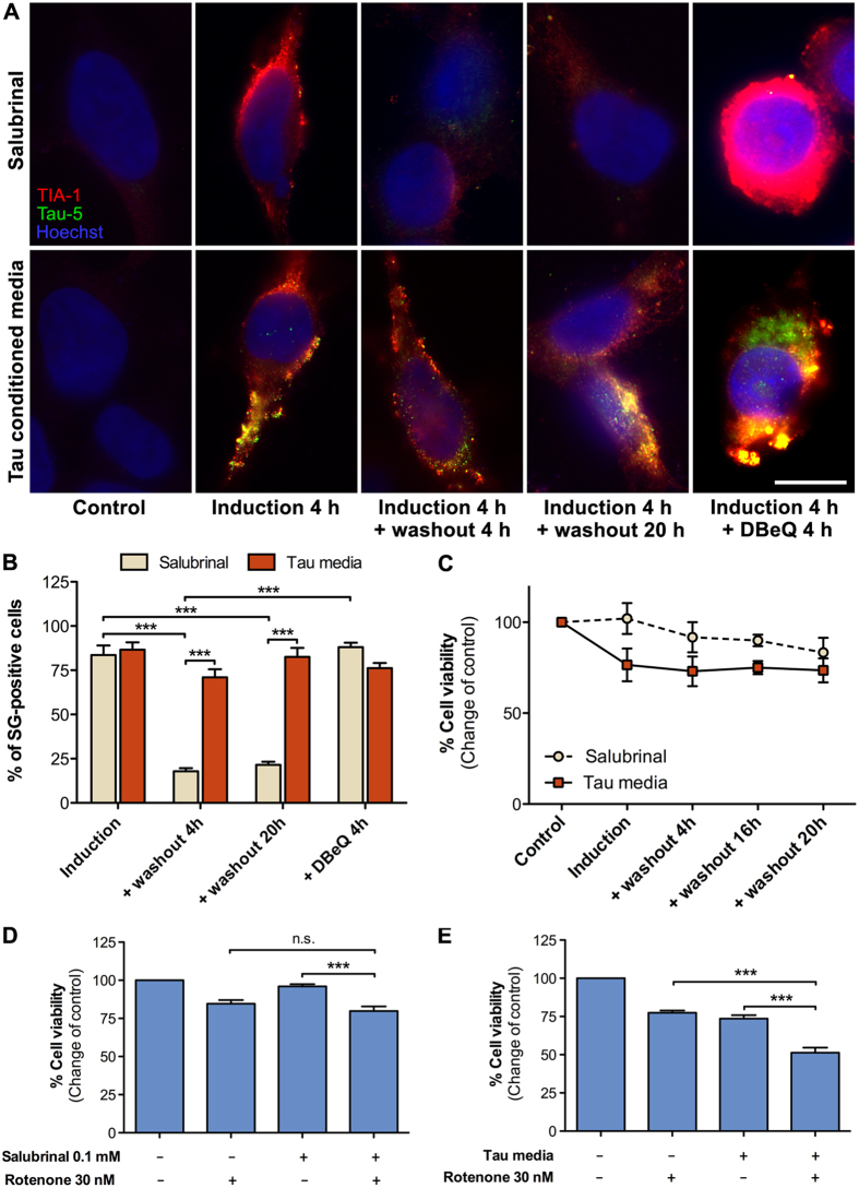
Stress granules are membrane-less RNA- and RNA-binding protein-containing complexes that are transiently assembled in stressful conditions to promote cell survival. Several stress granule-associated RNA-binding proteins have been associated with neurodegenerative diseases. In addition, a close link was recently identified between the stress granule core-nucleating protein TIA-1 and Tau. Tau is a central pathological protein in Alzheimer's disease and other tauopathies, and misfolded, aggregated Tau is capable of propagating pathology via cell-to-cell transmission. Here we show that following internalization hyperphosphorylated extracellular Tau associates with stress granules in a TIA-1 dependent manner. Cytosolic Tau normally only weakly interacts with TIA-1 but mutations mimicking abnormal phosphorylation promote this interaction. We show that internalized Tau significantly delays normal clearance of stress granules in the recipient cells sensitizing them to secondary stress. These results suggest that secreted Tau species may have properties, likely related to its hyperphosphorylation and oligomerization, which promote pathological association of internalized Tau with stress granules altering their dynamics and reducing cell viability. We suggest that stress granules and TIA-1 play a central role in the cell-to-cell transmission of Tau pathology.


Conflict of interest statement

H.J.H. is a co-founder and shareholder of Herantis Pharma plc, which is not related to this study. The other authors declare no conflict of interests.

Figures

Figure 1
Figure 1. Transfected and internalized extracellular Tau differ in their ability to associate with stress granules.
(A) Expression of non-tagged Tau, Tau-GLuc1/2 and TauE14-GLuc1/2 in HEK293T cells as detected by Western blot. The blot picture was cropped from a larger original image, maintaining all the stained bands. (B) HEK293T cells transiently transfected with the above-mentioned constructs and stained with Tau-5 (green) and TIA-1 (red) antibodies. Arsenite (0.5 mM for 30 min) was used as a positive control for induction of stress granules. (C) Quantitative analysis of stress granule formation. Stress granule-positive cells were counted among the Tau-transfected cells. Arsenite treatment promoted stress granule-formation in all cells while only some Tau-transfected, and more efficiently TauE14-transfected, cells contained stress-granules (n = 3). (D) Resazurin-based cell viability assay with HEK293T cells transiently transfected with the Tau constructs. Salubrinal and arsenite were used as positive controls for stress granule induction (n = 4). (E) LDH release assay in HEK293T cells transfected with the Tau constructs and treated with salubrinal and arsenite as positive controls (n = 4). Relative LDH release was calculated from the ratio of LDH in the media and total LDH (from media and the cells). (F) Protein-fragment complementation assay (PCA) in HEK293T cells transiently transfected with TIA-1-GLuc1, Tau-GLuc2 and TauE14-GLuc2 (n = 3). (G) PCA-based coplating experiment to study cell-to-cell transfer of Tau. Batches of HEK293T cells were separately transfected with a single PCA construct (TIA-1-GLuc1, Tau-GLuc2, TauE14-GLuc2 or GSK3β-GLuc2 as a control), followed by replating the cells in various combinations 24 h post-transfection (n = 3). Scalebar = 10 μm; average +/−SEM is shown; ***p < 0.001; **p < 0.01; *p < 0.05.
Figure 2
Figure 2. Internalized extracellular Tau localizes to stress granules.
(A) PCA from conditioned media collected from HEK293T transiently transfected with either mock plasmid, Tau-GLuc1, Tau-GLuc2 or Tau-GLuc1/Tau-GLuc2. Secreted Tau dimers accumulate in fresh serum-free media (n = 3). Media was conditioned for 24 h with HEK293T cells expressing different forms of Tau for the subsequent experiments. (B) Resazurin-based cell viability assay showing decreased viability of cells exposed to Tau-GLuc1/2-containing media compared to the control-media exposed cells (n = 6). (C) Relative LDH release for cells exposed to Tau-GLuc1/2-containing media show slightly decreased viability compared to the control cells (n = 4). (D) Naïve HEK293T cells were exposed to Tau-GLuc conditioned media for 6 h and stained with Tau-5 and (from left to right) Rab5, Rab7 and Lamp2 antibodies to mark early endosomes, late endosomes and lysosomes, respectively. (E) Fluorescent TAT-TAMRA peptide was used as a marker for macropinosomes and visualized in live-cell imaging together with Tau-BiFC containing media, showing some colocalization. Tau-BiFC media was produced collecting conditioned media from HEK293T cells transfected with Tau-GFP10C and Tau-GFP11C, two Tau constructs carrying complementary fragments of GFP. Similar to GLuc-based PCA, generation of GFP signal requires Tau dimerization. (F) Naïve HEK293T cells were exposed to untagged Tau conditioned media for 6 h and stained with Tau-5 and SG marker TIA-1. (G) Naïve HEK293T cells exposed to Tau-GLuc1/2 containing media for 6 h and costained with Tau-5 and SG markers TIA-1, eIF3η and TTP. (H) Quantitative analysis of SGs using different markers indicates that TIA-1 and eIF3η staining frequently localize with Tau while TTP colocalization with Tau is found in very few cells (n = 4). (I) Quantitative analysis of TIA-1-positive SG formation in cells conditioned with Tau-GLuc and Tau media compared to control media (n = 4). (J) Western blot of Tau-GLuc-transfected cell lysate and Tau-GLuc conditioned media, stained with AT8, Tau-5 and GAPDH antibodies. Media was concentrated by 250x using filter centrifugation before loading on gel. The blot picture was cropped from a larger image, maintaining all the stained bands. (K) Naïve HEK293T cells were exposed to Tau-GLuc conditioned media for 6 h and stained with AT8 and TIA-1 antibodies, showing colocalization of Ser199/Ser202/Thr205-phosphorylated Tau and TIA-1 in the recipient cells. Scalebar = 10 μm; average +/−SEM is shown; ***p < 0.001; **p < 0.005; *p < 0.05.
Figure 3
Figure 3. Internalized TauE14 and Tau(P301L) localize to stress granules.
(A) Naïve HEK293T cells were exposed to TauE14-GLuc media for 6 h and stained with Tau-5 and TIA-1 antibodies. (B) Similarly to panel A, cells were exposed to Tau(P301L)-GLuc conditioned media and stained with Tau-5 and TIA-1 antibodies. (C) Quantitative analysis of TIA-1-positive SGs in cells conditioned with Tau-GLuc media, TauE14-GLuc and Tau(P301L)-GLuc conditioned media compared to control media (n = 4). (D) Resazurin-based cell viability assay for cells exposed to Tau-GLuc, TauE14-GLuc and Tau(P301L)-GLuc conditioned media (n = 4). Scalebar = 10 μm; average +/−SEM is shown; ***p < 0.001; **p < 0.01.
Figure 4
Figure 4. Internalized Tau is recruited to stress granules in N2A cells.
(A) Naïve N2A cells exposed to Tau-GLuc conditioned media for 6 h and stained with Tau-5 and stress granule markers TIA-1, eIF3η and TTP. TIA-1 and eIF3η show clear colocalization with internalized Tau, while TTP does not show any. (B) Quantitative analysis of SG formation in cells exposed to Tau conditioned media compared to control media indicates that about 40% of N2A cells contained TIA-1-positive SGs following Tau uptake (n = 4). (C) Resazurin-based cell viability assay indicates that viability of N2A cells exposed to Tau-GLuc media was significantly decreased compared to cells exposed to control media (n = 8). Scalebar = 10 μm; average +/−SEM is shown; ***p < 0.001.
Figure 5
Figure 5. Tau localization to stress granules requires TIA-1.
(A) TIA-1 shRNA knockdown efficiency determined by qPCR following shRNA plasmid transfection of HEK293T cells. Levels of TIA-1 mRNA were normalized to GAPDH mRNA levels (n = 2). (B) TIA-1 was transiently knocked down in HEK293T cells (middle and right images) that were then exposed to Tau-GLuc-conditioned media and stained with Tau-5 and stress granule marker antibodies. The middle image shows typical cells with mostly cytosolic, non-punctate staining of Tau. The image on the right shows an example of a cell with SGs costaining with Tau and eIF3η. (C) Quantitative analysis of Tau-positive stress granule formation. SGs were present in the majority of cells exposed to Tau media when TIA-1 is normally expressed but were significantly decreased when TIA-1 was knocked down. Similar results were obtained with both TIA-1 and eIF3η staining (n = 3). (D) Resazurin-based cell viability assay showed that TIA-1 knockdown had no effect on cell viability per se but was able to improve viability in cells exposed to Tau-GLuc-conditioned media (n = 3). Scalebar = 10 μm; average +/− SEM is shown; ***p < 0.001; **p < 0.01; *p < 0.05.
Figure 6
Figure 6. Internalized Tau alters stress granule dynamics and sensitizes cells to stress.
(A) Tau-GLuc-conditioned media or 0.1 mM Salubrinal was applied to HEK293T cells for 4 h, followed by a washout with normal media. Cells were fixed at 4 and 20 h after the washout and stained with TIA-1 and Tau-5 antibodies. DBeQ (10 μM) was used as a positive control to inhibit stress granule disassembly after the treatments. (B) Quantitation of immunofluorescence images: stress granule-positive cells over total cells were counted for each condition (n = 3). (C) Cell viability assay for the washout experiment (n = 4). (D,E) Cell viability assay for cells treated either with salubrinal (D) or with Tau-conditioned media (E) followed by 4 hours of recovery in fresh media and further treatement with 30 nM Rotenone for 6 h. In the salubrinal control cells Rotenone showed minimal toxicity while in cells exposed to Tau-GLuc-conditioned media Rotenone significantly decreased cell viability (n = 4). Scalebar = 10 μm; average +/− SEM is shown; ***p < 0.001.

References

    1. Gerstberger S., Hafner M. & Tuschl T. A census of human RNA-binding proteins. Nat Rev Genet. 15, 829–845 (2014). - PMC - PubMed
    1. Neelamraju Y., Hashemikhabir S. & Janga S. C. The human RBPome: from genes and proteins to human disease. J Proteomics. 127, 61–70 (2015). - PubMed
    1. Kim H. J. et al. Mutations in prion-like domains in hnRNPA2B1 and hnRNPA1 cause multisystem proteinopathy and ALS. Nature 495, 467–473 (2013). - PMC - PubMed
    1. Liu-Yesucevitz L. et al. Tar DNA binding protein-43 (TDP-43) associates with stress granules: analysis of cultured cells and pathological brain tissue. PLoS One 5, e13250 (2010). - PMC - PubMed
    1. Sreedharan J. et al. TDP-43 mutations in familial and sporadic amyotrophic lateral sclerosis. Science 319, 1668–1672 (2008). - PMC - PubMed

Publication types