

Efficient double-quenching of electrochemiluminescence from CdS:Eu QDs by hemin-graphene-Au nanorods ternary composite for ultrasensitive immunoassay
- PMID: 27460868
- PMCID: PMC4962035
- DOI: 10.1038/srep30577
Efficient double-quenching of electrochemiluminescence from CdS:Eu QDs by hemin-graphene-Au nanorods ternary composite for ultrasensitive immunoassay
Abstract
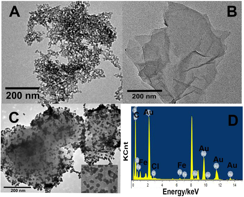
A novel ternary composite of hemin-graphene-Au nanorods (H-RGO-Au NRs) with high electrocatalytic activity was synthesized by a simple method. And this ternary composite was firstly used in construction of electrochemiluminescence (ECL) immunosensor due to its double-quenching effect of quantum dots (QDs). Based on the high electrocatalytic activity of ternary complexes for the reduction of H2O2 which acted as the coreactant of QDs-based ECL, as a result, the ECL intensity of QDs decreased. Besides, due to the ECL resonance energy transfer (ECL-RET) strategy between the large amount of Au nanorods (Au NRs) on the ternary composite surface and the CdS:Eu QDs, the ECL intensity of QDs was further quenched. Based on the double-quenching effect, a novel ultrasensitive ECL immunoassay method for detection of carcinoembryonic antigen (CEA) which is used as a model biomarker analyte was proposed. The designed immunoassay method showed a linear range from 0.01 pg mL(-1) to 1.0 ng mL(-1) with a detection limit of 0.01 pg mL(-1). The method showing low detection limit, good stability and acceptable fabrication reproducibility, provided a new approach for ECL immunoassay sensing and significant prospect for practical application.
Figures


Similar articles
-
Electrochemiluminescent quenching of quantum dots for ultrasensitive immunoassay through oxygen reduction catalyzed by nitrogen-doped graphene-supported hemin.Anal Chem. 2013 Jun 4;85(11):5390-6. doi: 10.1021/ac3036537. Epub 2013 May 22. Anal Chem. 2013. PMID: 23659573
-
A dual amplification strategy for ultrasensitive electrochemiluminescence immunoassay based on a Pt nanoparticles dotted graphene-carbon nanotubes composite and carbon dots functionalized mesoporous Pt/Fe.Analyst. 2014 Apr 7;139(7):1713-20. doi: 10.1039/c3an02084c. Analyst. 2014. PMID: 24519411
-
Electrochemiluminescence immunosensor based on graphene-CdS quantum dots-agarose composite for the ultrasensitive detection of alpha fetoprotein.Talanta. 2012 Jan 30;89:27-32. doi: 10.1016/j.talanta.2011.11.017. Epub 2011 Nov 18. Talanta. 2012. PMID: 22284455
-
Quenching study of Cu2S-MPA/NGODs composites in electrochemiluminescence detection by modulating resonance energy transfer and adsorption process.Bioelectrochemistry. 2024 Oct;159:108729. doi: 10.1016/j.bioelechem.2024.108729. Epub 2024 May 15. Bioelectrochemistry. 2024. PMID: 38772096 Review.
-
Immunosensing procedures for carcinoembryonic antigen using graphene and nanocomposites.Biosens Bioelectron. 2017 Mar 15;89(Pt 1):293-304. doi: 10.1016/j.bios.2015.11.053. Epub 2015 Nov 18. Biosens Bioelectron. 2017. PMID: 26620098 Review.
Cited by
-
A sandwich-type electrochemical aptasensor for the carcinoembryonic antigen via biocatalytic precipitation amplification and by using gold nanoparticle composites.Mikrochim Acta. 2019 Jun 26;186(7):473. doi: 10.1007/s00604-019-3542-2. Mikrochim Acta. 2019. PMID: 31243610
-
Visual Detection of Cucumber Green Mottle Mosaic Virus Based on Terminal Deoxynucleotidyl Transferase Coupled with DNAzymes Amplification.Sensors (Basel). 2019 Mar 14;19(6):1298. doi: 10.3390/s19061298. Sensors (Basel). 2019. PMID: 30875853 Free PMC article.
-
T4 DNA polymerase-assisted upgrade of a nicking/polymerization amplification strategy for ultrasensitive electrochemical detection of Watermelon mosaic virus.Anal Bioanal Chem. 2019 May;411(13):2915-2924. doi: 10.1007/s00216-019-01737-x. Epub 2019 Apr 17. Anal Bioanal Chem. 2019. PMID: 31016327
-
Current Technologies of Electrochemical Immunosensors: Perspective on Signal Amplification.Sensors (Basel). 2018 Jan 12;18(1):207. doi: 10.3390/s18010207. Sensors (Basel). 2018. PMID: 29329274 Free PMC article. Review.
References
-
- Stoeva S. I., Lee J. S., Smith J. E., Rosen S. T. & Mirkin C. A. Multiplexed detection of protein cancer markers with biobarcoded nanoparticle probes. J. Am. Chem. Soc. 128, 8378–8379 (2006). - PubMed
-
- Meng J. et al.. Carbon nanotubes conjugated to tumor lysate protein enhance the efficacy of an antitumor immunotherapy. Small. 4, 1364–1370 (2008). - PubMed
-
- Ferrari M. Cancer nanotechnology: opportunities and challenges. Nat. Rev. Cancer. 5, 161–171 (2005). - PubMed
-
- Braithwaite J. E., Yandle T. G., Nicholls M. G. & Lewis L. K. Interference by o-phenanthroline in the radioimmunoassay of angiotensin II in small volume blood samples. Clin Biochem. 45, 168–170 (2012). - PubMed
Publication types
MeSH terms
Substances
LinkOut - more resources
Full Text Sources
Other Literature Sources
