

ECM1 promotes migration and invasion of hepatocellular carcinoma by inducing epithelial-mesenchymal transition
- PMID: 27460906
- PMCID: PMC4962417
- DOI: 10.1186/s12957-016-0952-z
ECM1 promotes migration and invasion of hepatocellular carcinoma by inducing epithelial-mesenchymal transition
Abstract
Background: Extracellular matrix protein 1 (ECM1) is a glycoprotein involved in many biologic processes. To determine the expression of ECM1 in hepatocellular carcinoma (HCC), and to study the role of ECM1 in inducing epithelia-mesenchymal transition (EMT) to analyze the effect of ECM1 on the migration and invasion of HCC cells.
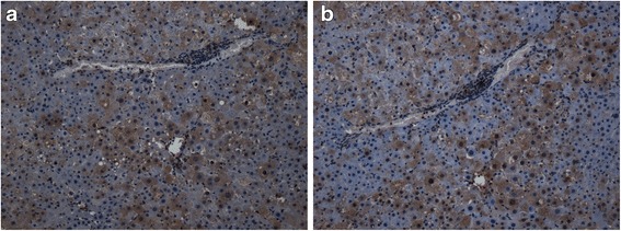
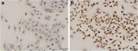
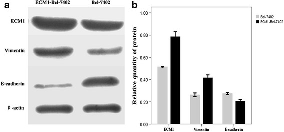
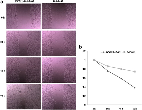
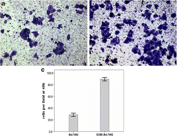
Methods: The expression of ECM1 in HCC specimens was examined by immunohistochemistry staining, and the correlations were analyzed between the expression of ECM1 and the clinicopathological data. The ECM1 expression plasmid was transfected into Bel-7402 cells to induce exogenous overexpression of ECM1 protein. The changes of the expression of ECM1, EMT-related protein (E-cadherin, Vimentin), in Bel-7402 cells were detected by Western blot after transfection of ECM1; the wound healing and invasion assay in vitro were used to determine the role of ECM1 gene transfection on the ability of migration and invasive potential of Bel-7402 cells.
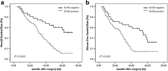
Results: Immumohistochemistry staining method displayed the ECM1 expression was positively associated with vascular invasion, TNM stage, and poor prognosis. A significant positive correlation was found between the expressions of ECM1 and Vimentin. After ECM1 overexpression, Western blot exhibited that the expression of E-cadherin was down-regulated and Vimentin expression was up-regulated in Bel-7402 cells; the wound healing and invasion assay showed that the migration and invasion potentials of Bel-7402 cells were significantly enhanced.
Conclusions: ECM1, which displayed a high expression in HCC specimens, was closely associated with clinicopathologic data and may promote migration and invasion of HCC cells by inducing EMT.
Keywords: Epithelia-mesenchymal transition; Extracellular matrix protein 1; Hepatocellular carcinoma; Invasion; Migration.
Figures
Similar articles
-
FoxM1 overexpression promotes epithelial-mesenchymal transition and metastasis of hepatocellular carcinoma.World J Gastroenterol. 2015 Jan 7;21(1):196-213. doi: 10.3748/wjg.v21.i1.196. World J Gastroenterol. 2015. PMID: 25574092 Free PMC article.
-
Long Noncoding RNA AK021443 Promotes Cell Proliferation and Migration by Regulating Epithelial-Mesenchymal Transition in Hepatocellular Carcinoma Cells.DNA Cell Biol. 2018 May;37(5):481-490. doi: 10.1089/dna.2017.4030. Epub 2018 Mar 14. DNA Cell Biol. 2018. PMID: 29638164
-
Overexpression of the long non-coding RNA SPRY4-IT1 promotes tumor cell proliferation and invasion by activating EZH2 in hepatocellular carcinoma.Biomed Pharmacother. 2017 Jan;85:348-354. doi: 10.1016/j.biopha.2016.11.035. Epub 2016 Nov 28. Biomed Pharmacother. 2017. PMID: 27899259
-
[The expression and clinopathological significance of miR-130b in human hepatocellular carcinoma].Xi Bao Yu Fen Zi Mian Yi Xue Za Zhi. 2016 Mar;32(3):387-92. Xi Bao Yu Fen Zi Mian Yi Xue Za Zhi. 2016. PMID: 26927562 Chinese.
-
The extracellular matrix protein 1: its molecular interaction and implication in tumor progression.Cancer Invest. 2008 May;26(4):375-84. doi: 10.1080/07357900701788148. Cancer Invest. 2008. PMID: 18443958 Review.
Cited by
-
Biomarkers of tumor invasiveness in proteomics (Review).Int J Oncol. 2020 Aug;57(2):409-432. doi: 10.3892/ijo.2020.5075. Epub 2020 May 28. Int J Oncol. 2020. PMID: 32468071 Free PMC article.
-
NTRK fusion promotes tumor migration and invasion through epithelial-mesenchymal transition and closely interacts with ECM1 and NOVA1.BMC Cancer. 2024 Dec 5;24(1):1502. doi: 10.1186/s12885-024-13271-w. BMC Cancer. 2024. PMID: 39639242 Free PMC article.
-
Bioinformatics analysis to reveal key biomarkers for early and late hepatocellular carcinoma.Transl Cancer Res. 2020 Jul;9(7):4070-4079. doi: 10.21037/tcr-19-2238. Transl Cancer Res. 2020. PMID: 35117777 Free PMC article.
-
Uveal Melanoma-Derived Extracellular Vesicles Display Transforming Potential and Carry Protein Cargo Involved in Metastatic Niche Preparation.Cancers (Basel). 2020 Oct 11;12(10):2923. doi: 10.3390/cancers12102923. Cancers (Basel). 2020. PMID: 33050649 Free PMC article.
-
Role of extra cellular proteins in gastric cancer progression and metastasis: an update.Genes Environ. 2020 May 15;42:18. doi: 10.1186/s41021-020-00157-z. eCollection 2020. Genes Environ. 2020. PMID: 32467737 Free PMC article. Review.
References
MeSH terms
Substances
LinkOut - more resources
Full Text Sources
Other Literature Sources
Medical
Research Materials
Miscellaneous
