. 2016 Oct 1:308:46-58.
doi: 10.1016/j.taap.2016.07.015. Epub 2016 Jul 25.

Activating transcription factor 4 underlies the pathogenesis of arsenic trioxide-mediated impairment of macrophage innate immune functions

Affiliations

Activating transcription factor 4 underlies the pathogenesis of arsenic trioxide-mediated impairment of macrophage innate immune functions

Ritesh K Srivastava et al. Toxicol Appl Pharmacol. .

Abstract

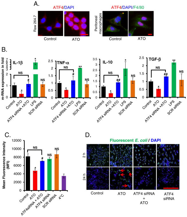
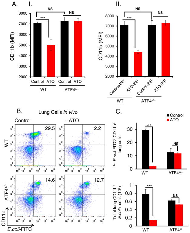
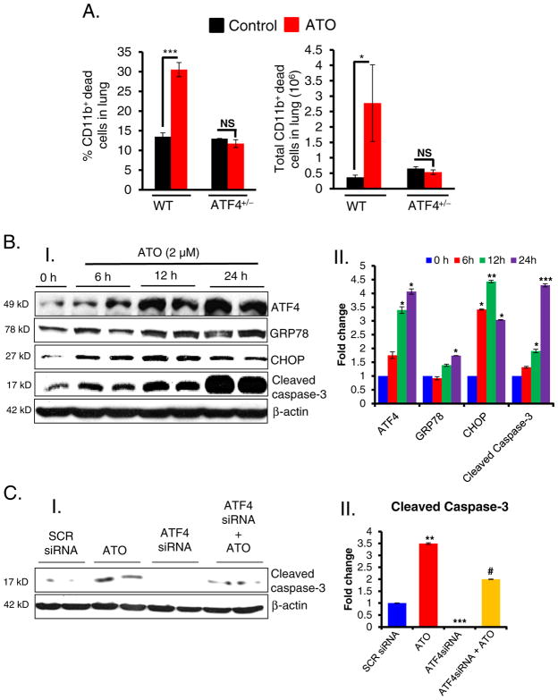
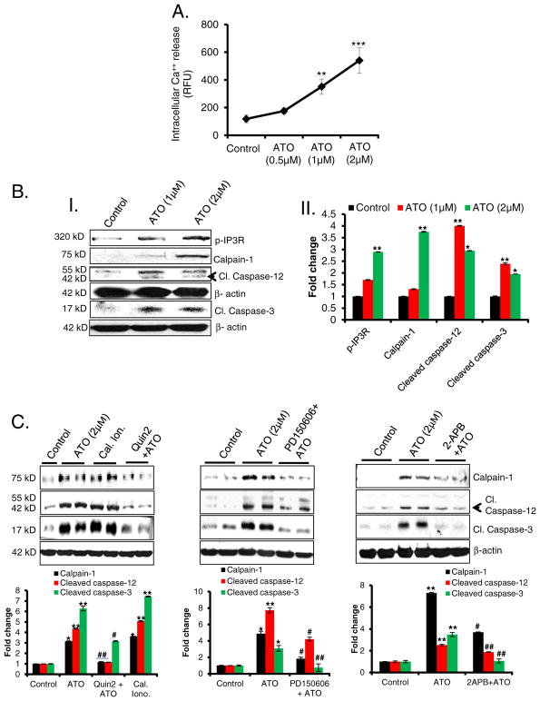
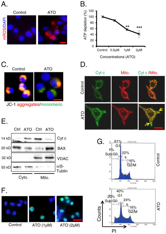
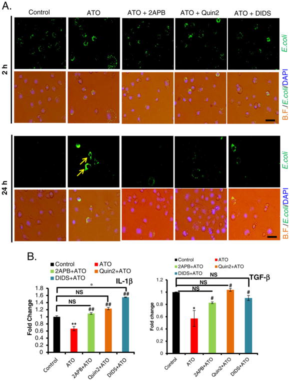
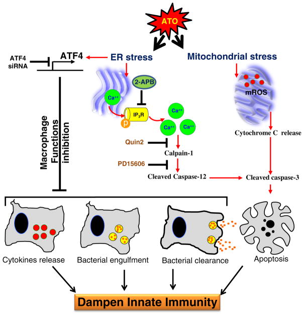
Chronic arsenic exposure to humans is considered immunosuppressive with augmented susceptibility to several infectious diseases. The exact molecular mechanisms, however, remain unknown. Earlier, we showed the involvement of unfolded protein response (UPR) signaling in arsenic-mediated impairment of macrophage functions. Here, we show that activating transcription factor 4 (ATF4), a UPR transcription factor, regulates arsenic trioxide (ATO)-mediated dysregulation of macrophage functions. In ATO-treated ATF4(+/+) wild-type mice, a significant down-regulation of CD11b expression was associated with the reduced phagocytic functions of peritoneal and lung macrophages. This severe immuno-toxicity phenotype was not observed in ATO-treated ATF4(+/-) heterozygous mice. To confirm these observations, we demonstrated in Raw 264.7 cells that ATF4 knock-down rescues ATO-mediated impairment of macrophage functions including cytokine production, bacterial engulfment and clearance of engulfed bacteria. Sustained activation of ATF4 by ATO in macrophages induces apoptosis, while diminution of ATF4 expression protects against ATO-induced apoptotic cell death. Raw 264.7 cells treated with ATO also manifest dysregulated Ca(++) homeostasis. ATO induces Ca(++)-dependent calpain-1 and caspase-12 expression which together regulated macrophage apoptosis. Additionally, apoptosis was also induced by mitochondria-regulated pathway. Restoring ATO-impaired Ca(++) homeostasis in ER/mitochondria by treatments with the inhibitors of inositol 1,4,5-trisphosphate receptor (IP3R) and voltage-dependent anion channel (VDAC) attenuate innate immune functions of macrophages. These studies identify a novel role for ATF4 in underlying pathogenesis of macrophage dysregulation and immuno-toxicity of arsenic.

Keywords: ATF4; Apoptosis; Arsenic; Ca(++) homeostasis; Macrophage functions; UPR.


Conflict of interest statement

Conflict of interest

The authors declare no conflict of interest.

Figures

Fig. 1
Fig. 1
ATO-inhibited cytokine production, bacterial engulfment and its clearance of engulfed bacteria in murine macrophage in ATF4-dependent manner. (A) Immunofluorescence staining showing migration and localization of ATF4 (red) from cytosol to nucleus in ATO (2 μM for 14 h)-treated Raw 264.7 cells (bar, 50 μm) and in peritoneal macrophages (F4/80, green) (bar, 25 μm) isolated from WT mice. Nuclei were stained with DAPI. (B) Histograms showing real time PCR analysis of mRNAs expression encoding the indicated cytokines in ATF4 siRNA or scrambled siRNA-transfected Raw 264.7 cells treated either with saline (control) or with ATO (2 μM for 14 h). Treatment of the cells with LPS (100 ng for 3 h) served as a positive control. (C) ATF4 siRNA- or scrambled siRNA-transfected Raw 264.7 cells were treated either with saline or with ATO (2 μM for 14 h), followed by incubation with fluorescently labeled rabbit-IgG coated latex beads with for 45 min at 37 °C. Histogram showing mean fluorescence intensity of engulfed fluorescent beads (recorded at excitation 485 nm and emission at 535 nm by microplate reader). Negative control cells received identical treatments as described above except were incubated at 4 °C instead of 37 °C. (D) Infection load of fluorescently labeled opsonized E. coli bioparticles in Raw 264.7 cells was recorded at 2 h and 24 h using fluorescent microscopy (bar, 200 μm). Arrows indicating the presence of E. coli bioparticles. Data are expressed as mean ± SEM. *P < 0.05, compared to control. #P < 0.05 and ##P < 0.01, compared to ATO-treated group. NS-nonsignificant compared to control.
Fig. 2
Fig. 2
ATO down regulates CD11b expression in ATF4-dependent manner. (A); (A–I) Histogram showing mean fluorescent intensity of CD11b expression determined by flow cytometry analysis of peritoneal macrophages, isolated from WT and ATF4+/−heterozygous mice. These macrophages were treated with either saline (control) or ATO (2 μM for 14 h). (A–II) Similar experiments were also performed by infecting (INF) macrophages with E. coli fluorescents conjugated bioparticles. For this study, following treatment with ATO (2 μM for 14 h), peritoneal macrophages were incubated with E. coli (bacterial: cell, 50:1 ratio) at 37 °C for 2 h and then cells were washed twice with PBS followed by flow cytometry analysis. (B) Flow cytometry analysis of lung infiltrates of WT and ATF4+/− heterozygous mice treated intraperitoneally with either saline (control) or ATO (50 μg/mouse in 200 μl PBS; daily for 10 days) and then intratracheally infected with E. coli-FITC. Representative dot plots showing percentages of CD11b + E. coli-FITC+ cells in the lung tissues. (C) Histogram showing percent E. coli FITC+CD11b+ cells (top panel) and absolute numbers of E. coli-FITC+ CD11b+ cells (lower panel) in lung tissues. Data are expressed as mean ± SEM. ***P < 0.001 compared to their respective controls. NS-nonsignificant.
Fig. 3
Fig. 3
ATO-induced apoptosis in murine macrophages. Histogram showing the percentage and total dead CD11b+ cells in the lung tissues of WT and ATF4+/−heterozygous mice. These mice were treated with either saline or ATO (50 μg/mouse in 200 μl PBS, intra-peritoneal; daily for 10 days) and then intratracheally infected with E. coli-FITC as referred to the Material & methods section. (B); (B–I) Western blot analysis of ATF4, GRP78, CHOP and cleaved caspse-3 in ATO-treated Raw 264.7 cells. (B–II) Histogram representing the densitometry analysis of western blots. (C); (C–I) Western blots analysis of cleaved caspase-3 in ATF4 knockdown cells treated with either saline or ATO (2 μM). Scrambled (SCR) siRNA was used as a negative control. (C–II) Histogram representing the densitometry analysis of western blots. Data are expressed as mean ± SEM. *P < 0.05, **P < 0.01, ***P < 0.001 compared to their respective controls. #P < 0.05 compared to ATO-treated group. NS-nonsignificant.
Fig. 4
Fig. 4
ATO-mediated apoptosis is regulated by Ca++/calpain-1/caspase-12-mediated apoptosis. (A) Line graph showing intracellular Ca++ release from ATO-treated Raw 264.7 cells at 14 h. Fluorescence intensity was recorded at excitation wavelength 494 nm and emission wavelength 516 nm using a microplate reader and expressed as mean of relative fluorescence unit (RFU). (B); (B–I) Western blot analysis for p-IP3R, calpain-1, cleaved caspase-12, and cleaved caspase-3 proteins in lysates prepared from ATO-treated (1 and 2 μM for 24 h) Raw 264.7 cells. (B–II) Histogram representing the densitometry analysis of western blots. (C) Western blot analysis for calpain-1, cleaved caspase-12 and cleaved caspase-3 proteins in ATO-treated Raw 264.7 cell lysate. These cells were pretreated with calcium chelator, Quin2 (20 μM for 3 h) or calpain inhibitor, PD150606 (10 μM for 3 h) or IP3R inhibitor, 2-APB (20 μM, 6 h). In these experiments calcium ionophore (cal. iono.), (2 μM for 3 h) was used as positive control to address whether the effects are indeed Ca++ regulated. Histograms representing the densitometry analysis of western blots. Data are expressed as mean ± SEM. *P < 0.05, **P < 0.01, ***P < 0.001 compared to control. #P < 0.05, ##P < 0.01, compared to ATO-treated group.
Fig. 5
Fig. 5
ATO-mediated apoptosis is regulated by mitochondrial signaling pathways. (A) Mitochondrial ROS (mROS) production in saline and ATO-treated (2 μM for 24 h) Raw 264.7 (bar, 30 μm). (B) Line graph showing ATP depletion in ATO-treated Raw 264.7 cells in comparison to saline-treated control cells. (C) Loss of MMP was observed using JC-1 dye in ATO-treated (2 μM for 24 h) cells under fluorescence microscope as assessed by the changes in the ratio of red (dye aggregates) to green (monomer) fluorescence (bar, 50 μm). (D) Immunofluorescence staining of ATO-induced release of cytochrome c (Cyt c) (green) in cytoplasm from mitochondria (red). Mitochondria were stained with mitotracker, a red dye (250 nM for 20 min) (bar, 50 μm). (E) Western blot analysis of ATO-induced alterations of Cyt c and BAX levels in cytoplasm and mitochondria. Fraction purity was confirmed by α/β-tubulin and VDAC proteins for cytoplasmic (Cyto.) and mitochondrial (Mito.) fractions respectively. (F) TUNEL assay was performed in ATO-treated (1 & 2 μM for 24 h) Raw 264.7 cells. Green fluorescence positive cells represent apoptotic cells (bar, 50 μm). (G) Apoptosis was also recorded by flow cytometry. Sub-G0 population of cells treated with ATO (2 μM for 24 h) represented apoptotic cells. Data are expressed as mean ± SE. **P < 0.01 and ***P < 0.001 show significance levels compared to control.
Fig. 6
Fig. 6
Treatment with 2-APB, Quin2 or DIDS attenuated ATO-induced impairment in macrophage functions disruption. (A) Overlay of microphotographs on bright filed (B.F.) and green channel (E. coli) with DAPI showing protection afforded by 2-APB (20 μM 6 h), Quin2 (20 μM, 3 h) and DIDS (200 μM, 6 h) against ATO-mediated (2 μM, 14 h) disruption of fluorescent E. coli bioparticles clearance (marked by arrows) at 24 h. Infection load of fluorescently labeled E. coli bioparticles were observed under the microscope at 2 h and 24 h after various treatments to Raw 264.7 cells (bar, 100 μm). (B) Histograms showing real time PCR analysis of cytokines IL-1β and TGF-β. Blocking IP3R or VDAC channel by 2-APB or DIDS, or chelating Ca++ by Quin2, significantly restored the level of diminished mRNA expression of cytokines in ATO-treated Raw 264.7 cells. Data are expressed as mean ± SE. *P < 0.05, **P < 0.01 compared to their respective controls. #P < 0.05, ##P < 0.01 compared to ATO-treated group. NS-nonsignificant.
Fig. 7
Fig. 7
Flow diagram depicting the mechanism by which ATO treatment blocks macrophage functions and induces apoptosis in ATF4-dependent manner. ATO activates ATF4, a UPR signaling transcription factor, in murine macrophages, which dysregulates multiple macrophage functions including cytokines release, bacterial engulfment and clearance of engulfed bacteria. In ATO-treated macrophages, sustained activation of ATF4 leads to apoptosis via multiple pathways. Ca++-dependent calpain-1/caspase-12-mediated apoptosis and mitochondrial-dependent apoptosis via release of cytochrome-c from mitochondria to cytoplasm have been recorded. Overall, these effects may lead to dampening of macrophage-dependent innate immune responses. The role of ATF4 in ATO-mediated macrophage dysregulation was ascertained by the genetic approaches where knocking down of ATF4 afforded significantly protection against ATO-mediated impairment of macrophage functions. Role of calcium homeostasis in this toxicity could be confirmed by the treatment of these macrophages with Ca++ channel blocker or Ca++ chelators which attenuated ATO-induced calpain-1/caspase-12-mediated apoptosis and perhaps other functions related to ATF4 activation.

Similar articles

Cited by

References

    1. Abhyankar LN, Jones MR, Guallar E, Navas-Acien A. Arsenic exposure and hypertension: a systematic review. Environ Health Perspect. 2012;120:494–500. - PMC - PubMed
    1. Ahmed S, Ahsan KB, Kippler M, Mily A, Wagatsuma Y, Hoque AM, Ngom PT, El Arifeen S, Raqib R, Vahter M. In utero arsenic exposure is associated with impaired thymic function in newborns possibly via oxidative stress and apoptosis. Toxicol Sci. 2012;129:305–314. - PubMed
    1. Aranyi C, Bradof JN, O’Shea WJ, Graham JA, Miller FJ. Effects of arsenic trioxide inhalation exposure on pulmonary antibacterial defenses in mice. J Toxicol Environ Health. 1985;15:163–172. - PubMed
    1. Attardi G, Ching E. Biogenesis of mitochondrial proteins in HeLa cells. Methods Enzymol. 1979;56:66–79. - PubMed
    1. Austyn JM, Gordon S. F4/80, a monoclonal antibody directed specifically against the mouse macrophage. Eur J Immunol. 1981;11:805–815. - PubMed