

Designer interface peptide grafts target estrogen receptor alpha dimerization
- PMID: 27462021
- PMCID: PMC5214063
- DOI: 10.1016/j.bbrc.2016.07.083
Designer interface peptide grafts target estrogen receptor alpha dimerization
Abstract
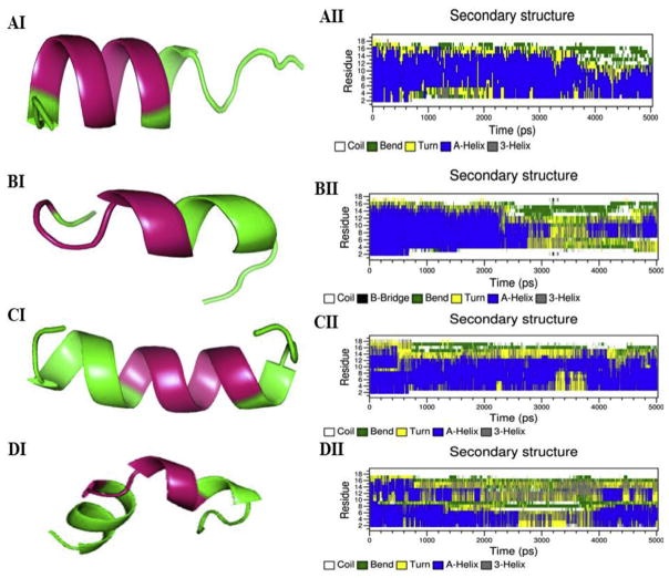
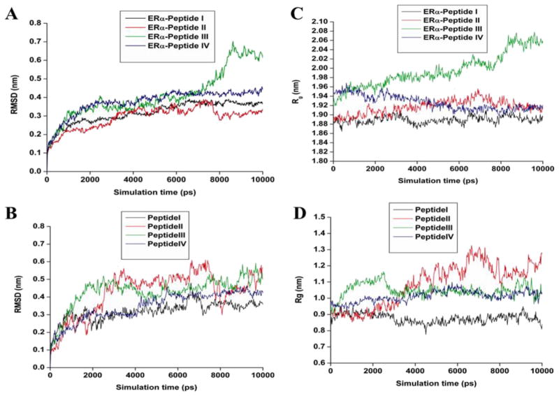
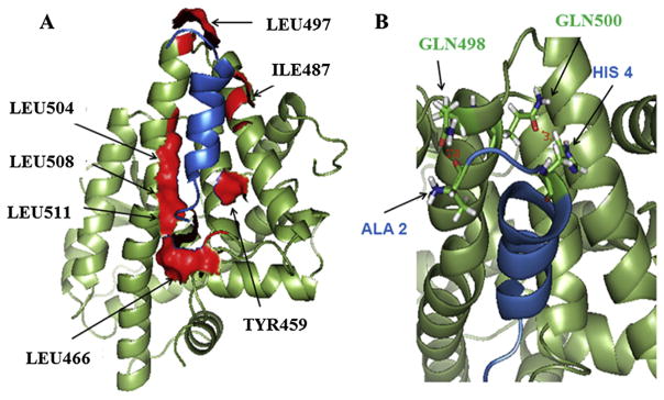
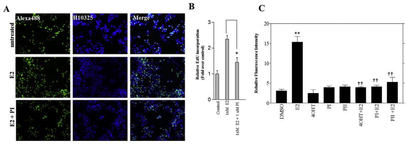
The nuclear transcription factor estrogen receptor alpha (ERα), triggered by its cognate ligand estrogen, regulates a variety of cellular signaling events. ERα is expressed in 70% of breast cancers and is a widely validated target for anti-breast cancer drug discovery. Administration of anti-estrogen to block estrogen receptor activation is still a viable anti-breast cancer treatment option but anti-estrogen resistance has been a significant bottle-neck. Dimerization of estrogen receptor is required for ER activation. Blocking ERα dimerization is therefore a complementary and alternative strategy to combat anti-estrogen resistance. Dimer interface peptide "I-box" derived from ER residues 503-518 specifically blocks ER dimerization. Recently using a comprehensive molecular simulation we studied the interaction dynamics of ERα LBDs in a homo-dimer. Based on this study, we identified three interface recognition peptide motifs LDKITDT (ERα residues 479-485), LQQQHQRLAQ (residues 497-506), and LSHIRHMSNK (residues 511-520) and reported the suitability of using LQQQHQRLAQ (ER 497-506) as a template to design inhibitors of ERα dimerization. Stability and self-aggregation of peptide based therapeutics poses a significant bottle-neck to proceed further. In this study utilizing peptide grafted to preserve their pharmacophoric recognition motif and assessed their stability and potential to block ERα mediated activity in silico and in vitro. The Grafted peptides blocked ERα mediated cell proliferation and viability of breast cancer cells but did not alter their apoptotic fate. We believe the structural clues identified in this study can be used to identify novel peptidometics and small molecules that specifically target ER dimer interface generating a new breed of anti-cancer agents.
Keywords: Antiestrogen resistance; Breast cancer; Cell proliferation; Estrogen receptor; Interface peptides.
Copyright © 2016 Elsevier Inc. All rights reserved.
Conflict of interest statement
None.
Figures
Similar articles
-
In silico design of peptidic inhibitors targeting estrogen receptor alpha dimer interface.Mol Divers. 2012 Aug;16(3):441-51. doi: 10.1007/s11030-012-9378-x. Epub 2012 Jun 30. Mol Divers. 2012. PMID: 22752657
-
Discovery at the interface: Toward novel anti-proliferative agents targeting human estrogen receptor/S100 interactions.Cell Cycle. 2016 Oct 17;15(20):2806-18. doi: 10.1080/15384101.2016.1220460. Epub 2016 Aug 11. Cell Cycle. 2016. PMID: 27580430 Free PMC article.
-
In silico discovery and validation of potent small-molecule inhibitors targeting the activation function 2 site of human oestrogen receptor α.Breast Cancer Res. 2015 Feb 25;17(1):27. doi: 10.1186/s13058-015-0529-8. Breast Cancer Res. 2015. PMID: 25848700 Free PMC article.
-
Recent advances in peptidomimetics antagonists targeting estrogen receptor α-coactivator interaction in cancer therapy.Bioorg Med Chem Lett. 2018 Sep 15;28(17):2827-2836. doi: 10.1016/j.bmcl.2018.05.062. Epub 2018 May 31. Bioorg Med Chem Lett. 2018. PMID: 30025900 Review.
-
The role of estrogen and estrogen receptors in chemoresistance.Curr Med Chem. 2011;18(30):4674-83. doi: 10.2174/092986711797379348. Curr Med Chem. 2011. PMID: 21867480 Review.
Cited by
-
Natural Anti-Estrogen Receptor Alpha Antibodies Able to Induce Estrogenic Responses in Breast Cancer Cells: Hypotheses Concerning Their Mechanisms of Action and Emergence.Int J Mol Sci. 2018 Jan 30;19(2):411. doi: 10.3390/ijms19020411. Int J Mol Sci. 2018. PMID: 29385743 Free PMC article. Review.
-
Flexible small molecular anti-estrogens with N,N-dialkylated-2,5-diethoxy-4-morpholinoaniline scaffold targets multiple estrogen receptor conformations.Cell Cycle. 2017 Aug 3;16(15):1465-1477. doi: 10.1080/15384101.2017.1339848. Epub 2017 Jul 19. Cell Cycle. 2017. PMID: 28723234 Free PMC article.
References
-
- Kumar V, Green S, Stack G, Berry M, Jim JR, Chambon P. Functional domains of the human estrogen receptor. Cell. 1987;51:941–951. - PubMed
-
- Hall JM, Couse JF, Korach KS. The multifaceted mechanisms of estradiol and estrogen receptor signaling. J Biol Chem. 2001;276:36869–36872. - PubMed
-
- Nadal A, Diaz M, Valverde MA. The estrogen trinity: membrane, cytosolic, and nuclear effects. News Physiol Sci. 2001;16:251–255. - PubMed
Publication types
MeSH terms
Substances
Grants and funding
LinkOut - more resources
Full Text Sources
Other Literature Sources
