. 2016 Sep 16;291(38):19813-25.
doi: 10.1074/jbc.M116.736736. Epub 2016 Jul 26.

Cell Damage in Light Chain Amyloidosis: FIBRIL INTERNALIZATION, TOXICITY AND CELL-MEDIATED SEEDING

Affiliations

Cell Damage in Light Chain Amyloidosis: FIBRIL INTERNALIZATION, TOXICITY AND CELL-MEDIATED SEEDING

Marta Marin-Argany et al. J Biol Chem. .

Abstract

Light chain (AL) amyloidosis is an incurable human disease characterized by the misfolding, aggregation, and systemic deposition of amyloid composed of immunoglobulin light chains (LC). This work describes our studies on potential mechanisms of AL cytotoxicity. We have studied the internalization of AL soluble proteins and amyloid fibrils into human AC16 cardiomyocytes by using real time live cell image analysis. Our results show how external amyloid aggregates rapidly surround the cells and act as a recruitment point for soluble protein, triggering the amyloid fibril elongation. Soluble protein and external aggregates are internalized into AC16 cells via macropinocytosis. AL amyloid fibrils are shown to be highly cytotoxic at low concentrations. Additionally, caspase assays revealed soluble protein induces apoptosis, demonstrating different cytotoxic mechanisms between soluble protein and amyloid aggregates. This study emphasizes the complex immunoglobulin light chain-cell interactions that result in fibril internalization, protein recruitment, and cytotoxicity that may occur in AL amyloidosis.

Keywords: amyloid; apoptosis; cardiomyocytes; cell internalization; endocytosis; fibril fragmentation; in vivo imaging; light chain amyloidosis; protein aggregation; toxicity.


Figures

FIGURE 1.
FIGURE 1.
LC soluble proteins internalize into AC16 cells. A, representative images of RFP-AC16 cells incubated with 1 μm OGAL-09 VL and OGκI FL proteins showing cell associated green fluorescence increasing every 4 h and localization in perinuclear regions. Because κI FL has more OG binding sites than AL-09 VL, κI FL appears more fluorescent than AL-09 VL. Green fluorescence signal was normalized for a correct quantification and comparison. B, quantification of soluble AL protein internalization over time. VL domains internalize faster than FL proteins. Also, both VL and FL AL-09 proteins internalize faster than the both κI germlines. C, quantification of decrease in protein-associated fluorescence emission. Internalized AL soluble protein fluorescence decreases rapidly after the OGLC-rich medium is replaced with OGLC-free medium. D, representative images of AC16 cells with decreasing amounts of intracellular OGκI FL protein fluorescence after 12 and 24 h of OGLC-rich medium wash. Green fluorescence intensities were normalized for each protein as a function of their degree of labeling. Samples were set up in triplicate in four independent assays (n = 4) with the average values and error bars as means ± S.E. *, two-tailed t test; p value < 0.05.
FIGURE 2.
FIGURE 2.
Macropinocytic pathway for soluble protein internalization. A–D, quantification of 1 μm soluble protein internalization (green bars) and AC16 viability (red bars) after 28 h of co-incubation in presence of 50 μm DYN, 10 μm MiTMAB, 50 μm GEN, and 1 μm CYT. All inhibitors decreased cell viability by ∼50%. DYN, MiTMAB, and GEN had no significant effect on protein internalization, whereas CYT reduced protein fluorescence by 75% in all cases. E, representative images of AC16 cells with intracellular OGκI FL protein after 28 h of incubation. DYN and GEN show similar green fluorescence intensity compared with no treatment. MiTMAB shows a slight reduction in fluorescence, which correlates with the decreased number of cells. CYT clearly shows a significant reduction of intracellular green signal, which indicates the inhibition of a macropinocytic mechanism. Samples were set up in triplicate in three independent assays (n = 3) with the average values and error bars as means ± S.E. *, two-tailed t test; p value < 0.05 with respect to the corresponding controls. #, two-tailed t test; p value < 0.05 between values at same condition.
FIGURE 3.
FIGURE 3.
LC fibrils interact with cell membranes and promote cell clustering. Shown is a sequence of representative images of AC16 cells incubated with 1 μm OGκI FL fibrils showing external aggregates surrounding the cardiomyocytes and promoting cell clustering increased over time.
FIGURE 4.
FIGURE 4.
LC fibrils internalize into AC16 through macropinocytosis. Representative images of AC16 cells after 28 h of co-incubation of 1 μm OGκI FL in the presence of 50 μm DYN, 10 μm MiTMAB, 50 μm GEN, or 1 μm CYT. In all cases, external aggregates surround the cardiomyocytes. A fraction of them appear to be internalized (yellow arrows) except for those incubated with CYT, where intracellular green fluorescence is practically nonexistent, indicating that fibril macropinocytic internalization is inhibited.
FIGURE 5.
FIGURE 5.
External LC amyloid fibrils act as a seeding point for soluble protein. A–C, representative images comparing AC16 cells after 24 h of incubation with 1 μm OGκI FL protein (A), 1 μm OGfibrils (B), or 1 μm unlabeled fibrils (C). D, competition experiments with 1 μm unlabeled κI FL fibrils co-incubated with 1 μm OGsoluble LC. Co-localization of OGsoluble LC with unlabeled fibrils causes the latter to become fluorescent, indicating a cell-mediated seeding. E, quantification of seeding effect describes how this behavior increases over time for each condition studied. The presence of the CL domain (FL proteins) does not affect the fibril elongation. Yellow arrows indicate internalized OGsoluble protein or OGfibrils, whereas white arrows indicate external aggregates attached to cell membrane. Samples were set up in triplicate in three independent assays (n = 3) with the average values and error bars as means ± S.E. *, two-tailed t test; p value < 0.05 with respect to the corresponding controls. #, two-tailed t test; p value < 0.05 between values at same condition.
FIGURE 6.
FIGURE 6.
LC fibrils inhibit AC16 cell growth. A and B, AC16 cells incubated with soluble proteins at 1 μm (A) and 12 μm (B) showed no effect on cell growth. C–E, AC16 cells incubated with 1 μm amyloid fibrils. Freshly prepared κI VL and Wil fibrils are highly toxic (C). Fibrils composed of AL-09 VL and AL-T05 increases their effect on cell growth fibrils following incubation at 4 °C (for 33 (D) and 46 days (E)) followed by a freeze/thaw cycle. FL fibrils slightly increase their cell effect over time. Red fluorescent counts were measured over time and represented the number of adherent RFP-AC16 cell per well. Images in insets show representative images of RFP-AC16 cells incubated with AL-09 VL fibrils (κI FL does not have an effect on cell growth and therefore was not a good representative for these images). Samples were set up in triplicate in four independent assays (n = 4) with the average values and error bars as means ± S.E.
FIGURE 7.
FIGURE 7.
Fresh and incubated LC fibrils show morphological changes. Transmission electron microscopy images show significant differences between freshly prepared fibrils (A) and fibrils incubated at 4 °C for 46 days, after a freeze/thaw cycle (B). Fresh fibrils form large complex clusters, whereas mature fibrils appear as smaller aggregates. All fibrils show similar morphological features except for the AL-T05 fresh fibrils, which form an individual fibril mesh instead of the dense matted morphology. All images were taken at 46,000×. The scale bars represent 200 nm.
FIGURE 8.
FIGURE 8.
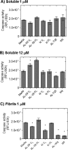
Caspase activity detected in cardiomyocytes cells. AC16 cells were incubated with 1 μm fibrils (A), 12 μm soluble protein (B), and 1 μm fibrils (C). Apoptosis reagents were added after 80 h of treatment and were incubated for 1 h at 37 °C before reading plate. Caspase activity is indicated as relative fluorescence units (RFU). Samples were set up in triplicate in two independent assays (n = 2) with the average values and error bars as means ± S.E. *, two-tailed t test; p value < 0.05 with respect to the corresponding media controls.
FIGURE 9.
FIGURE 9.
ThT detection of cytotoxic fibrillar species. A, ThT fluorescence decreased as a function of fibril concentration. B, cell toxicity experiments with a dilution series of Wil fibrils from 1 to 0.001 μm. Red counts were registered over time as a number of RFP-AC16 cells per well. AC16 growth rate decreases as Wil fibril concentration increases. C, plot of cell toxicity versus ThT signal. ThT fluorescence is barely above the buffer ThT baseline (black dashed line) when cells grow 50% with respect to the control. ThT fluorescence signal and percentage of cell growth intersect between 0.2 and 0.4 μm.

Similar articles

Cited by

References

    1. Baden E. M., Sikkink L. A., and Ramirez-Alvarado M. (2009) Light chain amyloidosis: current findings and future prospects. Curr. Protein Pept. Sci. 10, 500–508 - PMC - PubMed
    1. Falk R. H. (2005) Diagnosis and management of the cardiac amyloidoses. Circulation 112, 2047–2060 - PubMed
    1. Kumar S. K., Gertz M. A., Lacy M. Q., Dingli D., Hayman S. R., Buadi F. K., Short-Detweiler K., Zeldenrust S. R., Leung N., Greipp P. R., Lust J. A., Russell S. J., Kyle R. A., Rajkumar S. V., and Dispenzieri A. (2011) Recent improvements in survival in primary systemic amyloidosis and the importance of an early mortality risk score. Mayo Clin. Proc. 86, 12–18 - PMC - PubMed
    1. Glenner G. G., Cuatrecasas P., Isersky C., Bladen H. A., and Eanes E. D. (1969) Physical and chemical properties of amyloid fibers: II. Isolation of a unique protein constituting the major component from human splenic amyloid fibril concentrates. J. Histochem. Cytochem. 17, 769–780 - PubMed
    1. Olsen K. E., Sletten K., and Westermark P. (1998) Fragments of the constant region of immunoglobulin light chains are constituents of AL-amyloid proteins. Biochem. Biophys. Res. Commun. 251, 642–647 - PubMed

Publication types

MeSH terms