. 2016 Nov 1;214(9):1309-1318.
doi: 10.1093/infdis/jiw313. Epub 2016 Jul 26.

Relationship Between HIV Coinfection, Interleukin 10 Production, and Mycobacterium tuberculosis in Human Lymph Node Granulomas

Affiliations

Relationship Between HIV Coinfection, Interleukin 10 Production, and Mycobacterium tuberculosis in Human Lymph Node Granulomas

Collin R Diedrich et al. J Infect Dis. .

Abstract

Background: Human immunodeficiency virus type 1 (HIV)-infected persons are more susceptible to tuberculosis than HIV-uninfected persons. Low peripheral CD4+ T-cell count is not the sole cause of higher susceptibility, because HIV-infected persons with a high peripheral CD4+ T-cell count and those prescribed successful antiretroviral therapy (ART) remain more prone to active tuberculosis than HIV-uninfected persons. We hypothesized that the increase in susceptibility is caused by the ability of HIV to manipulate Mycobacterium tuberculosis-associated granulomas.

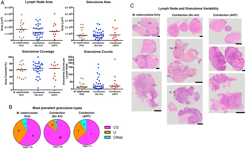
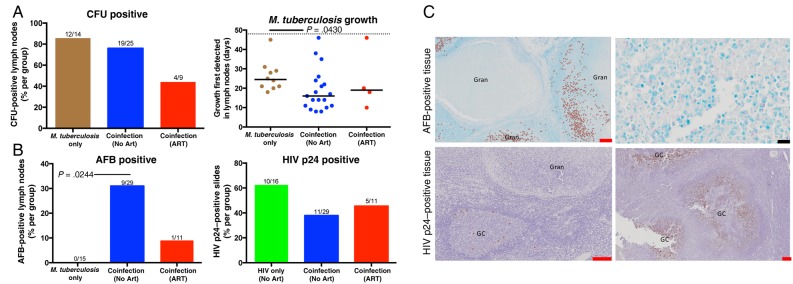
Methods: We examined 71 excised cervical lymph nodes (LNs) from persons with HIV and M. tuberculosis coinfection, those with HIV monoinfection, and those with M. tuberculosis monoinfection with a spectrum of peripheral CD4+ T-cell counts and ART statuses. We quantified differences in M. tuberculosis levels, HIV p24 levels, cellular response, and cytokine presence within granulomas.

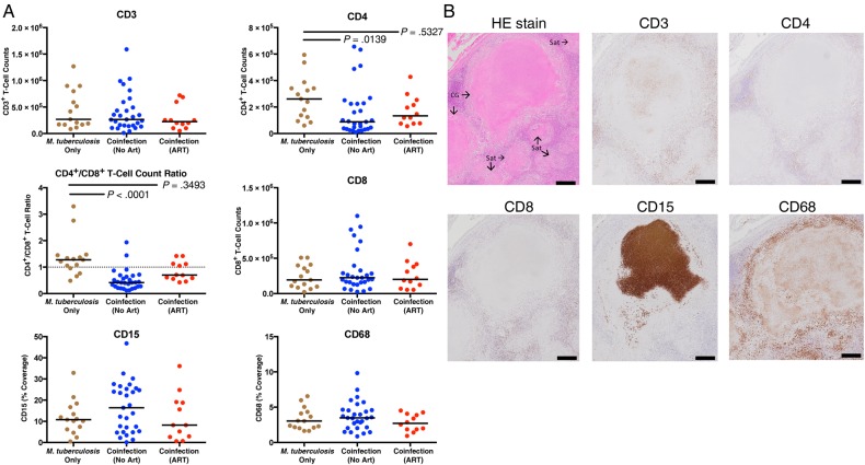
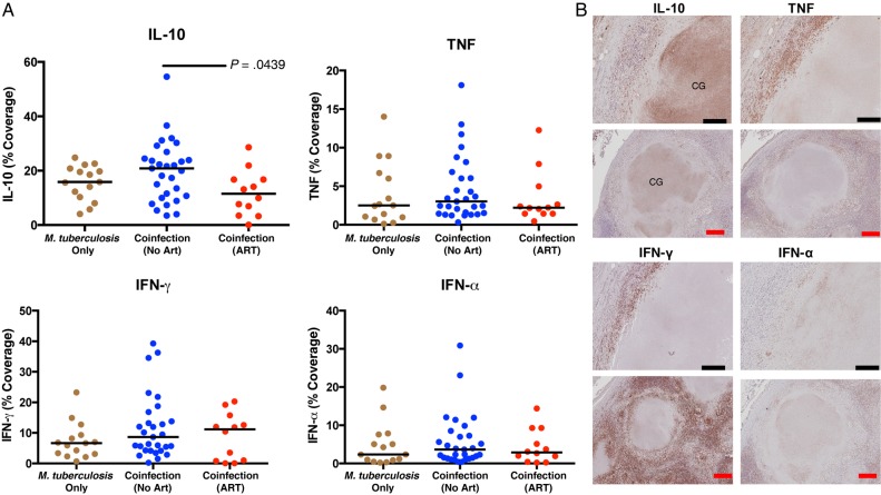
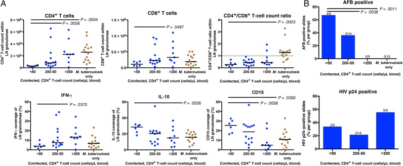
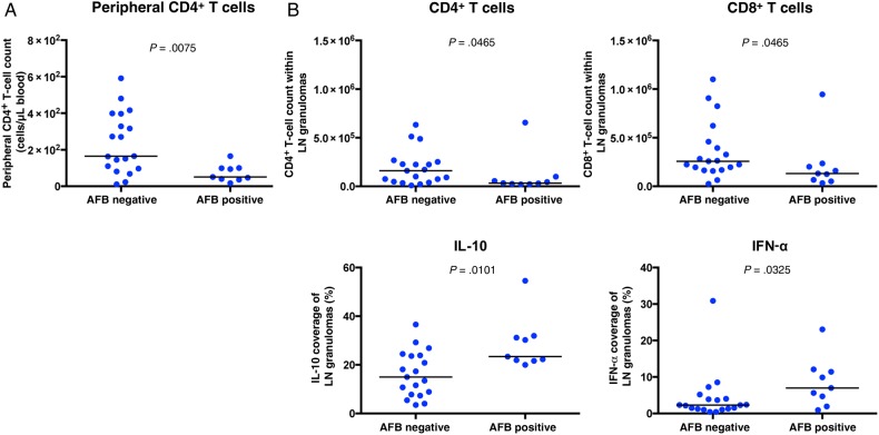
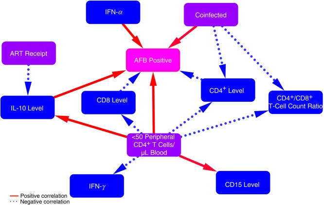
Results: HIV increased M. tuberculosis numbers and reduced CD4+ T-cell counts within granulomas. Peripheral CD4+ T-cell depletion correlated with granulomas that contained fewer CD4+ and CD8+ T cells, less interferon γ, more neutrophils, more interleukin 10 (IL-10), and increased M. tuberculosis numbers. M. tuberculosis numbers correlated positively with IL-10 and interferon α levels and fewer CD4+ and CD8+ T cells. ART reduced IL-10 production.

Conclusions: Peripheral CD4+ T-cell depletion correlated with increased M. tuberculosis presence, increased IL-10 production, and other phenotypic changes within granulomas, demonstrating the HIV infection progressively changes these granulomas.

Keywords: HIV; IL-10; antiretroviral therapy; coinfection; granuloma; histology; lymph node; mycobacterium; tuberculosis.


Figures

Figure 1.
Figure 1.
Persons coinfected with Mycobacterium tuberculosis and human immunodeficiency virus type 1 (HIV) do not have differences in lymph node size or granuloma area, counts, and type, compared with M. tuberculosis–monoinfected persons. A, No significant differences were observed in total cross-sectional areas of the excised lymph nodes and granulomas, percentage of lymph nodes covered in granulomas, or granuloma counts between M. tuberculosis–monoinfected persons and individuals coinfected with M. tuberculosis and HIV (who were or were not receiving antiretroviral therapy [ART]). B, The percentage of lymph nodes that contained the largest granulomas or unorganized inflammation within each infection group are displayed in each pie chart. C, Hematoxylin-eosin–stained slides demonstrating the heterogeneous nature of excised lymph nodes and their granulomas (black scale bar, 2 mm). A few granulomas and inflamed areas are labeled to demonstrate the high variability in granuloma types within individual lymph nodes. “Other” denotes suppurative (Sup), solid cellular (SC), and fibrotic (F) granulomas. CG, caseous granuloma; U, unorganized inflammation.
Figure 2.
Figure 2.
Lymph nodes from persons coinfected with Mycobacterium tuberculosis and human immunodeficiency virus type 1 (HIV) were more likely to contain higher bacterial loads, compared with M. tuberculosis–monoinfected persons. A, No difference in lymph node M. tuberculosis culture findings (ie, colony-forming units [CFU]) were observed among the 3 groups. Fractions above bars indicate positive lymph nodes over total lymph nodes (not all lymph nodes were cultured). Days to culture positivity (determined by the BACTEC MGIT system) was lowest within lymph nodes isolated from the coinfected recipients who were not receiving antiretroviral therapy (ART). The dotted line represents the culture-negative time limit (49 days). B, HIV was associated with an increased presence of acid-fast bacilli (AFB) that was not significantly reduced in the presence of ART. No AFB were observed in lymph nodes from HIV-uninfected persons. M. tuberculosis did not change the prevalence of HIV p24 among lymph nodes. C, AFB and HIV p24 staining within lymph nodes. AFB and HIV p24 are flagged in left panels. Significance was determined by the Kruskal–Wallis test with the Dunn multiple comparisons post hoc test (P = .05). Red and black scale bars represent 200 µM and 2 µM, respectively. GC, germinal center; Gran, granuloma.
Figure 3.
Figure 3.
Antiretroviral therapy (ART) partially restores the reduction of CD4+ T cells and CD4+/CD8+ T-cell count ratio observed within tuberculosis-diseased lymph nodes from persons coinfected with Mycobacterium tuberculosis and human immunodeficiency virus type 1 (HIV). A, HIV reduces CD4+ T-cell counts and CD4+/CD8+ T-cell count ratios, compared with M. tuberculosis–monoinfected persons. No significant reduction was observed in coinfected patients receiving ART. The Kruskal–Wallis test and Dunn multiple comparisons post hoc test were used to determine significance. Adjusted P values are displayed. B, Hematoxylin-eosin (HE) or cellular staining in consecutive lymph node sections that contain caseous granulomas (CG) with satellite solid cellular granulomas (Sat; black scale bar, 100 µm). CD3, CD4, and CD8 staining predominately occurred within lymphocyte layer of granulomas. Interestingly, circular CD3 staining occurred occasionally, within the center of caseous granulomas without associated nuclei. CD15 was most often localized within caseous centers, without nuclei, and sporadically within macrophage layer of granulomas. CD68 staining was within caseous granulomas without associating with nuclei and was most frequently present in the macrophage layer of granulomas.
Figure 4.
Figure 4.
Antiretroviral therapy (ART) reduces interleukin 10 (IL-10) presence within tuberculosis-diseased lymph nodes. A, Percentage of lymph node sections covered by cytokines are represented. Human immunodeficiency virus type 1 (HIV) coinfection did not change IL-10 presence in lymph nodes, compared with Mycobacterium tuberculosis monoinfection. ART significantly reduced IL-10 presence in persons coinfected with M. tuberculosis and HIV. HIV did not change tumor necrosis factor (TNF), interferon γ (IFN-γ), or interferon α (IFN-α) presence in these tissues. The Kruskal–Wallis test and Dunn multiple comparisons post hoc test were used to determine significance. Adjusted P values are displayed. B, Cytokine staining in consecutive lymph node sections that contain caseous granulomas (CGs; black scale bar, 20 µm; red scale bar, 100 µM). Staining revealed that IL-10 was most often present in the center of caseous granulomas. TNF and IFN-γ were predominately identified in macrophage and lymphocyte layer. IFN-α most often localized within macrophage layer.
Figure 5.
Figure 5.
As human immunodeficiency virus type 1 (HIV) infection progresses, tuberculosis-diseased tissue contain fewer CD4+ T cells and lower CD4+/CD8+ T-cell count ratios, along with increased acid-fast bacilli (AFB), neutrophil, and IL-10 presence. A, Lymph nodes from persons who were coinfected with M. tuberculosis and HIV, not receiving ART, and <50 peripheral CD4+ T cells/µL of blood contain fewer CD4+ T cells, lower CD4+/CD8+ T-cell count ratios, and more IL-10 and CD15 than lymph nodes from M. tuberculosis–monoinfected persons. Lymph nodes from persons with <50 peripheral CD4+ T cells contain fewer CD4+ and CD8+ T cells along with less interferon γ (IFN-γ), compared with lymph nodes from coinfected persons with >200 peripheral CD4+ T cells/µL of blood. The Kruskal–Wallis test and Dunn multiple comparisons post hoc test were used to determine the significance of differences from persons with <50 peripheral CD4+ T cells/μL of blood. B, Lymph nodes from coinfected persons who were not receiving ART and had fewer peripheral CD4+ T cells were more likely to be positive for AFB than coinfected patients with >200 peripheral CD4+ T cells/µL of blood and those with M. tuberculosis monoinfection. Fractions above bars indicate positive lymph nodes over total lymph nodes. Significance was determined by Kruskal–Wallis test with the Dunn multiple comparisons post hoc test (P = .05). Adjusted P values are displayed.
Figure 6.
Figure 6.
Acid-fast bacilli (AFB)–positive granulomas contained fewer CD4+ and CD8+ T cells along with increased interleukin 10 (IL-10) and interferon α (IFN-α) levels, compared with AFB-negative granulomas. A, Peripheral CD4+ T-cell counts were lower in coinfected persons with AFB-positive lymph node sections, compared with those with AFB-negative sections. B, AFB-positive lymph node sections contained fewer CD4+ and CD8+ T cells along with more IL-10 and IFN-α. No difference was observed in the expression of CD3, CD15, CD68, IFN-γ, or tumor necrosis factor (TNF) between AFB-positive and AFB-negative granulomas. The Mann–Whitney test was used to determine significance (P = .05).
Figure 7.
Figure 7.
Proposed network of how human immunodeficiency virus type 1 (HIV) and treatment status change phenotypic interactions within Mycobacterium tuberculosis granulomas. Red solid and blue dotted lines represent positive and negative correlations, respectively, between phenotypic changes within granulomas (blue and pink boxes) and clinical descriptions of persons (purple boxes). Arrows represent our hypotheses as to the cause of the phenotypic changes. Coinfection increases acid-fast bacilli (AFB) presence and reduces CD4+ T-cell counts within granulomas. As HIV infection progresses (to <50 peripheral CD4+ T cells/μL of blood), granulomas contain less CD4, CD8, and interferon γ (IFN-γ) and more CD15 and interleukin 10 (IL-10), which lead to increased M. tuberculosis growth. IL-10 and IFN-α lead to more M. tuberculosis growth within granulomas. Antiretroviral therapy (ART) receipt reduces IL-10 presence.

Comment in

Similar articles

Cited by

References

    1. World Health Organization. Global tuberculosis report 2014. Geneva: World Health Organization, 2015.
    1. World Health Organization. Consolidated strategic information guidelines for HIV in the health sector. Geneva: World Health Organization, 2015. - PubMed
    1. Lawn SD, Myer L, Edwards D, Bekker L-G, Wood R. Short-term and long-term risk of tuberculosis associated with CD4 cell recovery during antiretroviral therapy in South Africa. AIDS 2009; 23:1717–25. - PMC - PubMed
    1. Caruso AM, Serbina N, Klein E, Triebold K, Bloom BR, Flynn JL. Mice deficient in CD4 T cells have only transiently diminished levels of IFN-gamma, yet succumb to tuberculosis. J Immunol 1999; 162:5407–16. - PubMed
    1. Lin PL, Rutledge T, Green AM et al. . CD4 T cell depletion exacerbates acute Mycobacterium tuberculosis while reactivation of latent infection is dependent on severity of tissue depletion in cynomolgus macaques. AIDS Res Hum Retroviruses 2012; 28:1693–702. - PMC - PubMed

MeSH terms