

Case Reports
doi: 10.1159/000445795.
eCollection 2016 Jan-Apr.
Subretinal Fluid Drainage and Vitrectomy Are Helpful in Diagnosing and Treating Eyes with Advanced Coats' Disease
Affiliations
- PMID: 27462247
- PMCID: PMC4943774
- DOI: 10.1159/000445795
Item in Clipboard
Case Reports
Subretinal Fluid Drainage and Vitrectomy Are Helpful in Diagnosing and Treating Eyes with Advanced Coats' Disease
Case Rep Ophthalmol.
.
Abstract
Severe forms of Coats' disease are often associated with total retinal detachment, and a differential diagnosis from retinoblastoma is critically important. In such eyes, laser- and/or cryoablation is often ineffective or sometimes impossible to perform. We report a case of advanced Coats' disease in which a rapid pathological examination of subretinal fluid was effective for the diagnosis, and external subretinal drainage combined with vitrectomy was effective in preserving the eye.
Keywords: Coats' disease; Rapid pathological examination; Retinoblastoma; Vitrectomy.
Figures
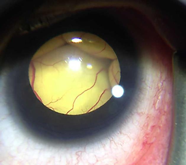
Anterior segment photograph at the first visit showing a totally detached retina in contact with the posterior surface of the lens. The peripheral retina could not be observed.
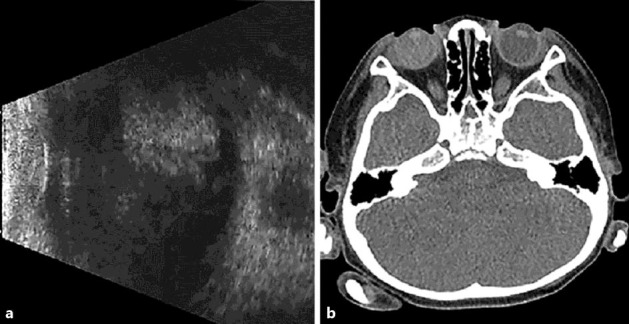
a B-mode echography of the right eye shows a high echo area in the vitreous cavity. b CT shows no evidence of calcification or tumor structure.
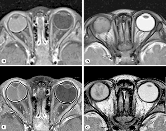
MRIs taken at two different periods. T1 (a) and T2 (b) images taken 3 months before the first visit show a hyperintense lesion and a slightly hypointense lesion in the subretinal space, respectively. Both T1 (c) and T2 (d) images taken at the first visit show hyperintense lesions in the subretinal space. Tumor structure is not observed in the eye.
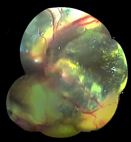
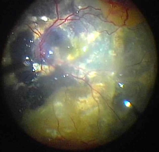
Fundus photograph of the right eye during the first operation under perfluoro-n-octane. Dilated retinal vessels, numerous microaneurysms, and intra- and subretinal exudates became visible.
Fundus photograph of the right eye during the second operation under perfluoro-n-octane. The amount of exudates were reduced.
Similar articles
-
Recent advances in the diagnosis and treatment of Coats' disease.Int Ophthalmol. 2019 Apr;39(4):957-970. doi: 10.1007/s10792-019-01095-8. Epub 2019 Mar 20. Int Ophthalmol. 2019. PMID: 30895419 Review.
-
Advanced Coats' disease.Trans Am Ophthalmol Soc. 1991;89:371-476. Trans Am Ophthalmol Soc. 1991. PMID: 1808814 Free PMC article. Review.
-
Risk of Tractional Retinal Detachment Following Intravitreal Bevacizumab Along with Subretinal Fluid Drainage and Cryotherapy for Stage 3B Coats' Disease.Middle East Afr J Ophthalmol. 2016 Apr-Jun;23(2):208-11. doi: 10.4103/0974-9233.175895. Middle East Afr J Ophthalmol. 2016. PMID: 27162454 Free PMC article.
-
Treatment of stage 3 Coats' disease by endolaser photocoagulation via a two-port pars plana nonvitrectomy approach.Graefes Arch Clin Exp Ophthalmol. 2015 Jul;253(7):999-1004. doi: 10.1007/s00417-015-2984-4. Epub 2015 Mar 21. Graefes Arch Clin Exp Ophthalmol. 2015. PMID: 25794987 Free PMC article.
-
High-density vitreous substitute in the management of advanced Coats' disease.Chang Gung Med J. 2002 Feb;25(2):128-32. Chang Gung Med J. 2002. PMID: 11952273
Cited by
-
Factors Predictive of Subretinal Fluid Resolution in Coats Disease: Analysis of 177 Eyes in 177 Patients at a Single Center.Asia Pac J Ophthalmol (Phila). 2019 Jul-Aug;8(4):290-297. doi: 10.1097/APO.0000000000000246. Asia Pac J Ophthalmol (Phila). 2019. PMID: 31356365 Free PMC article.
-
Recent advances in the diagnosis and treatment of Coats' disease.Int Ophthalmol. 2019 Apr;39(4):957-970. doi: 10.1007/s10792-019-01095-8. Epub 2019 Mar 20. Int Ophthalmol. 2019. PMID: 30895419 Review.
-
Coats' disease: characteristics, management, outcome, and scleral external drainage with anterior chamber maintainer for stage 3b disease.Medicine (Baltimore). 2020 Apr;99(16):e19623. doi: 10.1097/MD.0000000000019623. Medicine (Baltimore). 2020. PMID: 32311932 Free PMC article.
References
-
- Shields JA, Shields CL. Review: coats disease: the 2001 LuEsther T. Mertz lecture. Retina. 2002;22:80–91. - PubMed
-
- Ray R, Baranano DE, Hubbard GB. Treatment of Coats' disease with intravitreal bevacizumab. Br J Ophthalmol. 2013;97:272–277. - PubMed
-
- Grabowska A, Calvo JP, Fernandez-Zubillaga A, Rios JC, Gomez JA. A magnetic resonance imaging diagnostic dilemma: diffuse infiltrating retinoblastoma versus Coats' disease. J Pediatr Ophthalmol Strabismus. 2010;47:e1–e3. - PubMed
-
- Bhatnagar R, Vine AK. Diffuse infiltrating retinoblastoma. Ophthalmology. 1991;98:1657–1661. - PubMed
Publication types
LinkOut - more resources
Full Text Sources
Other Literature Sources
