. 2015 Aug 18:1:15021.
doi: 10.1038/celldisc.2015.21. eCollection 2015.

Targeting the γ-/β-secretase interaction reduces β-amyloid generation and ameliorates Alzheimer's disease-related pathogenesis

Affiliations

Targeting the γ-/β-secretase interaction reduces β-amyloid generation and ameliorates Alzheimer's disease-related pathogenesis

Jin Cui et al. Cell Discov. .

Abstract

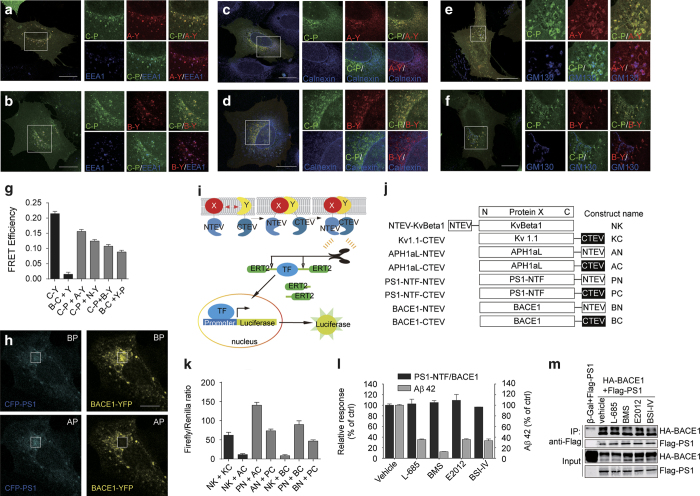
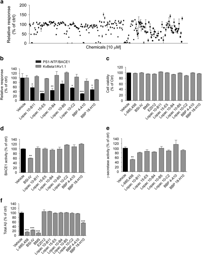
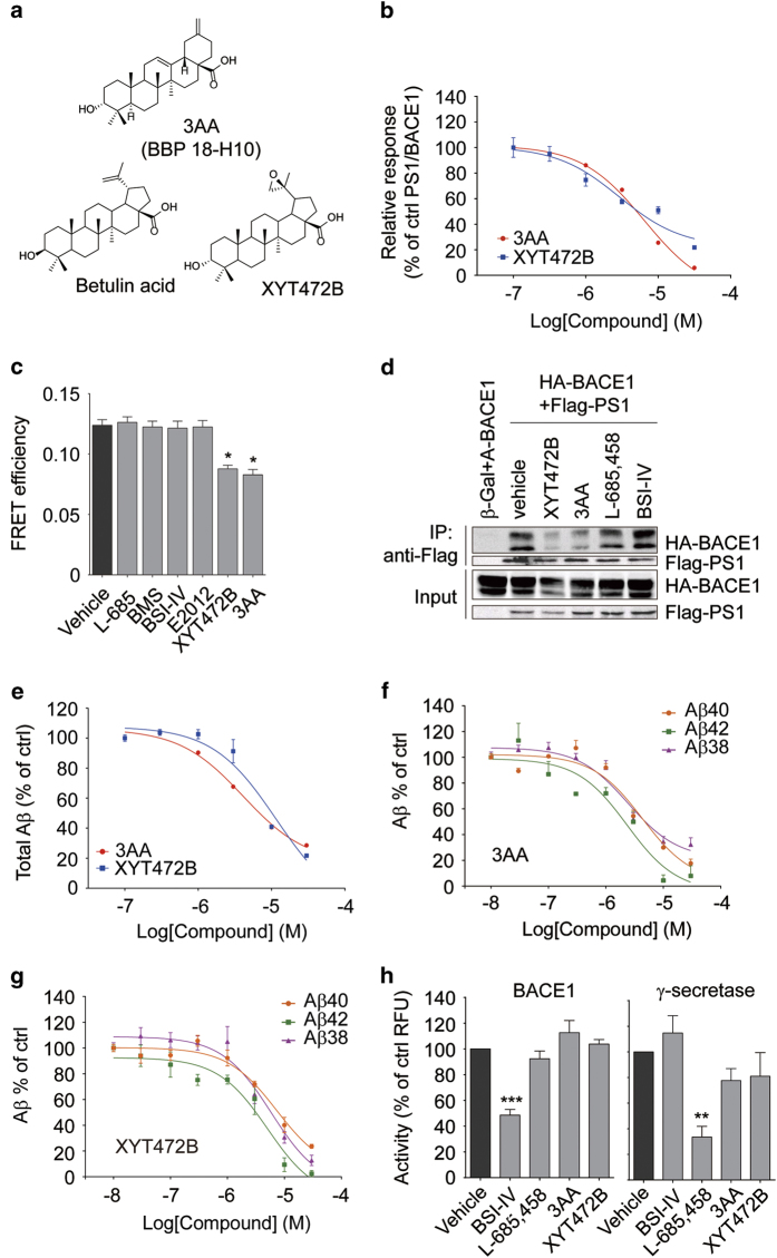
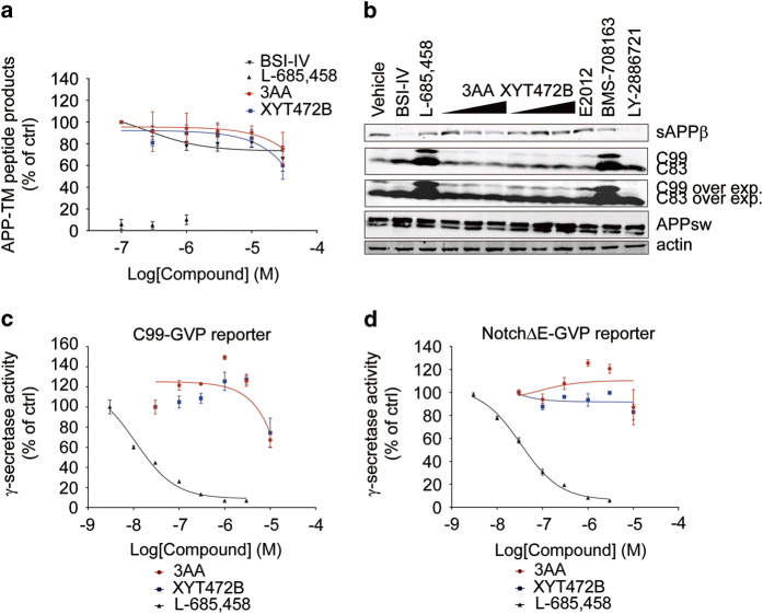
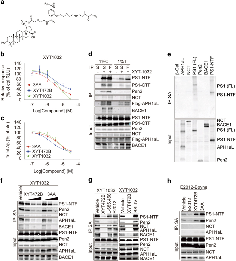
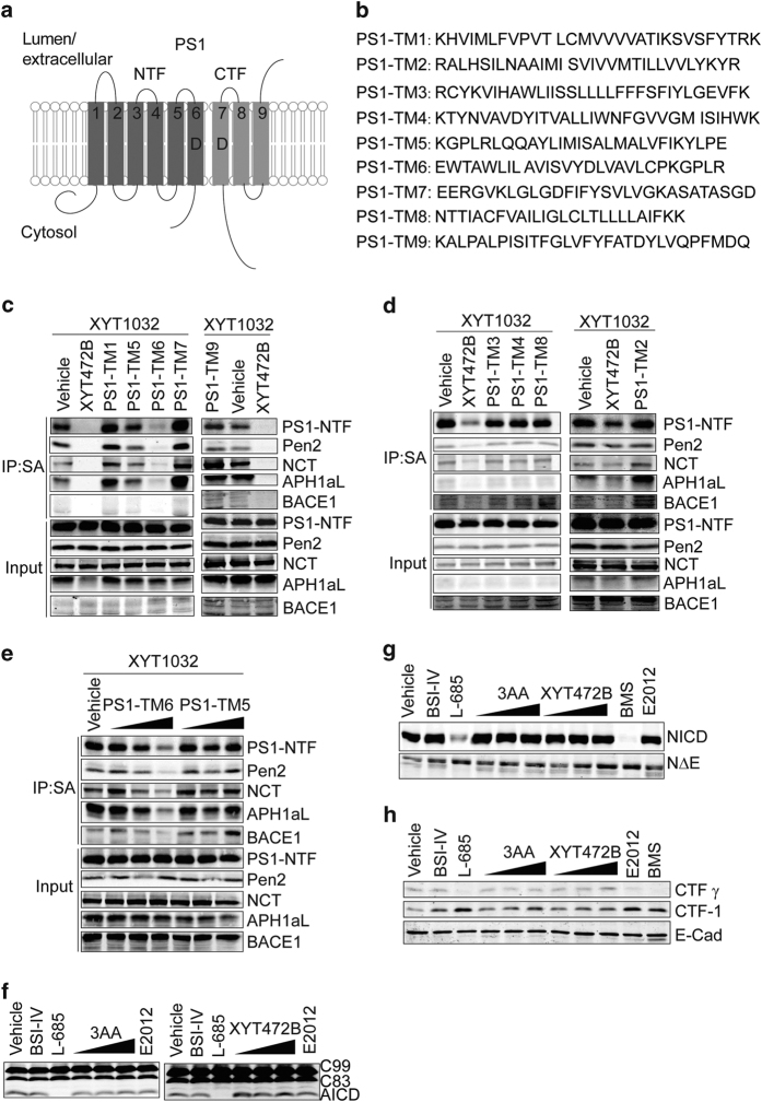
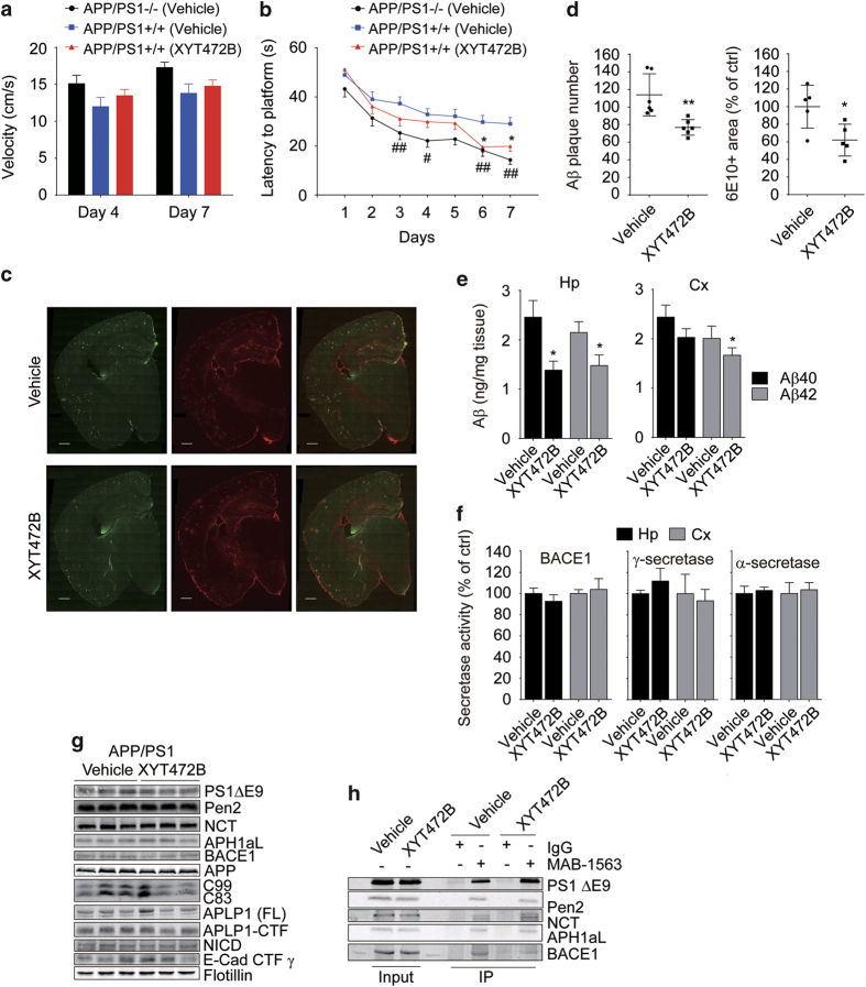
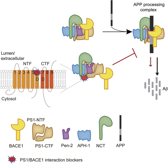
Despite decades of intense global effort, no disease-modifying drugs for Alzheimer's disease have emerged. Molecules targeting catalytic activities of γ-secretase or β-site APP-cleaving enzyme 1 (BACE1) have been beset by undesired side effects. We hypothesized that blocking the interaction between BACE1 and γ-secretase subunit presenilin-1 (PS1) might offer an alternative strategy to selectively suppress Aβ generation. Through high-throughput screening, we discovered that 3-α-akebonoic acid (3AA) interferes with PS1/BACE1 interaction and reduces Aβ production. Structural analogs of 3AA were systematically synthesized and the functional analog XYT472B was identified. Photo-activated crosslinking and biochemical competition assays showed that 3AA and XYT472B bind to PS1, interfere with PS1/BACE1 interaction, and reduce Aβ production, whereas sparing secretase activities. Furthermore, treatment of APP/PS1 mice with XYT472B alleviated cognitive dysfunction and Aβ-related pathology. Together, our results indicate that chemical interference of PS1/BACE1 interaction is a promising strategy for Alzheimer's disease therapeutics.

Keywords: Alzheimer’s disease; Aβ generation; PS1/BACE1 interaction.


Figures

Figure 1
Figure 1
Monitoring the cellular PS1/BACE1 interaction in FRET assay and Split-TEV assay. (af) Subcellular co-localization of CFP-PS1 (C-P) and APH1aL-YFP (A-Y), CFP-PS1 and BACE1-YFP (B-Y) with EEA1, calnexin and GM130 in the presence of the other γ-secretase subunits in U-2 OS cells. Forty-eight hours after transfection, cells overexpressing CFP-PS1, APH1aL-YFP, NCT and BACE1-YFP were fixed and counter-stained for EEA1, calnexin and GM130. Scale bar, 20 μm. White box indicates the enlarged area. (g) Acceptor photobleaching FRET analysis in HEK293 cells. A CFP-YFP (C-Y) fusion protein was used as a positive control, and BACE1-CFP (B-C)/YFP (Y) pair as a negative control. The intermolecular FRET efficiency of PS1 and BACE1, assayed using two fluorescent protein pairs: CFP-PS1 (C-P) and BACE1-YFP (B-Y), B-C and YFP-PS1 (Y-P), is lower than that of C-P/APH1aL-YFP (A-Y) and comparable with that of C-P/NCT-YFP (N-Y). n=20 cells per group. (h) Representative image of CFP-PS1/BACE1-YFP FRET in the presence of other γ-secretase components in HEK293 cells. Scale bar, 10 μm. White box indicates the photobleached area. (i) A schematic of the reporter-based split-TEV assay for monitoring protein–protein interaction and (j) the fusion-protein constructs used in this study. (k) Interaction of PS1 and BACE1 in HEK293MSR cells monitored by luciferase-based split-TEV assay. Luciferase reporter activities were measured 24 h after transfection. NTEV-KvBeta1 (NK)+Kv1.1-CTEV (KC) heterodimers and PS1-NTF-NTEV (PN)+APH1aL-CTEV (AC) were positive controls, NK+AC and NK+BACE1-CTEV (BC) were negative controls. PN+BC and BACE1-NTEV (BN)+PS1-NTF-CTEV (PC) showed high interaction signals, indicating an interaction between PS1-NTF and BACE1. (l) PS1-NTF/BACE1 split-TEV reporter activities and Aβ42 production in cells treated for 16 h with 1 μM chemicals indicated in the x-axis . (m) Co-immunoprecipitation of C-terminal Flag-tagged PS1 (Flag-PS1) and C-terminal HA-tagged BACE1 (HA-BACE1). HEK293T cells overexpressing HA-BACE1 and β-Gal or Flag-PS1 were treated with 3 μM chemicals for 16 h before cell lysis and immunoprecipitation. AP, after photobleaching; BP, before photobleaching.
Figure 2
Figure 2
Screening of PS1/BACE1 interaction-interfering chemicals. (a) Representative results of the screen for chemicals that interfere with PS1-NTF-TEV/BACE1-CTEV interaction. Two hours after transfection, HEK293MSR cells were treated with 10 μM chemicals for 16 h and the luciferase reporter activities were measured. (b) PS1-NTF/BACE1 and KvBeta1/Kv1.1 split-TEV reporter activities. Following transfection, cells were treated for 16 h with 10 μM candidate compounds. (c) Cell viability of HEK293/APPswe cells. HEK293/APPswe were treated with 1 μM L-685 458, BACE1 inhibitor-IV (BSI-IV), BMS-708163, E2012 or 10 μM candidate compounds for 8 h and subjected to cell viability test. (d) BACE1 and (e) γ-secretase activity in fluorogenic substrate assay. HEK293T cell membrane fractions were incubated with 10 μM chemicals. (f) Total Aβ production of HEK293/APPswe cells. Cells were treated with 1 μM L-685 458, BSI-IV or BMS, or 10 μM candidate chemicals for 8 h. **P<0.01 determined by one-way ANOVA.
Figure 3
Figure 3
3AA and its structural analog XYT472B reduce PS1/BACE1 interaction and Aβ production. (a) Chemical structures of 3-α-Akebonoic acid (3AA, BBP 18-H10), betulin acid and XYT472B. (b) 3AA and XYT472B reduce PS1-NTF/BACE1 interaction dose-dependently in split-TEV assay. Cells were treated with different concentrations of 3AA or XYT472B for 16 h. (c) 3AA and XYT472B reduce FRET efficiency of PS1 and BACE1. Cells (overexpressing fusion protein CFP-PS1, BACE1-YFP, APH1aL, NCT and Pen2) were treated with 3 μM chemicals for 16 h and subjected to acceptor photobleaching FRET analysis. (d) XYT472B and 3AA reduce PS1/BACE1 interaction in co-immunoprecipitation assay. HEK293T cells overexpressing C-terminal HA-tagged BACE1 and β-Gal or C-terminal Flag-tagged PS1 were treated with 3 μM chemicals for 16 h before cell lysis and immunoprecipitation. (e) 3AA and XYT472B reduce total Aβ production. HEK293/APPswe cells were treated with 3AA or XYT472B for 8 h and the culture media were collected for sandwich ELISA to quantify the total Aβ production. (f) 3AA and (g) XYT472B dose-dependently decrease Aβ40, Aβ42 and Aβ38 generation. HEK293/APPswe cells were treated with different concentrations of chemicals for 8 h before the supernatants were collected for ELISA quantification. (h) 3AA and XYT472B show no significant effects on BACE1 activity (left) or γ-secretase activity (right). HEK293T cell membrane fractions with 10 μM chemicals were incubated with fluorogenic substrates and the fluorescent signal from processed substrates were monitored and presented. *P<0.05, **P<0.01, ***P<0.001, determined by one-way ANOVA.
Figure 4
Figure 4
3AA and XYT472B do not inhibit secretase activities. (a) ELISA-based γ-secretase activity assay. HEK293T membrane fractions were incubated with chemicals and APP-TM peptides for 2 h before the ELISA analysis of the processed products. (b) Cellular APP processing was unaffected by 3AA or XYT472B treatment. HEK293/APPswe cells were incubated with chemicals for 4 h. Cell lysates were prepared and subjected to western blotting analysis of C99, C83 and APPswe. Culture media were collected for sAPPβ analysis. (c and d) Cellular γ-secretase activity was monitored and found unaltered in (c) C99-GVP and (d) NotchΔE-GVP reporter assays. Two hours after transfection, HEK293T cells were treated with chemicals. Luciferase reporter activities were measured 24 h after transfection.
Figure 5
Figure 5
Photoclickable analog XYT1032 reveals XYT472B and 3AA binding sites on PS1. (a) Chemical structure of XYT1032. (b and c) XYT1032 reduces PS1-NTF/BACE1 interaction in split-TEV assay (b) and total Aβ production (c). Transfected HEK293MSR cells were treated with chemicals for 16 h before DLR analysis of luciferase reporter activities, and HEK293/APPswe cells were treated for 8 h before the supernatants were collected for ELISA. (d) Labeling and affinity purification of γ-secretase subunits and BACE1 by XYT1032 in different detergents. Cells expressing PS1, Pen2, NCT, C-terminal Flag-tagged APH1aL and BACE1 were treated with XYT1032 (3 μM) and photo-activated click reaction were carried out. γ-secretase subunits and BACE1 were affinity purified by anti-Flag or straptavidin resin as indicated. (e) XYT1032 binds only to PS1. Cells expressing β-Gal, C-terminal Flag-tagged APH1aL, NCT, PS1 (FL), Pen2, BACE1 or PS1-NTF, respectively, were treated with XYT1032 (3 μM) and subjected to photo-activated click reaction and affinity purification. (f) XYT472B and 3AA compete with XYT1032 in labeling of PS1-NTF. Cells were treated with 3 μM XYT1032 in the absence or presence of 10 μM, 30 μM, 100 μM XYT472B or 3AA for 2 h. (g) Secretase inhibitors and modulator fail to compete with XYT1032 in binding to PS1-NTF. Cells were incubated with 3 μM XYT1032 in the absence or presence of 100 μM designated chemicals. (h) XYT472B and 3AA enhanced the labeling of PS1-NTF by E2012-BPyne. Cells were incubated with 1 μM E2012-BPyne in the absence or presence of 50 μM designated chemicals for 1 h. After chemical incubation, photo-activated crosslinking, click reaction and streptavidin affinity purification in 1% CHAPSO buffer were performed. C, CHAPSO; F, Flag; S, streptavidin; T, TritonX-100.
Figure 6
Figure 6
3AA and its analogs bind to the sixth transmembrane domain of PS1 without affecting γ-secretase activity. (a) A schematic of PS1 transmembrane domain-derived peptides. (b) Amino acid sequences of PS1-TM peptides. (c and d) Competition experiments of PS1 TM peptides to the photo-affinity labeling of PS1-NTF, other γ-secretase components and BACE1. Vehicle (DMSO), 50 μM XYT472B or PS1-TM peptides were pre-incubated in medium with XYT1032 for 15 min prior to the 2 h cellular treatment of transfected HEK293T cells. (e) PS1-TM6 peptide competes with PS1-NTF in the labeling of XYT1032. PS1-TM6 and PS1-TM5 peptide (10 μM, 20 μM and 50 μM) were pre-incubated with XYT1032 for 15 min in medium, and then added to the cells. After treatment, cells were lysed with 1% CHAPSO buffer and subjected to photo-activated crosslinking, click reaction and streptavidin purification. (f) 3AA and XYT472B exhibited little effect on AICD production in in vitro C99 assay. HEK293T cell membrane fractions were incubated with chemicals for 4 h. (g) Processing of NotchΔE in HEK293T cells was unaffected by 3AA- or XYT472B treatment. (h) 3AA and XYT472B do not affect E-Cadherin processing in vitro. The concentration of BSI-IV, L-685 458, E2012, BMS used in (f and h) was 10 μM.
Figure 7
Figure 7
XYT472B attenuates behavior deficits and Aβ pathology of APP/PS1 mice. (a) Swimming velocities of mice in all groups during the probe trials on day 4 and day 7. (b) XYT472B treatment ameliorates the cognitive deficits of APP/PS1 mice in Morris water maze. APP/PS1 mice show a slower learning curve compared with WT littermates with statistical significance on days 3, 4, 6 and 7. This impairment was partially improved by XYT472B treatment (with statistical significance on days 6 and 7). n=16 for each group. #P<0.05, ##P<0.01, *P<0.05, determined by two-way ANOVA. (c) Representative brain immunofluorescent images of vehicle- or XYT472B-treated mice. (d) Statistical analyses show that 6E10-positive Aβ plaques and GFAP-positive area were reduced by XYT472B treatment. Scale bar, 500 μm. (e) Reduced Aβ levels in hippocampus and prefrontal cortex of XYT472B-treated mice. Extracts from hippocampus or cortex were prepared and subjected to sandwich ELISAs for human Aβ40 and Aβ42. (f) BACE1, γ-secretase and α-secretase activities were unaltered by chronic treatment of XYT472B. Fluorogenic substrate assays were performed with the membrane fractions extracted from mouse hippocampi or cortices. (df) *P<0.05, **P<0.01, determined by unpaired Student’s t-test. (g) Chronic XYT472B treatment shows no effects on secretase expression or substrate processing. Extracts from hippocampus were analyzed by western blotting. (h) Chronic XYT472B treatment attenuates the interaction between BACE1 and PS1ΔE9 in vivo. Vehicle- or XYT472B-treated APP/PS1 mouse brains were homogenized, and purified membrane fractions were incubated with IgG or MAB-1563 antibody for immunoprecipitation. Cx, cortex; Hp, hippocampus.
Figure 8
Figure 8
A working model of 3AA and XYT472B. 3AA and XYT472B (PS1/BACE1 interaction blockers) bind to the sixth transmembrane domain of PS1, interfere with the β-/γ-secretase interaction and reduce Aβ production.

References

    1. Goedert M , Spillantini MG . A century of Alzheimer's disease. Science 2006; 314: 777–781. - PubMed
    1. Hardy J , Selkoe DJ . The amyloid hypothesis of Alzheimer's disease: progress and problems on the road to therapeutics. Science 2002; 297: 353–356. - PubMed
    1. Selkoe DJ . Deciphering the genesis and fate of amyloid beta-protein yields novel therapies for Alzheimer disease. J Clin Invest 2002; 110: 1375–1381. - PMC - PubMed
    1. Schenk D , Basi GS , Pangalos MN . Treatment strategies targeting amyloid beta-protein. Cold Spring Harb Perspect Med 2012; 2: a006387. - PMC - PubMed
    1. Karran E , Mercken M , De Strooper B . The amyloid cascade hypothesis for Alzheimer's disease: an appraisal for the development of therapeutics. Nat Rev Drug Discov 2011; 10: 698–712. - PubMed