. 2016 Jul 12:2:16017.
doi: 10.1038/celldisc.2016.17. eCollection 2016.

Nuclear localization of platelet-activating factor receptor controls retinal neovascularization

Affiliations

Nuclear localization of platelet-activating factor receptor controls retinal neovascularization

Vikrant K Bhosle et al. Cell Discov. .

Erratum in

Abstract

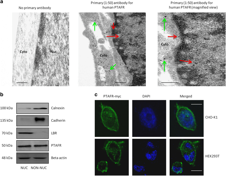
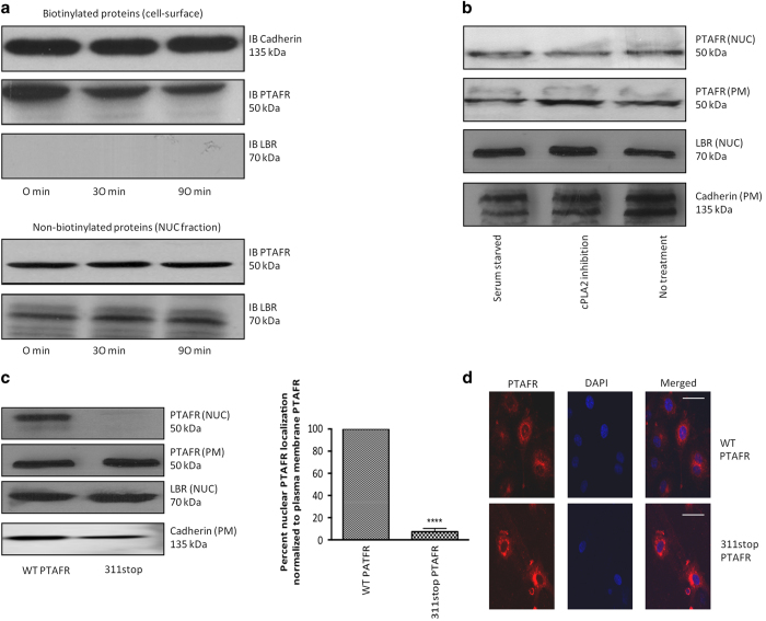
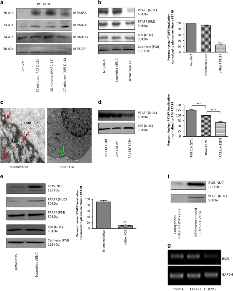
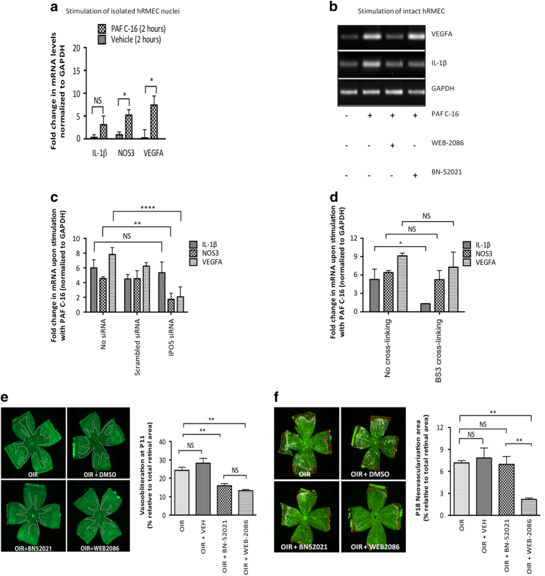
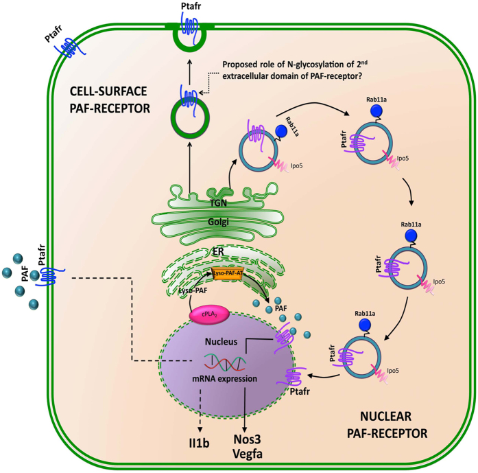
Platelet-activating factor (PAF) is a pleiotropic phospholipid with proinflammatory, procoagulant and angiogenic actions on the vasculature. We and others have reported the presence of PAF receptor (Ptafr) at intracellular sites such as the nucleus. However, mechanisms of localization and physiologic functions of intracellular Ptafr remain poorly understood. We hereby identify the importance of C-terminal motif of the receptor and uncover novel roles of Rab11a GTPase and importin-5 in nuclear translocation of Ptafr in primary human retinal microvascular endothelial cells. Nuclear localization of Ptafr is independent of exogenous PAF stimulation as well as intracellular PAF biosynthesis. Moreover, nuclear Ptafr is responsible for the upregulation of unique set of growth factors, including vascular endothelial growth factor, in vitro and ex vivo. We further corroborate the intracrine PAF signaling, resulting in angiogenesis in vivo, using Ptafr antagonists with distinct plasma membrane permeability. Collectively, our findings show that nuclear Ptafr translocates in an agonist-independent manner, and distinctive functions of Ptafr based on its cellular localization point to another dimension needed for pharmacologic selectivity of drugs.

Keywords: Ptafr; angiogenesis; importin; nuclear GPCR; rab GTPase.


Figures

Figure 1
Figure 1
Nuclear localization of PTAFR is cell-type-specific. (a) TEM on cultured hRMECs. The left panel indicates no primary antibody (negative control) and secondary anti-rabbit nanogold (1:500) showing the lack of nonspecific labeling. The other two panels were treated with respective primary (1:50) and secondary (1:500) antibodies. They show specific immunogold labeling, corresponding to PTAFR in perinuclear (green arrows), nuclear envelope and intranuclear regions (red arrows). The right panel is magnified to show intranuclear localization of the receptor. The TEM images are representative of three independent experiments. Scale bar=0.5 μm. (b) Purity of subcellular (NUC, nuclear; NON-NUC, non-nuclear) fractions of hRMECs. The images are representative of three western blots. Organelle-specific markers used are calnexin for endoplasmic reticulum (ER), cadherin for PM and lamin-B receptor (LBR) for nuclear envelope. Beta actin is found in both the cytoplasm and nucleoplasm and was used as a loading control [111]. PTAFR is also detected in both fractions of hRMECs. (c) Confocal microscopy on HEK293T and CHO-K1 cells stably transfected with PTAFR-myc receptor. Nuclei are stained with DAPI (4′,6-diamidino-2-phenylindole). PTAFR shows nuclear localization in CHO-K1 cells, but not in HEK293T cells. The images are representative of three replicates. Scale bar=20 μm.
Figure 2
Figure 2
Nuclear PTAFR does not arise from PM, and requires C-terminal internalization motif. (a) Biotinylation of hRMECs using PM-impermeable sulfo-NHS-SS-Biotin tag. Following stimulation of with PAF C-16 for indicated time-points, biotinylated proteins were separated by binding to streptavidin beads, whereas supernatant containing non-biotinylated intracellular proteins was further subfractionated into nuclear and non-nuclear parts. IB, immunoblot. The top two panels correspond to PM cadherin and PTAFR, respectively. Nuclear protein, LBR, is absent in biotinylated fraction. The last two panels confirm the presence of LBR and PTAFR in the nuclear fraction. All western blots are representative of three independent experiments. (b) Effect of AACOCF3 (cPLA2 inhibitor [98]) on subcellular localization of PTAFR in hRMECs. There is no difference in localization of PM or nuclear PTAFR following AACOCF3 treatment. LBR and cadherin are used as loading controls for nuclear (NUC) and PM fractions, respectively. (c) Subcellular fractionation of CHO-K1 cells transfected with either wild-type (WT) or 311stop PTAFR. The quantification of three independent western blots (representative blot is shown) using NCBI’s ImageJ software (Bethesda, MD, USA) indicates ~90% reduction in nuclear signal as compared with that at PM (normalized using LBR and cadherin as loading controls for the respective fractions). ****P<0.0001. (d) Confocal microscopy on CHO-K1 cells transfected with WT or 311stop PTAFR. The 311stop PTAFR is present at the cell surface and perinuclear regions, but not at the nucleus. The figures are representative of three replicates. Scale bar=50 μm.
Figure 3
Figure 3
RAB11A and IPO5 govern nuclear localization of PTAFR in hRMECs. (a) Co-immunoprecipitation (co-IP) of PTAFR with three major rabs controlling GPCR trafficking at indicated time-points following PAF C-16 stimulation. RAB5A (top row) co-IPs with only PTAFR following stimulation (early endocytosis [112]). RAB7A (second row) also co-IPs following stimulation and peaks at around 2 h (receptor targeted for degradation [113]). RAB11A (third row) co-IPs with PTAFR at all tested time-points, even in the absence of PAF C-16 stimulation (first column). The last row shows PTAFR as a loading control. (b) Knockdown of RAB11A using specific siRNA in hRMECs. The quantification of three westerns using the ImageJ software reveals ~75% reduction in nuclear immunoreactivity, as compared with that at PM. PM PTAFR is slightly affected (to much lesser extent), possibly due to recycling function of RAB11A. ****P<0.0001. (c) TEM on hRMECs transfected with either scrambled or specific RAB11A siRNAs. RAB11A knockdown specifically affects nuclear localization of PTAFR. Red arrows indicate nuclear labeling, whereas green arrows point labeling at PM. The TEM images are representative of three replicates. Scale bar=0.5 μm. (d) Overexpression of constitutively active (Q70L) or dominant-negative (S25N) RAB11A mutants in hRMECs (heterogeneous expression). The Q70L and S25N mutants resulted in ~125% and ~70% nuclear localization of PTAFR, respectively, as compared with non-transfected hRMECs with endogenous RAB11A levels (set at 100%) and normalized against LBR levels in all nuclear fractions. ****P<0.0001 and ***P<0.001 (e) siRNA-mediated knockdown of IPO5 in hRMECs. PM PTAFR is unaffected. The quantification of western blots reveals >90% reduction in nuclear immunoreactivity, as compared with that at PM. The values were normalized against LBR and cadherin in NUC and PM fractions, respectively. ****P<0.0001. (f) Co-transfection of HEK293T cells with PTAFR and IPO5. The overexpression of both proteins results in nuclear localization of PTAFR in HEK293T cells. All western blots are representative of three independent experiments. (g) Endogenous levels of IPO5 mRNA in hRMECs, CHO-K1 and HEK293T cells. HEK293T cells show negligible endogenous expression of IPO5.
Figure 4
Figure 4
Nuclear PTAFR has functions distinct from its cell surface counterpart and the former affects retinal neovascularization in OIR. *P<0.05, **P<0.01 and ****P<0.0001. (a) Stimulation of freshly isolated nuclei from hRMECs with PAF C-16 (100 nm for 30 min). PAF C-16 causes significant augmentation of NOS3 (P=0.015) and VEGFA (P=0.019), but not IL1B (P=0.071) levels. All values are represented as mean±s.d. The gene expression was analyzed using four independent replicates. RNA was isolated 120 min after stimulation in all conditions and gene expression was analyzed by qRT-PCR. (b) Stimulation of cultured hRMECs with PAF C-16 (100 nm for 30 min) with or without pre-treatment using indicated PTAFR antagonist for 30 min. The pre-treatment with membrane-permeable antagonist (100 nm WEB-2086) inhibits PAF-induced IL1B and VEGFA expression. The non-permeable antagonist (10 μm BN-52021), on the other hand, only attenuates PAF-induced IL1B, but not VEGFA mRNA levels. RNA was isolated 120 min after stimulation and analyzed by RT-PCR. (c) Effect of IPO5 knockdown on PAF-induced gene expression. IPO5 siRNA treatment significantly reduces PAF-induced augmentation of NOS3 (P=0.004) and VEGFA (P<0.0001) levels, but has no effect on IL1B (P=0.844) levels. (d) Effect of chemical crosslinking on PAF-induced gene expression. The crosslinking prevented upregulation of PAF-induced IL1B (P=0.013), but had no significant difference on NOS3 (P=0.997) and VEGFA (P=0.385) levels. (e) Retinal VO using the hyperoxia model of OIR in rats. The OIR resulted in central retinal VO at P11, as delineated by white margins in lectin-stained retinal flat-mounts (top left panel), and this was prevented by systemic administration of either Ptafr antagonists (five injections from P6 to P10) BN-52021 (P=0.006) and WEB-2086 (P=0.003) (two bottom panels), but not by the vehicle treatment (top right panel) (P=0.458); there was no statistically significant difference between the two antagonists (P>0.999). The Y axis in graph represents % of avascular retina at P11 relative to the total area (n=5–8 retinas per group, NS=not significant). (f) Retinal neovascularization using cycling model of OIR in rats. OIR-induced retinal neovascularization (top left panel) is reduced by the administration of membrane-permeable WEB-2086 (bottom right panel) (P=0.006), but not that of either impermeable BN-52021 (bottom left panel) (P>0.999) or the vehicle (top right panel) (P>0.999). The Y axis in graph represents % of retina with neovascularization (quantified with SWIFT-NV [110]) at P18 relative to the total area (n=5–8 retinas per group, NS, not significant).
Figure 5
Figure 5
Schematic diagram showing intracellular trafficking of PTAFR. Once synthesized in ER and glycosylated in TGN, Rab11a and Ipo5 together control (pathway highlighted by blue vesicles) nuclear localization of Ptafr, possibly directly from TGN. The nuclear PTAFR can be activated by the production of local PAF from membrane phospholipids by nuclear cPLA2 [72] and ER-localized Lyso-PAF-acetyltransferase (Lyso-PAF-AT) [73]. The nuclear Ptafr, in turn, activates the expression of proangiogenic genes such as Nos3, Vegfa (indicated by solid line), whereas PM Ptafr regulated the expression of proinflammatory cytokines such as Il1b (indicated by dotted line). The role of N-glycosylation in the second extracellular domain of the receptor has been proposed in the cell surface targeting of Ptafr from TGN [114].

References

    1. Barbaro JF, Zvaifler NJ. Antigen induced histamine release from platelets of rabbits producing homologous PCA antibody. Proc Soc Exp Biol Med 1966; 122: 1245–1247. - PubMed
    1. Ishii S, Nagase T, Tashiro F et al. Bronchial hyperreactivity, increased endotoxin lethality and melanocytic tumorigenesis in transgenic mice overexpressing platelet-activating factor receptor. EMBO J 1997; 16: 133–142. - PMC - PubMed
    1. Stafforini DM, Numao T, Tsodikov A et al. Deficiency of platelet-activating factor acetylhydrolase is a severity factor for asthma. J Clin Invest 1999; 103: 989–997. - PMC - PubMed
    1. Cundell DR, Gerard NP, Gerard C, Idanpaan-Heikkila I, Tuomanen EI. Streptococcus pneumoniae anchor to activated human cells by the receptor for platelet-activating factor. Nature 1995; 377: 435–438. - PubMed
    1. Harris ES, Rondina MT, Schwertz H, Weyrich AS, Zimmerman GA. Pathogenesis of sepsis and sepsis-induced acute lung injury. Acute Respiratory Distress Syndrome 2010; 2: 369–419.