

Effects of modified electroconvulsive therapy on the electroencephalogram of schizophrenia patients
- PMID: 27462511
- PMCID: PMC4942443
- DOI: 10.1186/s40064-016-2747-7
Effects of modified electroconvulsive therapy on the electroencephalogram of schizophrenia patients
Abstract
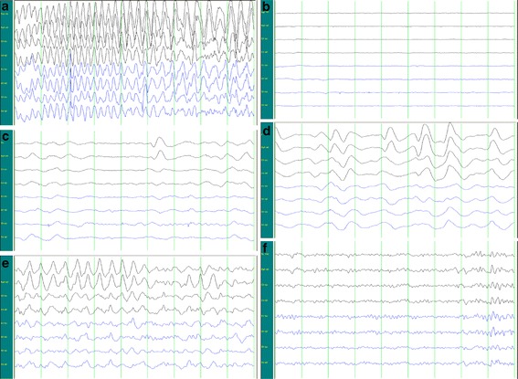
Background: This study aimed to investigate the modified electroconvulsive therapy (MECT) on the electroencephalogram (EEG) of schizophrenia patients. A total of 26 schizophrenia patients who received MECT were recruited. EEG recording was initiated at 30 min before 1st and 6th MECT and terminated on the 2nd day. Images without artifacts were selected for the analysis of δ, θ, α1, α2 and β bands. The wave energy at each frequency, index of waves at different bands from the same lead, index of waves at the same band from different leads, time of epileptic discharge, time of resting state, and time to the stable EEG were determined and compared.
Results: The energy of slow waves increased. α waves reduced, but θ waves increased in the frontotemporal area. The index of θ waves increased. After resting state, brainwaves first occurred in the frontal area. Significant difference was observed in the time to waves returning to normal (P = 0.05).
Conclusions: After MECT, the θ waves in the same lead increases, and its energy also elevates; α wave in the frontotemporal area reduces; there is transient reduction in cerebral function during MECT. After electric resting state, brainwaves mainly occur in the frontal area, and the time to brainwaves returning to normal reduces over time after MECT.
Keywords: Electroencephalogram; Modified electroconvulsive therapy; Schizophrenia.
Figures
Similar articles
-
The influence of EEG channels and features significance on automatic detection of epileptic waves in MECT.Comput Methods Biomech Biomed Engin. 2024 Sep;27(12):1633-1648. doi: 10.1080/10255842.2023.2252952. Epub 2023 Sep 5. Comput Methods Biomech Biomed Engin. 2024. PMID: 37668087
-
Effects of modified electroconvulsive therapy on the cognitive function and blood parameters in female patients with schizophrenia.Int J Clin Exp Med. 2015 Jan 15;8(1):1349-55. eCollection 2015. Int J Clin Exp Med. 2015. PMID: 25785136 Free PMC article.
-
Increased resting-state global functional connectivity density of default mode network in schizophrenia subjects treated with electroconvulsive therapy.Schizophr Res. 2018 Jul;197:192-199. doi: 10.1016/j.schres.2017.10.044. Epub 2017 Nov 6. Schizophr Res. 2018. PMID: 29117910
-
Sex differences in factors influencing hospital-acquired pneumonia in schizophrenia patients receiving modified electroconvulsive therapy.Front Psychiatry. 2023 Feb 14;14:1127262. doi: 10.3389/fpsyt.2023.1127262. eCollection 2023. Front Psychiatry. 2023. PMID: 36865072 Free PMC article.
-
The visual scoring of sleep and arousal in infants and children.J Clin Sleep Med. 2007 Mar 15;3(2):201-40. J Clin Sleep Med. 2007. PMID: 17557427 Review.
Cited by
-
Correlation between Post-Acute Electroconvulsive Therapy Alpha-Band Spectrum Power Increase and Improvement of Psychiatric Symptoms.J Pers Med. 2021 Dec 6;11(12):1315. doi: 10.3390/jpm11121315. J Pers Med. 2021. PMID: 34945787 Free PMC article.
-
How Electroconvulsive Therapy Works?: Understanding the Neurobiological Mechanisms.Clin Psychopharmacol Neurosci. 2017 Aug 31;15(3):210-221. doi: 10.9758/cpn.2017.15.3.210. Clin Psychopharmacol Neurosci. 2017. PMID: 28783929 Free PMC article. Review.
References
-
- Burnouf S, Martire A, Derisbourg M, Laurent C, Belarbi K, Leboucher A, Fernandez-Gomez FJ, Troquier L, Eddarkaoui S, Grosjean ME, Demeyer D, Muhr-Tailleux A, Buisson A, Sergeant N, Hamdane M, Humez S, Popoli P, Buee L, Blum D. NMDA receptor dysfunction contributes to impaired brain-derived neurotrophic factor-induced facilitation of hippocampal synaptic transmission in a Tau transgenic model. Aging Cell. 2013;12(1):11–23. doi: 10.1111/acel.12018. - DOI - PubMed
LinkOut - more resources
Full Text Sources
Other Literature Sources
