. 2016 Aug 4;536(7614):76-80.
doi: 10.1038/nature18944. Epub 2016 Jul 27.

A novel excitatory network for the control of breathing

A novel excitatory network for the control of breathing

Tatiana M Anderson et al. Nature. .

Abstract

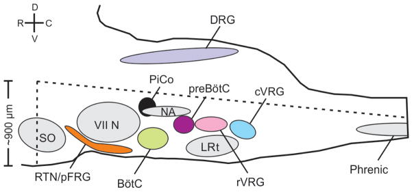
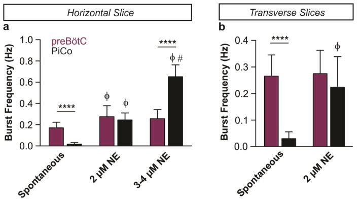
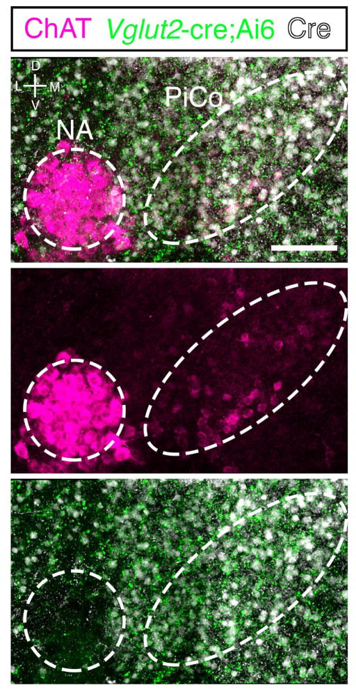
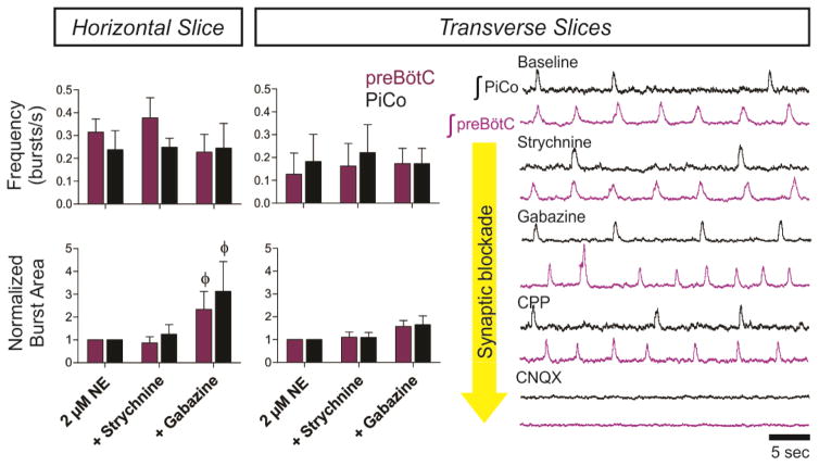
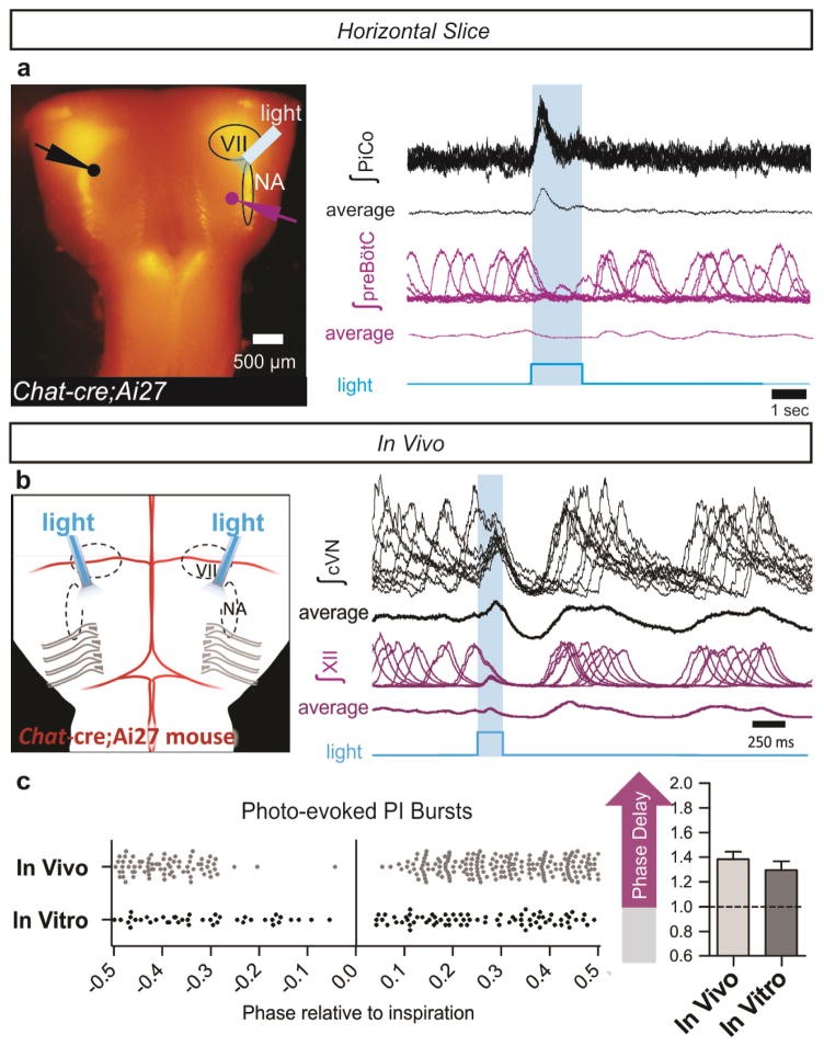
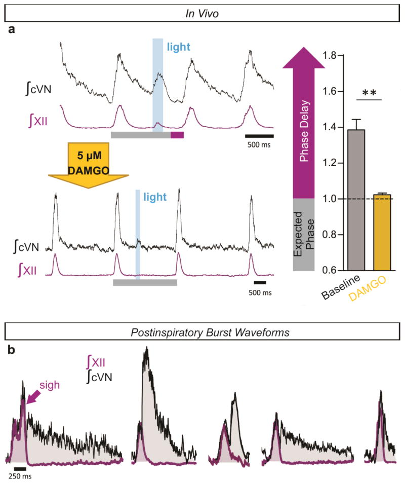
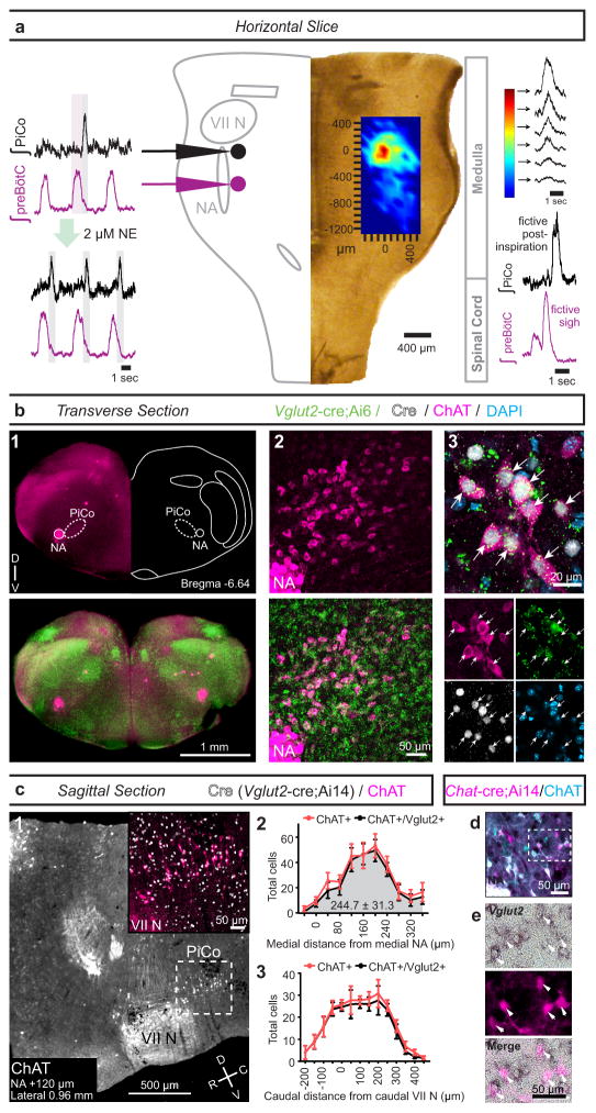
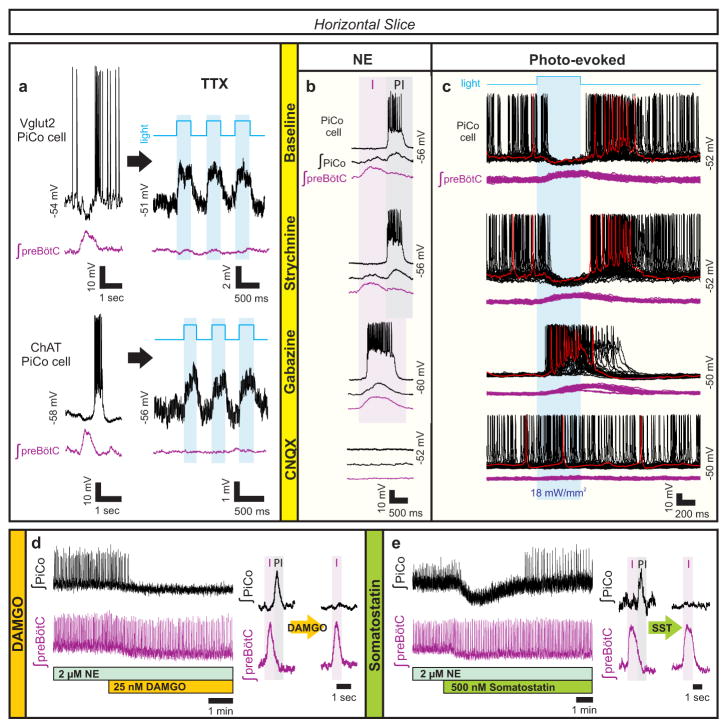
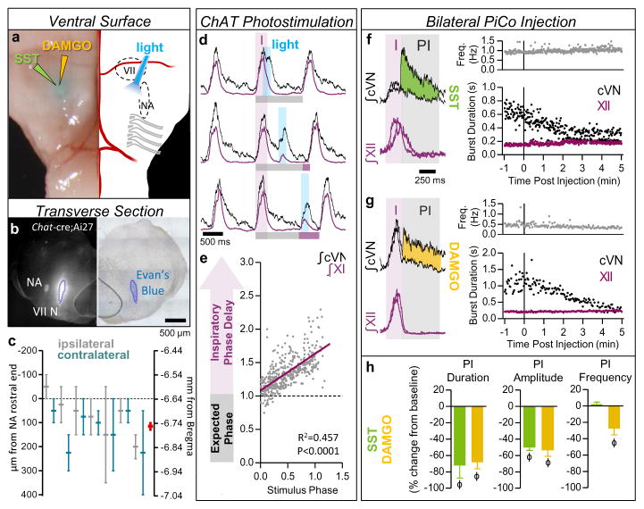
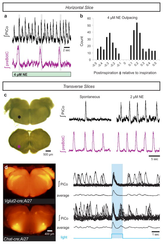
Breathing must be tightly coordinated with other behaviours such as vocalization, swallowing, and coughing. These behaviours occur after inspiration, during a respiratory phase termed postinspiration. Failure to coordinate postinspiration with inspiration can result in aspiration pneumonia, the leading cause of death in Alzheimer's disease, Parkinson's disease, dementia, and other neurodegenerative diseases. Here we describe an excitatory network that generates the neuronal correlate of postinspiratory activity in mice. Glutamatergic-cholinergic neurons form the basis of this network, and GABA (γ-aminobutyric acid)-mediated inhibition establishes the timing and coordination relative to inspiration. We refer to this network as the postinspiratory complex (PiCo). The PiCo has autonomous rhythm-generating properties and is necessary and sufficient for postinspiratory activity in vivo.The PiCo also shows distinct responses to neuromodulators when compared to other excitatory brainstem networks. On the basis of the discovery of the PiCo, we propose that each of the three phases of breathing is generated by a distinct excitatory network: the pre-Bötzinger complex, which has been linked to inspiration; the PiCo, as described here for the neuronal control of postinspiration; and the lateral parafacial region (pF(L)), which has been associated with active expiration, a respiratory phase that is recruited during high metabolic demand.


Conflict of interest statement

The authors declare no competing financial interests.

Figures

Extended Data Figure 1
Extended Data Figure 1. Schematic of the horizontal slice from a sagittal view that retains the medullary ventral respiratory column in the brainstem
Dotted lines represent approximate boundaries of the horizontal slice. Slice retains part of the superior olive (SO), and the entire retrotrapezoidal nucleus/para-facial respiratory group (RTN/pFRG), facial nucleus (VII N), Bötzinger Complex (BötC), Post-inspiratory Complex (PiCo), Nucleus ambiguus (NA), preBötzinger Complex (preBötC), lateral reticular nucleus (LRT), and the rostral and caudal ventral respiratory groups (rVRG and cVRG, respectively). The slice also retains a portion of the spinal cord and includes part of the phrenic motor nucleus (approximately cervical segment 3 and 4). The slice does not contain the dorsal portion of the medulla including the dorsal respiratory group (DRG). Legend: dorsal (D), ventral (V), rostral (R), caudal (C).
Extended Data Figure 2
Extended Data Figure 2. NE dose response of preBötC and PiCo rhythms in horizontal and transverse slices
The frequency of the PiCo rhythm (black) is highly sensitive to the application of low concentrations of NE while the preBötC rhythm (purple) stays relatively constant in both types of slice preparations. a, In horizontal slices the PiCo rhythm is slow under spontaneous conditions (n=10), the two rhythms have similar burst frequencies in 2 μM NE (n=6), and the PiCo rhythm significantly outpaces the preBötC rhythm under higher concentrations (n=4, 3–4 μM NE). b, Similarly, when isolated in transverse slices, the PiCo rhythm has a slow frequency under spontaneous conditions (n=10), and the preBötC and PiCo have similar frequencies at 2 μM NE (n=7). (mean ± s.e.m.) Two-way ANOVA followed by a Bonferroni post hoc test. ****P < 0.0001 comparing PiCo to preBötC, ϕ P<0.05 compared to baseline (Spon.), # P<0.05 compared to 2 μM NE.
Extended Data Figure 3
Extended Data Figure 3. NA neurons lack Vglut2-cre expression
High magnification view at the level of PiCo from a Vglut2-cre;Ai6 (ZsGreen1; green) mouse immunolabled with ChAT antibody (magenta) and Cre antibody (white). Note lack of green Vglut2-cre expression in the NA. Scale bar 100 μm.
Extended Data Figure 4
Extended Data Figure 4. Progressive synaptic blockade in horizontal and transverse slices
(Left) Graphs comparing frequency and normalized burst area between horizontal (n=5) and paired transverse slices (n=5) after the application of strychnine and gabazine. In both horizontal and paired transverse slices, PiCo and preBötC rhythms have nearly identical burst frequencies in the presence of gabazine (top graphs). The burst area of both rhythms also significantly increases with the application of gabazine in both slice preparations (bottom graphs). Two-way ANOVA followed by a Bonferroni post hoc test. ϕ P<0.05 compared to baseline (2 μM NE). (Right) Synaptic blockers were progressively perfused over paired transverse slices at 10 minute intervals. Both PiCo and preBötC rhythms persist in the presence of 1 μM strychnine, 10 μM gabazine, and 10 μM CPP. Population rhythms ceased in the presence of 20 μM CNQX, indicating that both rhythms are excitatory (n=5). The asterisk denotes a characteristic sigh in the preBötC trace.
Extended Data Figure 5
Extended Data Figure 5. Perievent interval between preBötC and PiCo bursts during inhibitory block in the horizontal slice
a, Peri-event interval, time between peak of preBötC and PiCo bursts, is constant in strychnine; however, gabazine initiates progressive synchronization between rhythms shown here in a representative experiment. b, Average peri-event intervals at baseline and after sequential application of strychnine and gabazine (n=6, mean ± s.e.m.). Repeated measures Friedman test followed by Dunn’s multiple comparisons post-hoc test. *P < 0.05.
Extended Data Figure 6
Extended Data Figure 6. Blocking muscarinic and nicotinic acetylcholine receptors does not abolish the PiCo rhythm
a, Raw population bursts from PiCo and contralateral preBötC with the progressive addition of 1 μM mecamylamine (nicotinic receptor antagonist), 10 μM atropine (muscarinic receptor antagonist), and 4 μM norepinephrine. b, The left two graphs show n=5 experiments in which atropine was applied first, and the right graphs illustrate n=3 experiments in which mecamylamine was applied first. Blockade of muscarinic receptors results in a larger decrease in PiCo burst frequency than blocking nicotinic receptors, while preBötC frequency does not change significantly (top graphs). Interestingly, blockade of muscarinic receptors increases the amplitude of PiCo bursts (bottom graphs). The PiCo rhythm persists after concurrent blockade of both types of acetylcholine receptors, and PiCo burst frequency rebounds to near baseline levels when an additional 2 μM NE is applied (total 4 μM NE; top graphs). (mean ± s.e.m.). Two-way ANOVA followed by a Bonferroni post hoc test. **P < 0.01, *P < 0.05 comparing preBötC to PiCo, ϕ P<0.05 compared to baseline (2 μM NE).
Extended Data Figure 7
Extended Data Figure 7. Synaptically isolated PiCo neurons decrease firing frequency in the presence of DAMGO
a, Top traces show intracellular recordings from PiCo cells with concurrent extracellular preBötC population activity from a horizontal slice under 1 μM NE baseline conditions. Bottom traces show the same recordings after blocking fast synaptic transmission (1 μM strychnine, 10 μM gabazine, 10 μM CPP, 20 μM CNQX) to synaptically isolate the PiCo neuron. Application of 10 nM DAMGO decreases the cell’s intrinsic firing frequency. b, Quantified data show that DAMGO significantly decreases action potential (AP) firing frequency of synaptically isolated PiCo neurons both in horizontal slices (black dots) and transverse PiCo slices (gray dots) (two-tailed paired t-test, *P < 0.05; n=5).
Extended Data Figure 8
Extended Data Figure 8. Differential PiCo and preBötC population responses to DAMGO and SST in horizontal and transverse slices
a, After the application of 25 nM DAMGO, preBötC burst frequency only slightly decreases (n=5), whereas PiCo bursting is nearly eliminated. b, Similar to results observed in horizontal slices, the PiCo rhythm is eliminated by 25 nM DAMGO in transverse slices that isolate PiCo and preBötC in the presence of 2 μM NE (n=5). Periodic large amplitude bursts in the bottom preBötC trace are fictive sighs. c, DAMGO dose response of normalized preBötC and PiCo burst frequency in transverse slices, illustrating the differential sensitivity of the PiCo and preBötC rhythms to DAMGO; burst frequency values are normalized to baseline frequency in 2 μM NE. (mean ± s.e.m., n=8 with minimum replicates of 4 for each location and concentration). d, The PiCo rhythm is selectively and transiently inhibited by the application of 500 nM SST whereas the preBötC rhythm persists in horizontal slices. Graph shows normalized average burst frequencies of both rhythms at baseline, 1.5–3.5 minutes after SST application, and 8–10 minutes after SST application (n=6). e, Similar to the horizontal slice, SST application results in a robust inhibition of PiCo bursting in paired transverse slices. f, Similar to d, complied normalized burst frequencies for n=6 transverse slices before and after SST application. (mean ± s.e.m.) Two-way ANOVA followed by a Bonferroni post hoc test. ****P < 0.0001 comparing PiCo to preBötC, ϕ P<0.05 compared to baseline.
Extended Data Figure 9
Extended Data Figure 9. Light stimulation of cholinergic cells evokes postinspiratory activity in horizontal slices and in vivo
a, Two population electrodes were placed at the level of PiCo (black dot and trace) and contralateral preBötC (purple dot and trace) in a Chat-cre;Ai27 horizontal slice. Under spontaneous conditions (no NE), cholinergic neurons expressing channelrhodopsin-2 were light activated with a fiber optic (labeled ‘light’) placed over PiCo ipsilateral to the preBötC electrode. PiCo population bursts were triggered upon the onset of a 1.5 second light pulse while no bursts were light evoked in the preBötC (n=6). Figure shows 10 traces overlaid for each electrode with averaged traces below from a representative experiment. b, Photo-stimulating PiCo in adult anesthetized Chat-cre;Ai27 mice reliably triggers cVN bursts. Figure shows 10 traces overlaid with averages below of cVN and XII activity during a 200 ms light stimulation of PiCo. c, Postinspiratory bursts can be photo-evoked both in vivo (n=6) and in vitro (n=6) at any phase except for during inspiration and just prior to inspiration (bottom left) due to the inspiratory phase delay that occurs when PiCo is stimulated (mean ± s.e.m., bottom right). NA= Nucleus ambiguus, VII= facial nucleus.
Extended Data Figure 10
Extended Data Figure 10. Elimination of phase delay by DAMGO and a diversity of postinspiratory waveforms in vivo
a, Injection of 5 μM DAMGO into PiCo eliminates the phase delay elicited by photostimulation of PiCo in Chat-cre;Ai27 mice. A representative experiment showing cVN and XII recordings during a 200 ms light pulse before and after injection of PiCo with DAMGO (left; gray bars = expected phase, purple bars = inspiratory phase delay) and the average inspiratory phase delay (right) (mean ± s.e.m., two-tailed paired t-test, **P<0.01; n=6). b, A diversity of postinspiratory vagal waveforms were recorded in vivo. Five examples of cVN (black) and XII (purple) recordings (overlaid) show that postinspiratory activity can vary from large decrementing patterns to small short bursts, potentially representing the neural basis for a variety of postinspiratory behaviors.
Figure 1
Figure 1. Horizontal slice and anatomy of PiCo
a, Population bursts PiCo (black), preBötC (purple). 2 μM NE stimulates PiCo bursts (n=23). Left, schematic; right, heat map of PiCo burst amplitude; legend (n=6). PiCo bursts follow fictive sighs. b1, Immunohistochemical labeling of PiCo dorsomedial to NA. b2, Higher magnification ChAT, Vglut2-cre;Ai6 (ZsGreen1) colocalization within PiCo. b3, Arrows: higher magnification triple-labeled ChAT+ ZsGreen1+ Cre+ PiCo cells (n=5). c1, Sagittal view, ChAT+ PiCo neurons (dashed-box). Inset, magnified dashed-box, colocalization of ChAT+, Cre+ PiCo neurons in Vglut2-cre;Ai14 mice. c2,3, Quantification of ChAT- and Vglut2-expressing PiCo cells mediolaterally from NA (244.7±31.3 average total # cells; n=5) c2, and rostrocaudally from VII N (n=4) c3, Bars, mean ± sem. d, ChAT+ PiCo neurons, Chat-cre;Ai14 mice. e, Magnified box (d) Arrowheads, Vglut2 mRNA in Chat-derived PiCo neurons.
Figure 2
Figure 2. Glutamatergic, cholinergic PiCo cells and role of synaptic inhibition
a, Intracellular PiCo recordings in Vglut2-cre;Ai27 (n=3) and Chat-cre;Ai27 (n=5) horizontal slices. Photo-stimulation after TTX depolarizes membrane potential. b, PiCo, preBötC in progressive synaptic block (n=5). Rhythms synchronized in gabazine, bursting abolished in CNQX. c, PiCo cell in Dbx1-cre;Ai27 horizontal slice with progressive synaptic block. PiCo cell inhibited during photo-evoked inspiratory burst in NE/strychnine, bursts with light-stimulation in gabazine, ceases bursting in CNQX (500 ms light, 40 sweeps, n=4). d, PiCo bursting eliminated by 25 nM DAMGO; representative bursts (n=5). e, PiCo inhibited by 500 nM SST; representative bursts (n=6).
Figure 3
Figure 3. Stimulation and inhibition of PiCo in vivo
a, Left, brightfield image; right, schematic; ventral sites for bilateral photo-stimulation, injection of SST/DAMGO in vivo. b, Representative dye injection at PiCo level. Left, Chat-cre;Ai27; right, brightfield. c, Dye-spread (n=7), centered at PiCo (0–200 μm caudal from rostral NA). Ipsilateral (grey), contralateral (teal) injections, bars ±max/min; pooled data in red, mean ± s.e.m. d, PiCo photo-stimulation in adult Chat-cre;Ai27 mice evokes vagal bursts and delays subsequent inspiration (grey bars = expected inspiratory phase, purple bars = inspiratory phase delay) e, Quantification of inspiratory phase delay (n=6); magnitude dependent on stimulus phase (slope: 0.549, linear regression analysis). f,g, Injection of SST or DAMGO progressively decreases cVN, not XII burst duration. h, Postinspiratory burst duration, amplitude, and frequency following injection of SST (n=3), DAMGO (n=4). Two-tailed paired t-test, ϕ P<0.05 compared to baseline; mean ± s.e.m.
Figure 4
Figure 4. PiCo is an autonomous, rhythm generating network
a, In 3–4 μM NE, PiCo outpaces the preBötC rhythm, but still bursts in the postinspiratory phase (gray bars) in horizontal slices. b, PiCo bursts occur in any phase except during inspiration (inspiratory peak=0; count=400 bursts; n=4). c, PiCo and preBötC isolated in transverse in vitro slices; PiCo bursting stimulated by 2 μM NE (n=33). d, Light stimulation evokes a burst in Vglut2-cre;Ai27 rostral transverse slices (91.3 ± 5.1% of stimulations, mean ± s.e.m., n=4) and Chat-cre;Ai27 slices (91.9 ± 1.8% of stimulations, mean ± s.e.m., n=6). 1.5 second light pulse. Top traces: 10 sweep overlay, bottom traces: sweep average.

References

    1. Richter DW, Smith JC. Respiratory rhythm generation in vivo. Physiology (Bethesda) 2014;29:58–71. doi: 10.1152/physiol.00035.2013. - DOI - PMC - PubMed
    1. Troche MS, Brandimore AE, Godoy J, Hegland KW. A framework for understanding shared substrates of airway protection. J Appl Oral Sci. 2014;22:251–260. - PMC - PubMed
    1. Smith JC, Ellenberger HH, Ballanyi K, Richter DW, Feldman JL. Pre-Botzinger complex: a brainstem region that may generate respiratory rhythm in mammals. Science (New York, NY) 1991;254:726–729. - PMC - PubMed
    1. Huckstepp RT, Cardoza KP, Henderson LE, Feldman JL. Role of parafacial nuclei in control of breathing in adult rats. J Neurosci. 2015;35:1052–1067. doi: 10.1523/JNEUROSCI.2953-14.2015. - DOI - PMC - PubMed
    1. Pagliardini S, et al. Active expiration induced by excitation of ventral medulla in adult anesthetized rats. J Neurosci. 2011;31:2895–2905. doi: 10.1523/JNEUROSCI.5338-10.2011. - DOI - PMC - PubMed

Publication types