

Correlation of Thyroid Imaging Reporting and Data System [TI-RADS] and fine needle aspiration: experience in 1,000 nodules
- PMID: 27462883
- PMCID: PMC4943343
- DOI: 10.1590/S1679-45082016AO3640
Correlation of Thyroid Imaging Reporting and Data System [TI-RADS] and fine needle aspiration: experience in 1,000 nodules
Abstract
Objective: To correlate the Thyroid Imaging Reporting and Data System (TI-RADS) and the Bethesda system in reporting cytopathology in 1,000 thyroid nodules.
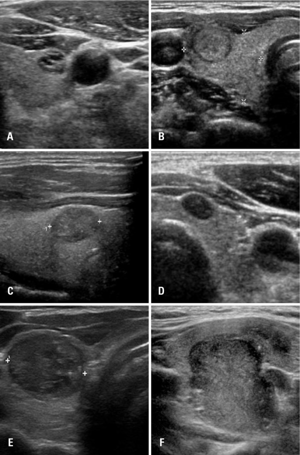
Methods: A retrospective study conducted from November 2011 to February 2014 that evaluated 1,000 thyroid nodules of 906 patients who underwent ultrasound exam and fine needle aspiration.
Results: A significant association was found between the TI-RADS outcome and Bethesda classification (p<0.001). Most individuals with TI-RADS 2 or 3 had Bethesda 2 result (95.5% and 92.5%, respectively). Among those classified as TI-RADS 4C and 5, most presented Bethesda 6 (68.2% and 91.3%, respectively; p<0.001). The proportion of malignancies among TI-RADS 2 was 0.8%, and TI-RADS 3 was 1.7%. Among those classified as TI-RADS 4A, proportion of malignancies was 16.0%, 43.2% in 4B, 72.7% in 4C and 91.3% among TI-RADS 5 (p<0.001), showing clear association between TI-RADS and biopsy results.
Conclusion: The TI-RADS is appropriate to assess thyroid nodules and avoid unnecessary fine needle aspiration, as well as to assist in making decision about when this procedure should be performed.
Objetivo: Apresentar a correlação entre o Thyroid Imaging Reporting and Data System (TI-RADS) e o sistema Bethesda, para relatar citopatologia em 1.000 nódulos tireoidianos.
Métodos: Estudo retrospectivo realizado no período de novembro de 2011 a fevereiro de 2014, que avaliou 1.000 nódulos tireoidianos de 906 pacientes submetidos a exame de ultrassonografia e à punção aspirativa por agulha fina.
Resultados: Observou-se associação significativa entre o TI-RADS e o resultado da classificação de Bethesda (p<0,001). A maioria dos indivíduos com TI-RADS 2 ou 3 teve resultado citológico Bethesda 2 (95,5% e 92,5%, respectivamente). Entre aqueles classificados TI-RADS 4C e 5, a maioria teve resultado Bethesda 6 (68,2% e 91,3%, respectivamente; p<0,001). A proporção de malignidades em TI-RADS 2 foi 0,8% e em TI-RADS 3 foi 1,7%. Entre TI-RADS 4A, foi de 16,0%, 43,2% em 4B, 72,7% em 4C e em 5 foi de 91,3% (p<0,001), mostrando clara associação entre o TI-RADS e os resultados da biópsia.
Conclusão: O TI-RADS é apropriado para avaliar nódulos da tireoide e evitar punção aspirativa por agulha fina desnecessária, além de auxiliar na decisão sobre quando este procedimento deve ser realizado.
Conflict of interest statement
Conflict of interest: none.
Figures

References
-
- Frates MC, Benson CB, Charboneau JW, Cibas ES, Clark OH, Coleman BG, Cronan JJ, Doubilet PM, Evans DB, Goellner JR, Hay ID, Hertzberg BS, Intenzo CM, Jeffrey RB, Langer JE, Larsen PR, Mandel SJ, Middleton WD, Reading CC, Sherman SI, Tessler FN, Society of Radiologists in Ultrasound Management of thyroid nodules detected at US: Society of Radiologists in US consensus conference statement. Radiology. 2005;237(3):794–800. - PubMed
-
- Hoang JK, Lee WK, Lee M, Johnson D, Farrell S. US features of thyroid malignancy: pearls and pitfalls. Radiographics. 2007;27(3):847–860. discussion 861-5. Review. - PubMed
-
- Horvath E, Majlis S, Rossi R, Franco C, Niedmann JP, Castro A, et al. An ultrasonogram reporting system for thyroid nodules stratifying cancer risk for clinical management. J Clin Endocrinol Metab. 2009;94(5):1748–1751. - PubMed
-
- Kwak JY, Han KH, Yoon JH, Moon HJ, Son EJ, Park SH, et al. Thyroid imaging reporting and data system for US features of nodules: a step in establishing better stratification of cancer risk. Radiology. 2011;260(3):892–899. - PubMed
-
- Chammas MC, Gerhard R, Oliveira IR, Widman A, Barros N, Durazzo M, et al. Thyroid nodules: evaluation with power Doppler and duplex Doppler ultrasound. Otolaryngol Head Neck Surg. 2005;132(6):874–882. - PubMed
MeSH terms
LinkOut - more resources
Full Text Sources
Other Literature Sources
