. 2016 Jul 27;12(7):e1006192.
doi: 10.1371/journal.pgen.1006192. eCollection 2016 Jul.

Tracking the Fragile X Mental Retardation Protein in a Highly Ordered Neuronal RiboNucleoParticles Population: A Link between Stalled Polyribosomes and RNA Granules

Affiliations

Tracking the Fragile X Mental Retardation Protein in a Highly Ordered Neuronal RiboNucleoParticles Population: A Link between Stalled Polyribosomes and RNA Granules

Rachid El Fatimy et al. PLoS Genet. .

Abstract

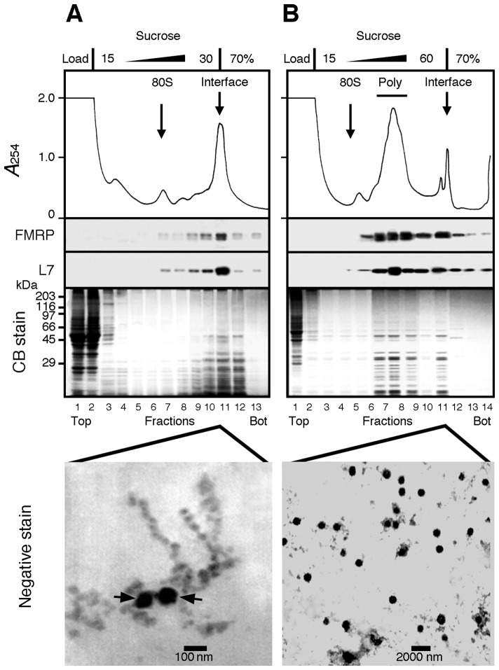
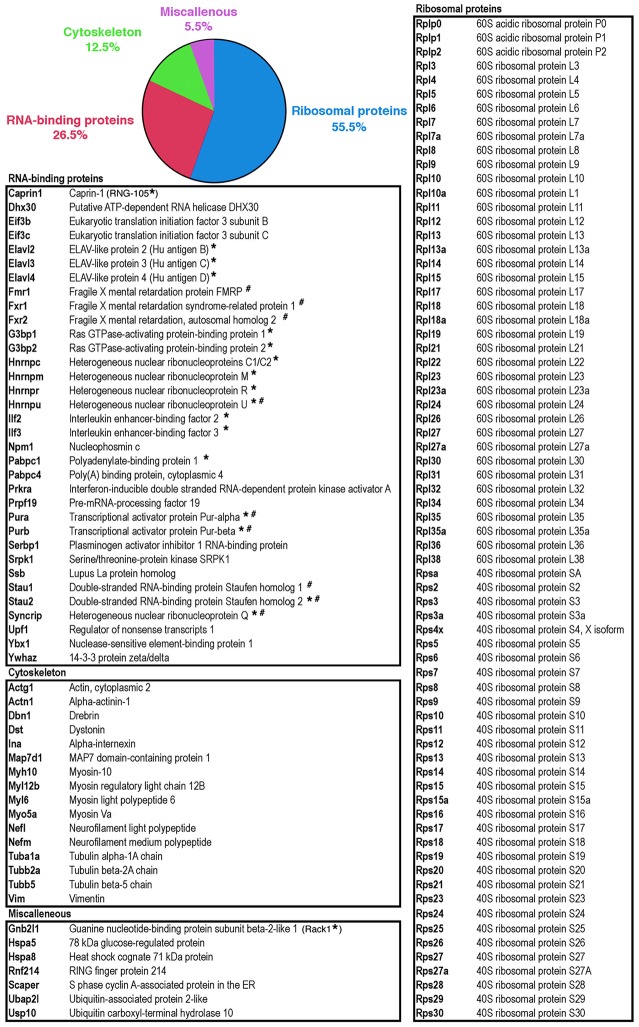
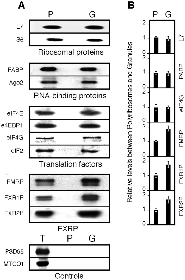
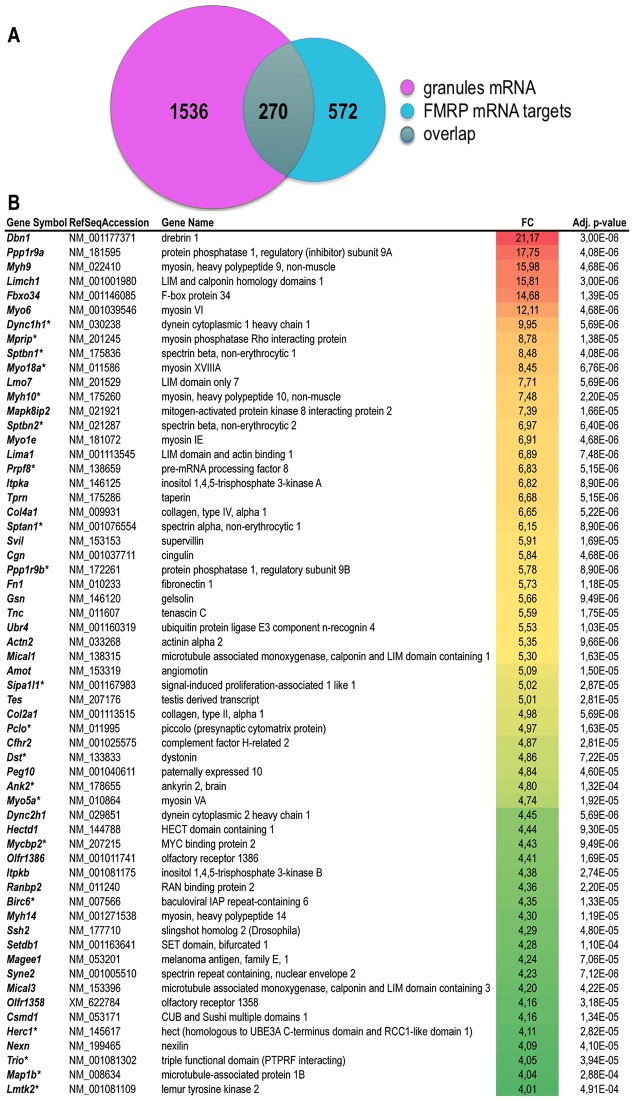
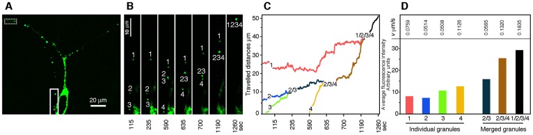
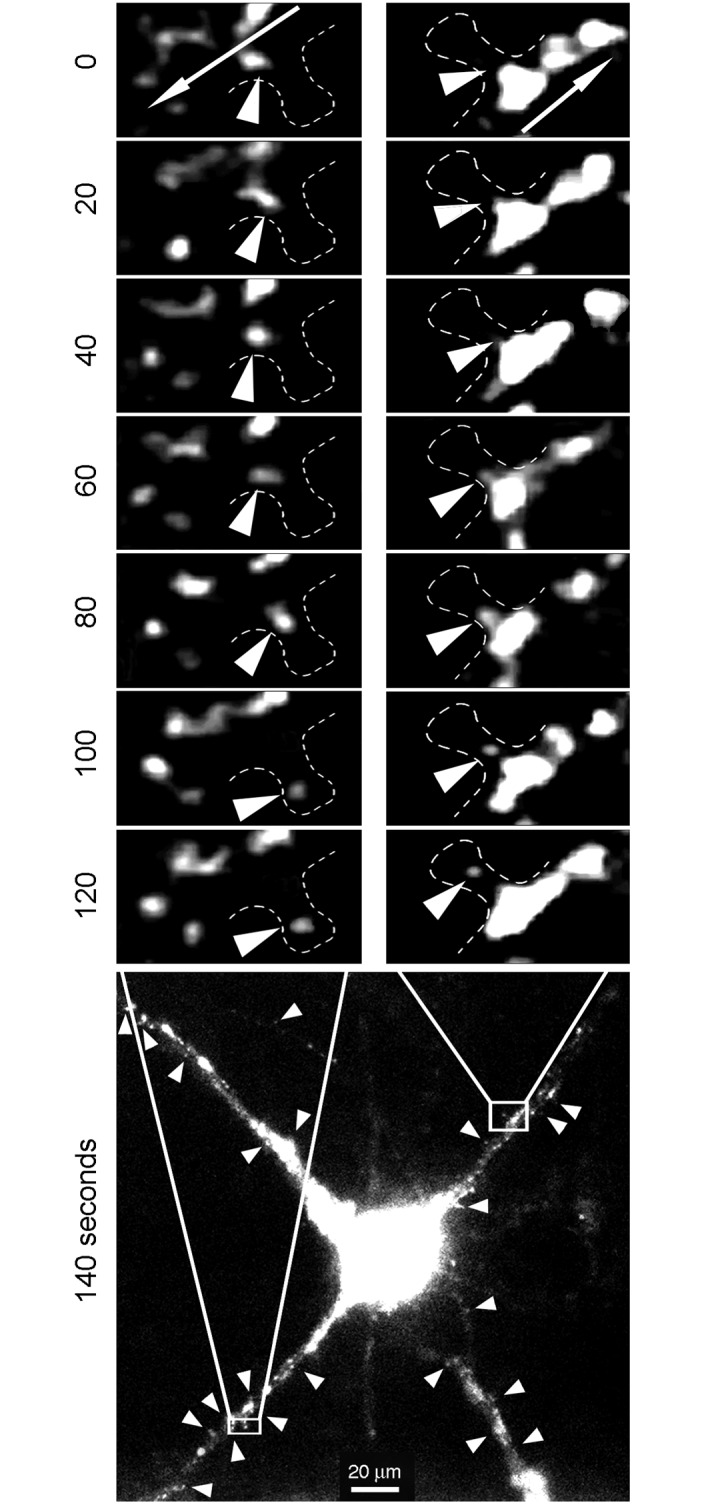
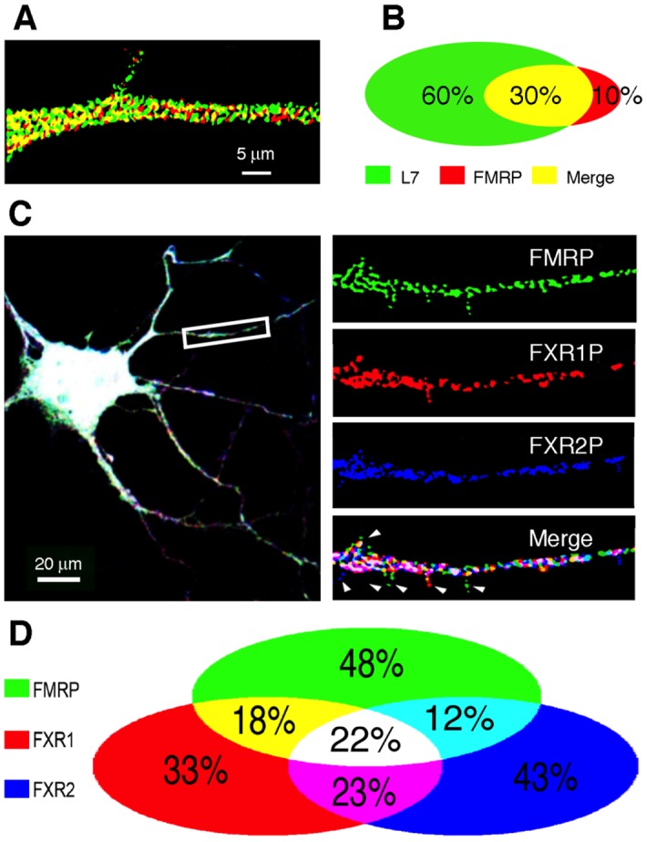
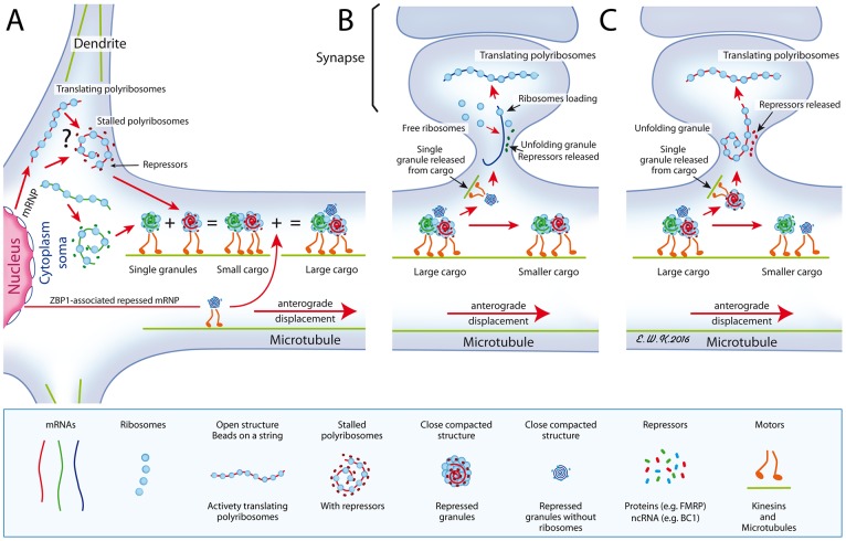
Local translation at the synapse plays key roles in neuron development and activity-dependent synaptic plasticity. mRNAs are translocated from the neuronal soma to the distant synapses as compacted ribonucleoparticles referred to as RNA granules. These contain many RNA-binding proteins, including the Fragile X Mental Retardation Protein (FMRP), the absence of which results in Fragile X Syndrome, the most common inherited form of intellectual disability and the leading genetic cause of autism. Using FMRP as a tracer, we purified a specific population of RNA granules from mouse brain homogenates. Protein composition analyses revealed a strong relationship between polyribosomes and RNA granules. However, the latter have distinct architectural and structural properties, since they are detected as close compact structures as observed by electron microscopy, and converging evidence point to the possibility that these structures emerge from stalled polyribosomes. Time-lapse video microscopy indicated that single granules merge to form cargoes that are transported from the soma to distal locations. Transcriptomic analyses showed that a subset of mRNAs involved in cytoskeleton remodelling and neural development is selectively enriched in RNA granules. One third of the putative mRNA targets described for FMRP appear to be transported in granules and FMRP is more abundant in granules than in polyribosomes. This observation supports a primary role for FMRP in granules biology. Our findings open new avenues for the study of RNA granule dysfunctions in animal models of nervous system disorders, such as Fragile X syndrome.


Conflict of interest statement

The authors have declared that no competing interests exist.

Figures

Fig 1
Fig 1. Attempts to separate polyribosomes from heavy sedimenting granules.
A) Brain cytoplasmic extract prepared without detergent was analysed by sedimentation velocity throughout a 5–30% (w/v) sucrose density gradient layered over a 70% (w/v) sucrose cushion. The collected fractions were analysed by SDS-PAGE followed by Coomassie blue staining and immunoblotting to detect the ribosomal protein L7 and FMRP. A major UV-absorbing peak is observed at the 30–70% sucrose interface that contained L7 and FMRP. Electron micrographs of this fraction revealed that both beads on a string like structures of polyribosomes and dense amorphous granule-like structures (arrows) were recovered at the interface. B) Total polyribosomes from brain cytoplasmic extract were first concentrated by ultracentrifugation, resuspended, and then analysed by sedimentation velocity throughout a 15–60% (w/v) sucrose density gradient layered over a 70% (w/v) sucrose pad. All collected fractions were analysed by Coomassie blue staining after SDS-PAGE, and by immunoblotting to detect the ribosomal protein L7 and FMRP. While polyribosomes were detected in the middle of the gradient, electron micrographs revealed that the sucrose interface fraction contained 100–800 nm diameter granule-like structures. Note the extended scale in B as compared to that in panel A.
Fig 2
Fig 2. Heavy sedimenting structures are not bone fide polyribosomes.
Aliquots of concentrated polyribosomes were analysed by sedimentation velocity through 15–60% (w/v) sucrose density gradients. A) In the presence of MgCl2, FMRP was detected at the level of polyribosomes (Poly) and the pellet fraction (Pel). After incubation with 30 mM EDTA (B) or treatment with 10 μg/ml RNase A (C), a clear displacement of the ribosomal L7 protein and FMRP towards the top of the gradient was observed, while both proteins were still detectable in the pellet (Pel). Conversely, in the presence of 0.4 M NaCl (D) or the anionic detergent deoxycholate (DOC, E), the majority of FMRP was found in the loading volume that did not penetrate the gradient, while the polyribosomal UV profile remained unchanged. In these conditions no UV-absorbing materials as well as no FMRP or L7 were detected in the pellet fraction.
Fig 3
Fig 3. Ribosomes are the basic units of the granules.
A) Concentrated samples of polyribosomes were analysed by centrifugation through linear 15–60% (w/v) sucrose gradient, and fractions were collected with continuous monitoring at 254 nm. Reducing the time of centrifugation to 45 min, allowed polyribosomes to be separated from granules that sediment at the bottom of the gradient. B) Isolated polyribosomes and granules were observed by electron microscopy after negative staining. While polyribosomes present an open structure similar to beads on a string, granules display a densely compacted morula-like structure. C) Shown are two granules of two different sizes. The diameter of each unit composing the granules is similar to the reported size of 25 nm for ribosomes. D) Size distribution of granules according to their number of visible units as revealed by negative staining. Quantification of ribosomes present in each granule shows that their number varies from 5 to 20, with a mean average of 9 to14 ribosomes. E) 3D model of granules from top and back views suggests that the number of ribosomes observed in flatten EM preparations is under estimated. F and F’) Immunogold labelling of ribosomal protein L7 and S6 (15 nm, arrow heads), and G) FMRP (5 nm, arrow heads) in granules; double arrow heads point to granules free of FMRP gold signals. H and H’) RNAse and EDTA treated granules show slightly altered structures. I) Control analysis without primary antibodies showing a single contaminant signal (arrow). Ultrathin sections were obtained on materials embedded in LR-White resin.
Fig 4
Fig 4. Protein analyses of the enriched and purified granules fractions.
Distribution of proteins in polyribosomes (P) and granules (G) fractions: A) Following centrifugation in sucrose gradient and, B) After isopynic centrifugation in Metrizamide gradients. Coomassie blue staining of proteins separated by SDS-PAGE and their corresponding scans. Immunoblot analyses (I.B.) for both preparations were performed in parallel in the same conditions with identical exposure time to the X ray films.
Fig 5
Fig 5. List of proteins detected in granules by mass-spectrometry.
The pie chart reflects the main functional categories derived from the Gene Ontology analysis (S2 Table). Proteins labelled with * and # were previously identified in RNA granules [, respectively].
Fig 6
Fig 6. Comparative immunoblot analyses of selected proteins from polyribosomes and granules fractions.
A) Immunoblot analyses of the steady state levels of selected proteins in polyribosomes (P) and in granules (G) and in total extract (T). B) Quantification of the signals. Mean values ± SEM of ratios calculated from 4 independent analyses.
Fig 7
Fig 7. Overlap between mRNA present in granules and FMRP putative mRNA targets.
A) Venn diagram presenting intersection between the list of mRNA present in granules listed in S4 Table (n = 1806) to the list of 842 putative FMRP mRNA targets identified by Darnell et al. [33]. B) List of mRNA selectively enriched in granules as compared to polyribosomes. Enrichment is calculated as the ratio in average probe intensity (fold-of-change, FC) in granules as compared to polyribosomes preparation. Only transcripts corresponding to probes displaying an enrichment above 4 (FC>2) with a significant adjusted pvalue (pval<0.002) are presented. In case of redundant probes targeting a single mRNA, data are provided for the probe providing the highest level of variation. The color code indicates the highest (red) to the lowest (green) folds of change detected. The asterisk (*) indicates putative FMRP mRNA targets identified by Darnell et al. [33].
Fig 8
Fig 8. Traficking and merging of puncta into cargoes.
A) GFP-FMRP is distributed in the somatodendritic compartment of a neuron in culture (DIV 7) transfected with the Syn-promoter driven GFP-FMRP expression vector. B) Insert at higher magnification, showing the movements of 4 independent puncta that finally merge into a large cargo. C) Trajectories and distance travelled by individual puncta across a 20 min period. D) Quantification of fluorescence intensity of each independent or merged puncta shown in B) and their respective speed.
Fig 9
Fig 9. Translocation of small puncta into spines.
Neurons (10–12 DIV) were transfected with the Syn-promoter driven GFP-FMRP expression vector. Time-lapse video microscopy showing small puncta emerging from large cargo-like structures and moving out of the dendrite main axis to reach the spine head. Arrows in the top insets indicate the anterograde flow movements. Arrow heads in the bottom image point to puncta emerging from the anterograde flow.
Fig 10
Fig 10. FMRP only partially co-localizes with the ribosomal protein L7 and members of the FXR family.
A) Double-immunofluorescence of FMRP (red) and L7 (green) showing that the majority of L7 does not co-localize with FMRP, while a minority of the latter (B) is free of L7. C) Co-localization of the three FXR protein members in dendritic granules of primary hippocampal neurons. Triple immunofluorescence of FMRP, FXR1P, and FXR2P, showing that all three members are present in dendritic granules, but do not always co-localize. Arrowheads point to granules containing a single FXR protein, presumably at the spines. D) Quantification and distribution of the three FXR protein members in dendritic granules shown in (C).
Fig 11
Fig 11. A proposed model for the formation of cargoes.
A) Shown in the cytoplasm is the place of birth of granules that derive from stalled polyribosomes, in a close compacted structure. Alternatively (shown by an interrogation mark), polyribosomes might interact with yet unknown repressors (non coding RNA or proteins) to form granules that are transported to dendrites along microtubules, using motor proteins. Also, shown is a ZBP1-associated repressed RNP emerging from the nucleus. On their way, granules merge to form large cargoes. B) As large cargoes are too voluminous to cross the spine neck, individual small granules, in this instance the ZBP1-RNP devoid of ribosomes, emerge from cargoes and penetrate the narrow spine neck to join free ribosomes present in the post-synaptic area. C) A small granule containing the whole translation apparatus is translocated to the spine. In each instances, the close structure unfolds upon stimuli after releasing repressor molecules, either protein or RNA, to allow translation of its carried mRNA. Red lines with arrows indicate movements of the structures.

Similar articles

Cited by

References

    1. Ramón y Cajal S. 1894. The Croonian lecture: la fine structure des centres nerveux. Proceedings of the Royal Society of London 55: 444–468.
    1. Abbott LF, Nelson SB. 2000. Synaptic plasticity: taming the beast. Nat Neurosci 3: 1178–1183. - PubMed
    1. Sutton MA, Schuman EM. 2006. Dendritic protein synthesis, synaptic plasticity, and memory. Cell 127: 49–58. - PubMed
    1. Citri A, Malenka RC. 2008. Synaptic plasticity: multiple forms, functions, and mechanisms. Neuropsychopharmacol 33: 18–41. - PubMed
    1. Gerrow K, Triller A. 2010. Synaptic stability and plasticity in a floating world. Curr Opin Neurobiol 20: 631–639. 10.1016/j.conb.2010.06.010 - DOI - PubMed

Publication types

MeSH terms

Substances