

Potential implications of Apolipoprotein E in early brain injury after experimental subarachnoid hemorrhage: Involvement in the modulation of blood-brain barrier integrity
- PMID: 27463015
- PMCID: PMC5302894
- DOI: 10.18632/oncotarget.10821
Potential implications of Apolipoprotein E in early brain injury after experimental subarachnoid hemorrhage: Involvement in the modulation of blood-brain barrier integrity
Abstract
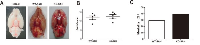
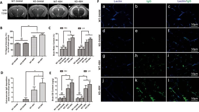
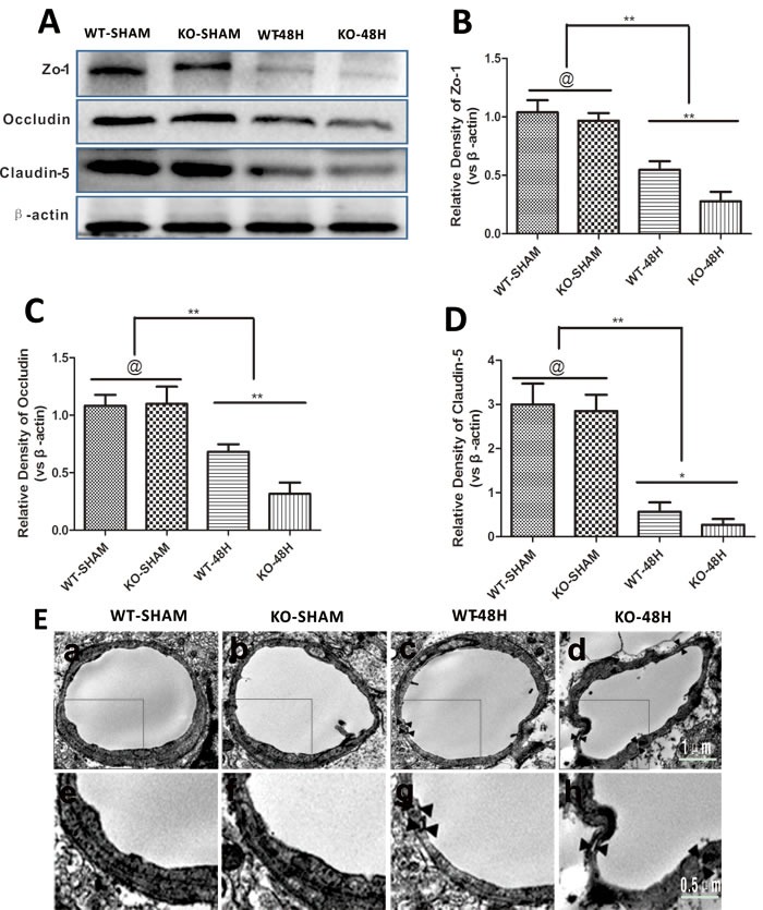
Apolipoprotein E (Apoe) genetic polymorphisms have been implicated in the long term outcome of subarachnoid haemorrhage (SAH), but little is known about the effect of Apoe on the early brain injury (EBI) after SAH. This study investigated the potential role of APOE in EBI post-SAH. Multiple techniques were used to determine the early BBB disruption in EBI post-SAH in a murine model using wild-type (WT) and Apoe-/- (KO) mice. Progressive BBB disruption (Evans blue extravasation and T2 hyperintensity in magnetic resonance imaging) was observed before the peak of endogenous APOE expression elevation at 48h after SAH. Moreover, Apoe-/- mice exhibited more severe BBB disruption charcteristics after SAH than WT mice, including higher levels of Evans blue and IgG extravasation, T2 hyperintensity in magnetic resonance imaging, tight junction proteins degradation and endothelial cells death. Mechanistically, we found that APOE restores the BBB integrity in the acute stage after SAH via the cyclophilin A (CypA)-NF-κB-proinflammatory cytokines-MMP-9 signalling pathway. Consequently, although early BBB disruption causes neurological dysfunctions after SAH, we capture a different aspect of the effects of APOE on EBI after SAH that previous studies had overlooked and open up the idea of BBB disruption as a target of APOE-based therapy for EBI amelioration research in the future.
Keywords: Apolipoprotein E; Pathology Section; blood-brain barrier; early brain injury; neuroinflamamation; subarachnoid hemorrhage.
Conflict of interest statement
The authors declare no conflicts of interest.
Figures


Similar articles
-
Inhibition of Blood-Brain Barrier Disruption by an Apolipoprotein E-Mimetic Peptide Ameliorates Early Brain Injury in Experimental Subarachnoid Hemorrhage.Transl Stroke Res. 2017 Jun;8(3):257-272. doi: 10.1007/s12975-016-0507-1. Epub 2016 Oct 31. Transl Stroke Res. 2017. PMID: 27796945
-
Deficiency of tenascin-C and attenuation of blood-brain barrier disruption following experimental subarachnoid hemorrhage in mice.J Neurosurg. 2016 Jun;124(6):1693-702. doi: 10.3171/2015.4.JNS15484. Epub 2015 Oct 16. J Neurosurg. 2016. PMID: 26473781
-
White Matter Injury After Subarachnoid Hemorrhage: Role of Blood-Brain Barrier Disruption and Matrix Metalloproteinase-9.Stroke. 2015 Oct;46(10):2909-15. doi: 10.1161/STROKEAHA.115.010351. Epub 2015 Sep 15. Stroke. 2015. PMID: 26374478 Free PMC article.
-
An apolipoprotein E receptor mimetic peptide decreases blood-brain barrier permeability following intracerebral hemorrhage by inhibiting the CypA/MMP-9 signaling pathway via LRP1 activation.Int Immunopharmacol. 2024 Dec 25;143(Pt 3):113007. doi: 10.1016/j.intimp.2024.113007. Epub 2024 Oct 31. Int Immunopharmacol. 2024. PMID: 39486173 Review.
-
Underlying Mechanisms and Potential Therapeutic Molecular Targets in Blood-Brain Barrier Disruption after Subarachnoid Hemorrhage.Curr Neuropharmacol. 2020;18(12):1168-1179. doi: 10.2174/1570159X18666200106154203. Curr Neuropharmacol. 2020. PMID: 31903882 Free PMC article. Review.
Cited by
-
Genetic underpinnings of cerebral edema in acute brain injury: an opportunity for pathway discovery.Neurosci Lett. 2020 Jun 21;730:135046. doi: 10.1016/j.neulet.2020.135046. Epub 2020 May 26. Neurosci Lett. 2020. PMID: 32464484 Free PMC article. Review.
-
Apolipoprotein E Deficiency Exacerbates Spinal Cord Injury in Mice: Inflammatory Response and Oxidative Stress Mediated by NF-κB Signaling Pathway.Front Cell Neurosci. 2018 May 23;12:142. doi: 10.3389/fncel.2018.00142. eCollection 2018. Front Cell Neurosci. 2018. PMID: 29875635 Free PMC article.
-
COG133 Attenuates the Early Brain Injury Induced by Blood-Brain Barrier Disruption in Experimental Subarachnoid Hemorrhage.J Healthc Eng. 2022 Jan 7;2022:4404039. doi: 10.1155/2022/4404039. eCollection 2022. J Healthc Eng. 2022. Retraction in: J Healthc Eng. 2023 Dec 6;2023:9793101. doi: 10.1155/2023/9793101. PMID: 35035834 Free PMC article. Retracted.
-
Apolipoprotein E Deficiency Aggravates Neuronal Injury by Enhancing Neuroinflammation via the JNK/c-Jun Pathway in the Early Phase of Experimental Subarachnoid Hemorrhage in Mice.Oxid Med Cell Longev. 2019 Dec 26;2019:3832648. doi: 10.1155/2019/3832648. eCollection 2019. Oxid Med Cell Longev. 2019. PMID: 31949876 Free PMC article.
-
Apolipoprotein E Exerts a Whole-Brain Protective Property by Promoting M1? Microglia Quiescence After Experimental Subarachnoid Hemorrhage in Mice.Transl Stroke Res. 2018 Dec;9(6):654-668. doi: 10.1007/s12975-018-0665-4. Epub 2018 Sep 17. Transl Stroke Res. 2018. PMID: 30225551
References
-
- Grunwald IQ, Kuhn AL, Schmitt AJ, Balami JS. Aneurysmal SAH: current management and complications associated with treatment and disease. The Journal of invasive cardiology. 2014;26:30–37. - PubMed
-
- Hop JW, Rinkel GJ, Algra A, van Gijn J. Case-fatality rates and functional outcome after subarachnoid hemorrhage: a systematic review. Stroke. 1997;28:660–664. - PubMed
-
- Johnston SC, Selvin S, Gress DR. The burden, trends, and demographics of mortality from subarachnoid hemorrhage. Neurology. 1998;50:1413–1418. - PubMed
-
- Huang J, van Gelder JM. The probability of sudden death from rupture of intracranial aneurysms: a meta-analysis. Neurosurgery. 2002;51:1101–1105. discussion 1105-1107. - PubMed
-
- Young AM, Karri SK, Helmy A, Budohoski KP, Kirollos RW, Bulters DO, Kirkpatrick PJ, Ogilvy CS, Trivedi RA. Pharmacologic Management of Subarachnoid Hemorrhage. World neurosurgery. 2015;84:28–35. - PubMed
MeSH terms
Substances
LinkOut - more resources
Full Text Sources
Other Literature Sources
Research Materials
Miscellaneous
