. 2016 Jul 27;11(7):e0158757.
doi: 10.1371/journal.pone.0158757. eCollection 2016.

Identification of a Monoclonal Antibody That Attenuates Antiphospholipid Syndrome-Related Pregnancy Complications and Thrombosis

Affiliations

Identification of a Monoclonal Antibody That Attenuates Antiphospholipid Syndrome-Related Pregnancy Complications and Thrombosis

Chieko Mineo et al. PLoS One. .

Abstract

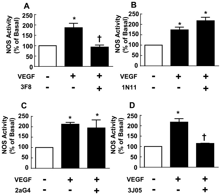
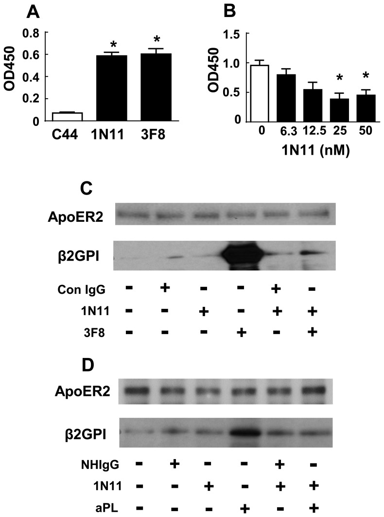
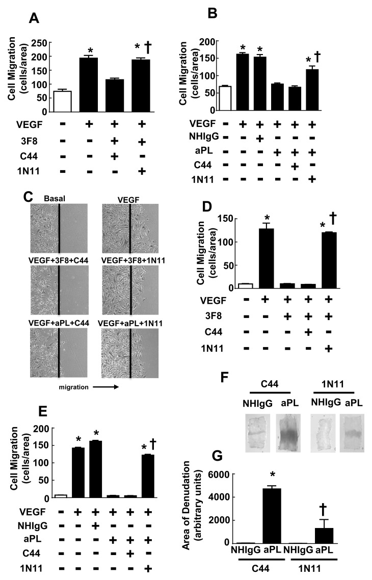
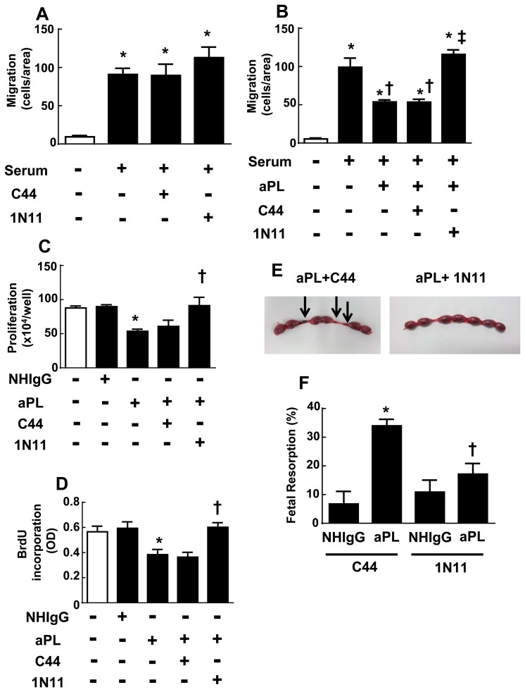
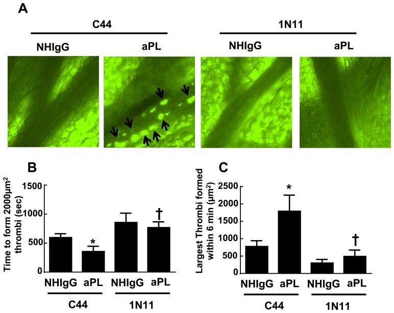
In the antiphospholipid syndrome (APS), patients produce antiphospholipid antibodies (aPL) that promote thrombosis and adverse pregnancy outcomes. Current therapy with anticoagulation is only partially effective and associated with multiple complications. We previously discovered that aPL recognition of cell surface β2-glycoprotein I (β2-GPI) initiates apolipoprotein E receptor 2 (apoER2)-dependent signaling in endothelial cells and in placental trophoblasts that ultimately promotes thrombosis and fetal loss, respectively. Here we sought to identify a monoclonal antibody (mAb) to β2-GPI that negates aPL-induced processes in cell culture and APS disease endpoints in mice. In a screen measuring endothelial NO synthase (eNOS) activity in cultured endothelial cells, we found that whereas aPL inhibit eNOS, the mAb 1N11 does not, and instead 1N11 prevents aPL action. Coimmunoprecipitation studies revealed that 1N11 decreases pathogenic antibody binding to β2-GPI, and it blocks aPL-induced complex formation between β2-GPI and apoER2. 1N11 also prevents aPL antagonism of endothelial cell migration, and in mice it reverses the impairment in reendothelialization caused by aPL, which underlies the non-thrombotic vascular occlusion provoked by disease-causing antibodies. In addition, aPL inhibition of trophoblast proliferation and migration is negated by 1N11, and the more than 6-fold increase in fetal resorption caused by aPL in pregnant mice is prevented by 1N11. Furthermore, the promotion of thrombosis by aPL is negated by 1N11. Thus, 1N11 has been identified as an mAb that attenuates APS-related pregnancy complications and thrombosis in mice. 1N11 may provide an efficacious, mechanism-based therapy to combat the often devastating conditions suffered by APS patients.


Conflict of interest statement

Competing Interests: RAB, XH and PET were consultants for Peregrine Pharmaceuticals, Inc., and their efforts on the project were partially funded by Peregrine Pharmaceuticals, Inc. YMS served as a speaker for Janssen for Xarelto, which is an oral anticoagulant, and for Alexion for Soliris, which is being studied for catastrophic APS. JES was a consultant for Alexion. These competing interests do not alter the authors' adherence to PLOS ONE policies on sharing data and materials. The authors have no additional declarations regarding possible competing interests.

Figures

Fig 1
Fig 1. Effect of monoclonal antibodies on eNOS activation by VEGF.
Bovine aortic endothelial cells were incubated with vehicle, monoclonal antibody (mAb) 3F8 (A, 2μg/ml), 1N11 (B, 10 μg/ml), 2aG4 (C, 10 μg/ml) or 3J05 (D, 10 μg/ml) for 30 min, and NOS activity was then determined in their continued absence or presence by quantifying [14C]-L-arginine to [14C]-L-citrulline conversion without (basal) or with added VEGF (100 ng/ml) over 15 min. (N = 3–6, mean±SEM, *p<0.05 vs. no VEGF, †p<0.05 vs. no mAb.).
Fig 2
Fig 2. Effect of monoclonal antibodies (mAb) on eNOS inhibition by 3F8 or aPL.
The impact of mAb’s 2aG4 (A, B, 200 μg/ml) or 1N11 (C-I, 200 μg/ml) on the antagonism of VEGF stimulation of eNOS by either 3F8 (A, C, H, 2 μg/ml) or aPL (B,D-G, I 100 μg/ml) was determined in bovine aortic endothelial cells (A-G) or human aortic endothelial cells (H, I). This was accomplished by quantifying [14C]-L-arginine to [14C]-L-citrulline conversion without (basal) or with added VEGF (100 ng/ml) over 15 min. Antibodies were present during 30 min preincubations and during the NOS activity incubations. Studies of 1N11 action on eNOS antagonism by aPL were performed with aPL from 4 different patients. (N = 3–6, mean±SEM, *p<0.05 vs. no VEGF, †p<0.05 vs. VEGF alone, ‡p<0.05 vs. no 1N11.).
Fig 3
Fig 3. Effect of 1N11 on 3F8 binding to β2-GPI, and on β2-GPI-ApoER2 complex formation induced by 3F8 or aPL.
(A) Binding of biotinylated monoclonal antibodies (C44, 1N11 and 3F8) to immobilized human β2-GPI (A) on phosphatidylserine-coated 96-well plates was evaluated (N = 4, mean±SEM, *p<0.05 vs. C44 control.) (B) Increasing concentrations of unlabeled 1N11 were added concurrently with biotinylated 3F8 (10 nM), and 3F8 binding to human β2-GPI was quantified (N = 6–8, *p<0.05 vs. no 1N11). (C) Endothelial cells were incubated in the absence or presence of control IgG or 3F8 (1 μg/ml), without or with 1N11 added (200 μg/ml) for 30 min, ApoER2 was immunoprecipitated, and the presence of ApoER2 and β2-GPI in the immunoprecipitates was evaluated by immunoblotting. (D) Coimmunprecipitation experiments were also performed evaluating β2-GPI and ApoER2 interaction in the absence or presence of NHIgG or aPL (100 μg/ml), without or with 1N11 added. Findings in C and D were confirmed in 2 independent experiments.
Fig 4
Fig 4. Effect of 1N11 on cultured endothelial cell migration impairment by 3F8 or aPL, and on aPL blunting of carotid artery reendothelialization in vivo.
(A) Bovine aortic endothelial cells (BAEC) were incubated without or with 3F8 (2 μg/ml) and either C44 or 1N11 (200 μg/ml), and cell migration induced by VEGF (100 ng/ml) over 16h was assessed by scratch assay. (B) BAEC were incubated with NHIgG or aPL (100 μg/ml) in the presence of C44 or 1N11 (200 μg/ml), and cell migration induced by VEGF (100 ng/ml) over 16h was assessed by scratch assay. (In A and B, N = 8, mean±SEM, *p<0.05 vs. no VEGF, †p<0.05 vs. no 1N11). (C-E) Human aortic endothelial cells (HAEC) were incubated with or without 3F8 (2 μg/ml) or with NHIgG versus aPL (100 μg/ml) in the presence of C44 or 1N11 (200 μg/ml), and cell migration induced by VEGF (100 ng/ml) over 16h was assessed by scratch assay. Representative images of migrating cells are shown in C. (In D and E, N = 5–10, mean±SEM, *p<0.05 vs. no VEGF, †p<0.05 vs. no 1N11). (F, G) Male C57Bl/6 mice (10–12 weeks old) were coinjected with NHIgG or aPL (100 μg/mouse) and either C44 or 1N11 (100 μg/mouse) 24 h before, on the day of carotid artery thermal injury, and 24 and 48 h after injury. Reendothelialization was assessed by evaluating intimal layer Evans blue dye incorporation 72h postinjury. (F) Representative images of the carotid artery intimal surface. (G) Summary data for the area of remaining denudation at 72 h, expressed in arbitrary units. (N = 4–5, *p<0.05 vs. NHIgG, †p<0.05 vs. C44).
Fig 5
Fig 5. Effect of 1N11 on aPL impairment of trophoblast migration and proliferation, and aPL-induced fetal resorption.
(A) The migration of HTR-8 SVneo trophoblasts was evaluated during 16h incubations in the absence or presence of serum (5%), without or with C44 or 1N11 (200 μg/ml) added. (N = 4–8. Mean±SEM, *p<0.05 vs. no serum.) (B) Serum-stimulated migration of HTR-8 SVneo trophoblasts was evaluated without or with added aPL (100 μg/ml), in the absence or presence of C44 or 1N11 (200 μg/ml). (N = 8–16, *p<0.05 vs. no serum. †<0.05 vs. no aPL, ‡p<0.05 vs. C44) (C,D) HTR-8 SVneo trophoblasts were incubated with NHIgG or aPL (100 μg/ml) in the presence of C44 or 1N11 (200 μg/ml) for 24 h, and cell number (C) or BrdU incorporation (D) was quantified. (N = 3–6, *p<0.05 vs. NHIgG, †p<0.05 vs. C44.) (E, F) Pregnant Balb/c mice (8–10 weeks old) were injected IP with aPL or NHIgG (10 mg/mouse) at day 8 and 12 of pregnancy, C44 or 1N11 (0.5 mg/mouse) was administered daily from day 8 to day 14, and fetal resorptions (as indicated by arrows in E) were evaluated at day 15. Fetal resorption rates (number of resorption sites/number of surviving fetuses + number of resorption sites) were calculated (N = 8–9, *p<0.05 vs. NHIgG, †p<0.05 vs. C44.).
Fig 6
Fig 6. Effect of 1N11 on thrombus formation induced by aPL in mice.
(A) Male C57BL/6 mice (5–6 weeks old) received NHIgG or aPL (100 μg/mouse) and either C44 or 1N11 (100 μg/mouse) by intraperitoneal injection. Twenty-four hours later thrombus formation (indicated by arrows) in mesenteric arterioles was evaluated by intravital microcopy. (B) Time required to form 2000 μm2 or larger thrombi. (C) Size of largest thrombi formed within 6 min. (N = 6–9, Mean±SEM, *p<0.05 vs. NHIgG, †p<0.05 vs. C44.) The movies corresponding to the images shown in (A) are available at https://figshare.com/s/7f9e28e40bff379c1035.

Similar articles

Cited by

References

    1. Meroni PL, Borghi MO, Raschi E, Tedesco F. Pathogenesis of antiphospholipid syndrome: understanding the antibodies. Nat.Rev.Rheumatol. 2011;7: 330–339. 10.1038/nrrheum.2011.52 - DOI - PubMed
    1. Ruiz-Irastorza G, Crowther M, Branch W, Khamashta MA. Antiphospholipid syndrome. Lancet. 2010;376: 1498–1509. 10.1016/S0140-6736(10)60709-X - DOI - PubMed
    1. Miyakis S, Lockshin MD, Atsumi T, Branch DW, Brey RL, Cervera R et al. International consensus statement on an update of the classification criteria for definite antiphospholipid syndrome (APS). J Thromb.Haemost. 2006;4: 295–306. - PubMed
    1. Giannakopoulos B, Krilis SA. The pathogenesis of the antiphospholipid syndrome. New England Journal of Medicine. 2013;368: 1033–1044. 10.1056/NEJMra1112830 - DOI - PubMed
    1. Levine JS, Branch DW, Rauch J. The antiphospholipid syndrome. N.Engl.J Med. 2002;346: 752–763. - PubMed

MeSH terms

Substances