. 2016 Jul 22;353(6297):389-94.
doi: 10.1126/science.aaf8818.

Accurate design of megadalton-scale two-component icosahedral protein complexes

Affiliations

Accurate design of megadalton-scale two-component icosahedral protein complexes

Jacob B Bale et al. Science. .

Abstract

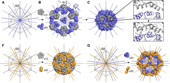
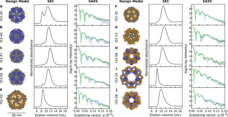
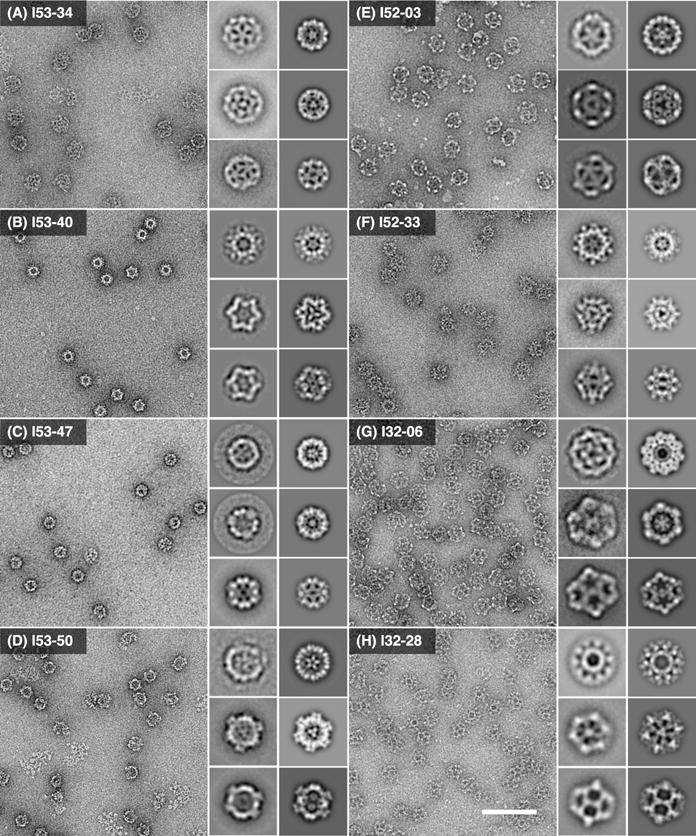
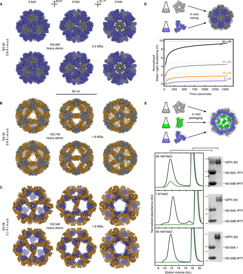
Nature provides many examples of self- and co-assembling protein-based molecular machines, including icosahedral protein cages that serve as scaffolds, enzymes, and compartments for essential biochemical reactions and icosahedral virus capsids, which encapsidate and protect viral genomes and mediate entry into host cells. Inspired by these natural materials, we report the computational design and experimental characterization of co-assembling, two-component, 120-subunit icosahedral protein nanostructures with molecular weights (1.8 to 2.8 megadaltons) and dimensions (24 to 40 nanometers in diameter) comparable to those of small viral capsids. Electron microscopy, small-angle x-ray scattering, and x-ray crystallography show that 10 designs spanning three distinct icosahedral architectures form materials closely matching the design models. In vitro assembly of icosahedral complexes from independently purified components occurs rapidly, at rates comparable to those of viral capsids, and enables controlled packaging of molecular cargo through charge complementarity. The ability to design megadalton-scale materials with atomic-level accuracy and controllable assembly opens the door to a new generation of genetically programmable protein-based molecular machines.


Figures

Fig. 1
Fig. 1. Overview of the design method and target architectures
(AE) A schematic of the design process illustrated with the I53 architecture. (A) An icosahedron is outlined with dashed lines, with the five-fold symmetry axes (grey) going through each vertex and three-fold symmetry axes (blue) going through each face of the icosahedron. (B)12 pentamers (grey) and 20 trimers (blue) are aligned along the 5-fold and 3-fold symmetry axes, respectively. Each oligomer possesses two rigid body DOFs, one translational (r) and one rotational (ω) that are systematically sampled to identify configurations (C) with a large interface between the pentamer and trimer (D) suitable for protein-protein interface design; only the backbone structure and beta carbons of the oligomers are taken into account during this procedure. (E) Amino acid sequences are designed at the new interface to stabilize the modeled configuration. (F) The I52 architecture comprises 12 pentamers (grey) and 30 dimers (orange) aligned along the five-fold and two-fold icosahedral symmetry axes. (G) The I32 architecture comprises 20 trimers (blue) and 30 dimers (orange) aligned along the three-fold and two-fold icosahedral symmetry axes.
Fig. 2
Fig. 2. Experimental characterization by size exclusion chromatography and small-angle X-ray scattering
Computational design models (left), SEC chromatograms (middle), and SAXS profiles (right) are shown for (A) I53-34, (B) I53-40, (C) I53-47, (D) I53-50, (E) I52-03, (F) I52-32, (G) I52-33, (H) I32-06, (I) I32-19, and (J) I32-28. Design models (shown to scale relative to the 30 nm scale bar) are viewed down one of the 5-fold symmetry axes with ribbon-style renderings of the protein backbone (pentamers are shown in grey, trimers in blue, and dimers in orange). Co-expressed and purified designs yield dominant SEC peaks near the expected elution volumes for the target 120-subunit complexes and X-ray scattering intensities (grey dots) that match well with profiles calculated from the design models (green). Alternative configurations of the designs, generated by translating and/or rotating the oligomeric building blocks in the design models about their aligned symmetry axes by +/− 10 Å and/or 20 degrees, respectively, generally fit worse with the SAXS data than the original design models (the range of values obtained from fitting the alternative configurations is shown with light blue shading).
Fig. 3
Fig. 3. Characterization of the designed materials by electron microscopy
Left: raw negative stain electron micrographs of co-expressed and purified (A) I53-34, (B) I53-40, (C) I53-47, (D) I53-50, (E) I52-03, (F) I52-33, (G) I32-06, and (H) I32-28. All raw micrographs shown to scale relative to 100 nm scale bar in panel (H). Insets: experimentally computed class averages (roughly corresponding to the five-fold, three-fold, and 2-fold icosahedral symmetry axes; left) along with back projections calculated from the design models (right). Each inset box width corresponds to 55 nm.
Fig. 4
Fig. 4. Crystal structures, assembly dynamics, and packaging
Design models (top) and X-ray crystal structures (bottom) of (A) I53-40, (B) I52-32, and (C) I32-28. Views shown to scale along the 3-fold, 2-fold, and 5-fold icosahedral symmetry axes. Pentamers shown in grey, trimers blue, and dimers orange. R.m.s.d.s are between crystal structures and design models over all backbone atoms in all 120 subunits. (D) In vitro assembly dynamics of I53-50. (Top) Schematic illustration. (Bottom) Normalized static light scattering intensity (detector voltage, solid lines) plotted over time after mixing independently expressed and purified variants of the I53-50 trimer and pentamer in a 1:1 molar ratio at final concentrations of 8, 16, 32, or 64 μM each (blue, orange, grey, and black lines, respectively). Intensities measured from SEC-purified assembly at 8, 16, 32, or 64 μM concentrations indicated with dashed horizontal lines and used as the expected endpoint of each assembly reaction. The midpoint of each reaction is marked with a dashed vertical line. (E) Encapsulation of supercharged GFP in a positively charged I53-50 variant. (Top) Schematic illustration. (Bottom) Superose 6 chromatograms and SDS-PAGE analysis of packaging/assembly reactions performed in buffer containing: (Top Panel) 65 mM NaCl, (Middle Panel) 1 M NaCl, or (Bottom Panel) 65 mM NaCl with a trimer variant without mutations to positively charged residues. In each case, the same buffer used in the packaging/assembly reaction was also used during SEC. Absorbance measurements at 280 nm (black) and 488 nm (green) are shown. Each SEC chromatogram was normalized relative to the 280 nm peak near 12 mL elution volume. Locations of 37, 25, 20, and 15 kDa molecular weight markers on SDS-PAGE gels are indicated by horizontal lines.

References

    1. Lai YT, King NP, Yeates TO. Principles for designing ordered protein assemblies. Trends Cell Biol. 2012;22:653–661. - PubMed
    1. King NP, Lai YT. Practical approaches to designing novel protein assemblies. Curr Opin Struct Biol. 2013;23:632–638. - PubMed
    1. Padilla JE, Colovos C, Yeates TO. Nanohedra: using symmetry to design self assembling protein cages, layers, crystals, and filaments. Proc Natl Acad Sci U S A. 2001;98:2217–2221. - PMC - PubMed
    1. Ringler P, Schulz GE. Self-assembly of proteins into designed networks. Science. 2003;302:106–109. - PubMed
    1. Raman S, Machaidze G, Lustig A, Aebi U, Burkhard P. Structure-based design of peptides that self-assemble into regular polyhedral nanoparticles. Nanomedicine. 2006;2:95–102. - PubMed

Publication types

MeSH terms